Monthly Archives: November 2018

Schistosoma haematobium Infection and the Gynecological and Obstetric Effects on Girls and Women: A Short Note

DOI: 10.31038/IGOJ.2018131

Short Commentary

Schistosoma haematobium (blood fluke) has a complex life cycle, involving freshwater snails as intermediate hosts and humans as definitive host.

Humans are passive victims of this parasite, not as occurs with other parasites that have insects as vectors that are actively introduced when the insects have a blood meal, when humans enter into fresh water they are infected from snails parasitized with S. haematobium in the form of numerous infective cercariae. These cercariae swim in the water and penetrate the human skin.. After penetration, the cercariae are known as schistosomulae. These migrate and develop into mature adult schistosome worms in and around the vesical plexus, and occasionally in the rectal region, the mesenteric portal system and ectopic sites [1]. Then, the parasite can have several negative effects on female health, and here our objective is to alert medical practitioners of the gynecological and obstetric effects on girls and women. Considering the mode of transmission of S. haematobium, we must recognize the fact that the high risk of infection occurs in regions where the human population is exposed to snails where there is a lack of a water canalization system, and basic sanitation. In general, these are in conditions of severe poverty. In Africa where these situations are frequent schistosomiasis caused by S. haematobium is endemic, and girls and women are more exposed to infection because of their domestic work, bathing and fishing and agricultural, activities that in several African countries are especially attributed to girls and women.. Accordingly [2] women suffer considerably from female genital schistosomiasis that causes infertility, preterm labor, anemia, menstrual disorders, and dyspareunia. Also infected pregnant women have low birth weight infants, and an increased infant and maternal mortality rates. In Africa approximately 10 million women have schistosomiasis in pregnancy [3–5]. Schistosomiasis has been detected in the placenta and in newborns thus confirming congenital infection. Data suggest that infected women have a higher rate of spontaneous abortions and a higher risk for ectopic pregnancies [6,7].

Concerning schistosomiasis treatment [8], Praziquantet (PZQ) was made available in 1979 and has been the mainstay of schistosomiasis control programs for decades. PZQ was never studied in pregnant and lactating women and was, therefore, designated as a “pregnancy class B” drug. In this class, the drugs are presumed to be safe based on animal studies but lack safety data from studies in pregnant women. In practice, this has led to the withholding of treatment for pregnant and lactating women in most schistosomiasis endemic countries.

In [8] we have a good and recent article on PZQ for the treatment of schistosomiasis during human pregnancy. The authors cite in their conclusion: “based on recent evidence of the safety of praziquantel in human pregnancy, we recommend that, in areas with endemic schistosomiasis, women of reproductive age, including pregnant and breastfeeding women, be treated with PZQ either individually, in local antenatal clinics, or in mass drug administration”.

Considering our personal knowledge acquired during our scientific activities on S. haematobium / schistosomiasis carried out in Guinea Bissau [9], we found that many girls and women had a history of menstrual disorders, infertility and spontaneous abortions.

Final conclusion: in view of the negative effects of S. haematobium on physical health here related and, certainly, on psychological health of girls and women, treatment with PZD may be ministered to pregnant and lactating girls and women.

Keywords

Schistosoma haematobium; Girls; Women; Gynecology; Obstetric; Praziquantel

References

  1. Eddington GM, Nwabuelo I, Junaid TA (1975) The pathology of schistosomiasis in Ibadan Nigeria, with special reference to the appendix, brain, pancreas and genital organs. Trans R Soc trop Med Hyg 69: 153–156.
  2. Nour NM (2010) Schistosomiasis: health effects on women. Rev Obstet Gynecol winter; 3(1): 28–32.
  3. Friedman JF, Mital P, Kanzaria HK, Olds GR, Kurtis JD (2007) Schistosomiasis and pregnancy. Trends in Parasitology 23(4): 159–164.
  4. Slegrist D, Slegrist-Obimpeh P (1992) Schistosoma haematobium infection in pregnancy. Acta Trop 50: 317/322.
  5. Helling-Giese G, Kjetland EF, Gundersen SG et al (1996). Schistosomiasis in woman: manifestations in the upper reproductive tract. Acta Trop 62: 225–238.
  6. Bahrain S, Alatassi H, Slone SP, O’Connor DM (2006) Tubal gestation and schistosomiasis: a case report. J Reprod Med 51: 595–598.
  7. Laxma VV, Adamson B, Mahmood T (2008). Recurrent ectopic pregnancy due to Schistosoma haematobium. J Obstet Gynaecol 461–462.
  8. Friedman JF, Olveda RM, Mirochnich MH, Bustinduy AL, Elliott (2018). Praziquantel for the treatment has of schistosomiasis during human pregnancy. Bull World Health Organ. Jan 1; 96(1): 59–61.
  9. Grácio MA, Nhaque AT, Rollinson D (1987-1991). Schistosomiasis in Guinea Bissau, contract TS2 -0205- Science and Technology for Development, Second program, European Commission, vol 2 – Parasitology: 239–247.

Off-Pump Myocardial Revascularization from the Beginning till Now

DOI: 10.31038/JCCP.2018114

Editorial

Surgical myocardial revascularization was introduced in the cardiovascular surgical practice late, just after two decades of experience in congenital heart disease and valvular dysfunctions. The landmark of direct myocardial revascularization was the possibility to access the coronary arteries through coronary angiography developed by Mason Jones in Cleveland Clinic in early sixties [1].

The possibility to identify the presence and severity of coronary obstructions was the birth of modern coronary surgery and more than this, to evaluate and select really effective surgical techniques.

Of several indirect surgical procedures made in the past only the Vineberg technique showed some efficiency [2].

The modern era of direct myocardial revascularization started with the saphenous vein bypass grafts, with massive information coming mainly from Cleveland Clinic regarding diagnosis, selection of patients and technical challenges [3].  Nevertheless, Garret utilized years before a bypass graft instead of endarterectomy in 1962 and published a successful follow-up in 1973. It is important to remind that at that time he didn´t have information of cinecoronarygraphy and did this option during a valve operation [4].

Although coronary arteries are located on the surface of the heart and it is not necessary to open cardiac chambers, surgeons started to use cardiopulmonary bypass to make the graft anastomosis due to development of myocardial protection methods, familiarity with extracorporeal circuit and mainly due to perform a difficult anastomosis in a beating field.

Nevertheless, the idea to make a coronary artery anastomosis in a beating heart was applied before the concept of doing a saphenous vein graft bypass under extracorporeal circulation and cardiac arrest.

Goetz et al 1961 [5] did a direct myocardial revascularization utilizing the mammary artery and mechanical suture with tantalum ring.

Vasilii Kolesov and Potashov [6,7] published in Russia (1965) and later in United States (1967) their experience with mammary artery to left anterior descending coronary artery in a beating heart through a left thoracotomy and in few cases with mechanical suture. It is interesting to observe that he didn´t have informations about the coronary arteries and made the operation only in clinical basis.

Years later Trapp & Bisarya [8] in Canada and in the same year Ankeney [9] in Western Reserve – Cleveland independently reported the first series of patients with acceptable results. This alternative of myocardial revascularization did not have acceptance due to technical difficulties and the concept that it was not possible to occlude a coronary artery even for a few minutes without causing a myocardial infarction. Due to this fact complicated distal perfusion devices were necessary introducing more difficulties to perform coronary anastomosis.

Only years later Buffolo et al [10] and independently Benetti [11] published a consecutive series of patients with saphenous or mammary artery grafts to left anterior descending, diagonal and right coronary arteries, calling attention to feasibly and safety of this alternative of myocardial revascularization. Technical maneuvers were described to facilitate anastomosis as position of the table, anesthesia expertise and utilization of pharmacological “stabilizers” like verapamil to reduce heart rate, oxygen consumption and arterial pressure.

During the next decade some papers tried to demonstrate the advantages of myocardial revascularization without pump, but did not have wide acceptance by local and international community [12–18].

Despite the evidence of the feasibility with good results, the confort to perform a conventional coronary artery bypass in an arrested heart and the concerns about the quality of anastomosis led to only isolated experiences for many years, and sometimes with strong criticisms in editorials like: “Look ma no hands!” or “myocardial revascularization without pump, a benefit or a risk for the patient”.

The development and persistence in performing off-pump coronary operation by pioneer groups culminated with the concept proposed by Benetti [19] to make a mammary-artery-LAD anastomosis in a beating heart through a mini-left thoracotomy. This idea was presented in a meeting in Rome in 1994 and received the name of MIDCAB (Minimally Invasive Direct Coronary Artery Bypass). The clinical experience was disseminated all around the world by Benetti and popularized by Calafiore et al [20], among others, with the name of “Last operation” (left anterior small thoracotomy).

This idea was strongly attractive and was in our opinion the key to the interest in beating heart surgery. Many groups started to learn how to operate without pump and discovered that was really possible to achieve good quality anastomosis even more, utilizing sternotomy.

Curiously in 1982, we collected only 5 direct publications in off-pump coronary surgery, in 1992,18 and in 1998 an explosion of 18.423 papers in the subject.

With the widespread application of beating heart coronary surgery many important contributions were made like the concept of hybrid approach and the use of stabilizers [21–23].

In the hybrid approach, we use a mammary-artery-left anterior descending anastomosis (LAD) with minimal left thoracotomy (MIDCAB) and before or after, percutaneous angioplasty to other coronary arteries combining the five star treatment of the LAD with a minimally invasive treatment of multivessel coronary artery disease [24–26].

The stabilizers by compression or suction like OCTOPUS® facilitate the anastomosis making a regional “cardiac arrest”.   It can be used with devices like “STARFISH”® that put the “apex cordis” up to permit visibility of marginal branches of circumflex artery. The use of the stabilizers was very important to get better quality anastomosis and now they are essential in off pump coronary surgery.

In the following decade, we can observe a lot of contributions  and randomized controlled trials comparing off-pump and on-pump surgery regarding mortality, morbidity, inflammatory response, patency rates, stroke, blood transfusion, costs, results in high risk patients among others [27–32].

Off-pump coronary surgery was studied under various aspects: advantages and criticisms and opinion makers like Thomas Salerno, Michael Mack, David Taggart, R. Ascione, Gianni Angelini, John Puskas, Antonio Calafiore [33–39] among others established the main concepts, advantages and disadvantages of this alternative of myocardial revascularization.

The different results regarding patency rates and benefits of the off-pump technique in our opinion is mainly due to improper training, selection of cases and surgical skills.

Although some groups are now performing 90% or even 100% of the operations off-pump, we believe that is difficult to achieve a 100% application of the technique. In our experience, patients with hypertrophic left ventricles, diffuse athermanous coronary arteries that sometimes need endarterectomy intramyocardial coronary arteries among others need the conventional approach, and the key to avoid unacceptable conversion rates is the proper preoperative selection of patients.

In retrospective analysis of our experience, we think that off-pump myocardial revascularization have strong evidences of advantages regarding reduction in mortality rates, reduction in stroke, lesser major postoperative complications, shorter hospital stay and lower costs.  The controversies that we observe in the literature regarding mortality and morbidity are mainly due to excess of enthusiasm trying to apply the technique to all patients, untrained surgeons and selection of patients.

It is very difficult to demonstrate advantages in low risk patients [40]. The differences appears in patients with co-morbidities; worse the patient, better the outcome.

Recent large international randomized trials did show advantages of off-pump coronary surgery compared with conventional revascularization for high risk patients with important co-morbidities like previous CVA, renal insufficiency, pulmonary insufficiency or porcelain aorta. Off-pump surgery in the hands of well trained teams offers a reduced risk regarding mortality and mobility [41–43]

In recent years it was demonstrated that it is possible to perform myocardial revascularization using a robotic system with closed chest in a selected group of patients (TECAB) Totally Endoscopic Coronary Artery Bypass.

This technique using port access and robotics represents the maximum application of a minimally invasive concept using the technology of the new millennium and the applicability and results will be analysed in the next years.

Comparative studies between off-pump coronary surgery and angioplasties will be necessary to stablish new strategies in the management of coronary artery disease because the parameters we have now compares conventional surgery and percutaneous intervention in the majority of randomized controlled trials.

In summary, in our long and large experience with CABG without CPB, the indications for operation with this method has been identified; the method can be used in approximately 25% of patients undergoing coronary revascularization. Arterial conduits can be used, and the patency rate is similar to that of conventional techniques. The mortality rate is acceptably low, and complication rates were lower compared with conventional techniques. In selected cases, the procedure is cost-effective due to lower use of hospital resources in the operating room, intensive care unit, and ward. The continuing use of this technique of coronary artery surgery is therefore justified.

References

  1. Sones FM Jr, Shirey EK (1962) Cine coronary arteriography. Mod Concepts Cardiovasc Dis 31: 735–738. [crossref]
  2. Vineberg AM (1946) Development of an anastomosis between the coronary vessels and a transplanted internal mammary artery. Can Med Assoc J  55: 117–119.
  3. Favaloro RG (1968) Saphenous vein autograft replacement of severe segmental coronary artery occlusion: operative technique. Ann Thorac Surg Apr;5: 334–339.
  4. Garrett HE, Dennis EW, DeBakey ME (1996) Aortocoronary bypass with saphenous vein graft. Seven-year follow-up. 1973. JAMA 276: 1519–1520. [crossref
  5. Goetz RH, Rohman M, Haller JD, et al. (1961) Internal mammary-coronary artery anastomosis. A nonsuture method employing tantalum rings. J Thorac Cardiovasc Surgm41: 378–386
  6. Kolesov VI, Potashov LV (1965) [Surgery of coronary arteries]. Eksp Khir Anesteziol 10: 3–8. [crossref]
  7. Kolesov VI (1967) Mammary artery-coronary artery anastomosis as method of treatment for angina pectoris.  J Thorac Cardiovasc Surg 54: 535–544
  8. Trapp WG, Bisarya R (1975) Placement of coronary artery bypass graft without pump oxygenator. Ann Thorac Surg 19: 1–9. [crossref]
  9. Ankeney JL (1975) Editorial: To use or not to use the pump oxygenator in coronary bypass operations. Ann Thorac Surg 19: 108–109. [crossref]
  10. Buffolo E, Andrade JCS, Succi JF, et al (1982) Revascularização direta do miocárdio sem circulação extracorpórea. descrição da técnica e resultados iniciais. Arq Bras Cardiol  38: 365–373
  11. Benetti FJ (1985) Direct coronary surgery with saphenous vein bypass without either cardiopulmonary bypass or cardiac arrest J Cardiovasc Surg (Torino) 26: 217–22.
  12. Buffolo E, Andrade JC, Succi J, et al (1985) Direct myocardial revascularization without cardiopulmonary bypass. Thorac Cardiovasc Surg 33: 26–29
  13. Buffolo E, Andrade JC, Branco JN, et al (1990) “Myocardial revascularization without extracorporeal circulation. seven-year experience in 593 cases.”.Eur J Cardiothorac Surg 4: 504–507
  14. Buffolo E, De Andrade CS, Branco JN, et al (1996) Coronary artery bypass grafting without cardiopulmonary bypass. Ann Thorac Surg 61: 63–66
  15. Archer R, Ott DA, Parravicini R, Cooley DA, Reul GJ, et al. (1984) Coronary artery revascularization without cardiopulmonary bypass. Tex Heart Inst J 11: 52–57. [crossref]
  16. Laborde F, Abdelmequid I, Piwnica A (1989) Aortocoronary bypass without extracorporeal circulation: why and when? Eur J Cardiothorac Surg 3: 152–154
  17. Rivetti LA, Gandra SM (1997) Initial experience using an intraluminal shunt during revascularization of the beating heart. Ann Thorac Surg 63: 1742–1747
  18. Lima RC, Soares M A, Lobo FJG, et al (2003) Resultados cirúrgicos na revascularização do miocárdio sem circulação extracorpórea. analise de 3.410 pacientes.  Rev. Bras. Cir. Cardiovascular18: 261–267
  19. Benetti FJ, Ballester C, Sani G, Doonstra P, Grandjean J (1995) Video assisted coronary bypass surgery. J Card Surg 10: 620–625. [crossref]
  20. Calafiore AM, Giammarco GD, Teodori G, et al (1996) Left anterior descending coronary artery grafting via left anterior small thoracotomy without cardiopulmonary bypass. Ann Thorac Surg 61: 1658–1663
  21. Borst C, Jansen EW, Tulleken CA, et al  (1996) Coronary artery bypass grafting without cardiopulmonary bypass and without interruption of native coronary flow using a novel anastomosis site restraining device (“octopus”) J Am Coll Cardiol. 27: 1356–1364
  22. Izzat MB, Yim AP (1997) Cardiac stabilizer for minimally invasive direct coronary artery bypass. Ann Thorac Surg 64: 570–571. [crossref]
  23. Shennib H, Lee AG, Akin J (1997) Safe and effective method of stabilization for coronary artery bypass grafting on the beating heart. Ann Thorac Surg 63: 988–992. [crossref]
  24. Angelini GD, Wilde P. Salerno T. A. et al (1966) Integrated left small thoracitomy and angioplasty for multivessel coronary artery revascularization Lancet 347: 757–758
  25. Wittwer T, Cremer J, Boonstra P, Grandjean J, Mariani M, et al. (2000) Myocardial “hybrid” revascularisation with minimally invasive direct coronary artery bypass grafting combined with coronary angioplasty: preliminary results of a multicentre study. Heart 83: 58–63. [crossref]
  26. Riess FC, Bader R, Kremer P, Kühn C, Kormann J, et al. (2002) Coronary hybrid revascularization from January 1997 to January 2001: a clinical follow-up. Ann Thorac Surg 73: 1849–1855. [crossref]
  27. Cleveland JC Jr, Shroyer AL, Chen AY, et al (2001)  Off-pump coronary artery bypass grafting decreases risk-adjusted mortality and morbidity. Ann Thorac Surg 72: 1282–1288.
  28. Al-Ruzzeh S, Ambler G, Asimakopoulos G, et al (2003) Off-pump coronary artery bypass (OPCAB) surgery reduces risk-stratified morbidity and mortality: a United Kingdom multi-center comparative analysis of early clinical outcome.  Circulation 108: 115–120
  29. Sergeant P, Wouters P, Meyns B, et al (2004) Opcab versus early mortality and morbidity: an issue between clinical relevance and statistical significance. Eur J Cardiothorac Surg 25: 779–785
  30. Brasil LA, Gomes WJ, Salomão R, Buffolo E (1998) Inflammatory response after myocardial revascularization with or without cardiopulmonary bypass. Ann Thorac Surg 66: 56–59
  31. Ascione R, Lloyd CT, Underwood MJ, et al (2000) Inflammatory response after coronary revascularization with or without cardiopulmonary bypass. Ann Thorac Surg 69: 1198–1204.
  32. Edmunds LH Jr (1998) Inflammatory response to cardiopulmonary bypass. Ann Thorac Surg 66: 12–16. [crossref]
  33. Mack MJ (2000) Beating heart surgery for coronary revascularization: is it the most important development since the introduction of heart lung machine? Ann Thorac Surg 70: 1779–1781
  34. Salerno T, Ricci M, Karamanoukian HL et al (2001) Beating heart coronary artery surgery” Futura Publishing Company Inc 2001
  35. Mack MJ, Pfister A, Bachand D, et al (2004) Comparison of coronary bypass surgery with and without cardiopulmonary bypass in patients with multivessel disease. J Thorac Cardiovasc Surg 127(1): 167–173
  36. Calafiore AM, Di Mauro M, Canosa C, et al (2003) Myocardial revascularization with and without cardiopulmonary bypass: advantages, disadvantages and similarities.” Eur J Cardiothorac Surg 24: 953–960
  37. Angelini GD, Taylor FC, Reeves BC, Ascione R (2002) Early and midterm outcome after off-pump and on-pump surgery in Beating Heart Against Cardioplegic Arrest Studies (BHACAS 1 and 2): a pooled analysis of two randomised controlled trials. Lancet 359: 1194–1199. [crossref]
  38. Puskas JD, Williams WH, Duke PG, et al (2003) Off-pump coronary artery bypass grafting provides complete revascularization with reduced myocardial injury, transfusion requirements, and length of stay: a prospective randomized comparison of two hundred unselected patients undergoing off-pump versus conventional coronary artery bypass grafting. J Thorac Cardiovasc Surg 125: 797–808
  39. Taggart DP1, Westaby S (2001) Neurological and cognitive disorders after coronary artery bypass grafting. Curr Opin Cardiol 16: 271–276. [crossref]
  40. Gerola LR, Buffolo E, Jasbik W, Botelho B, Bosco J, et al. (2004) Off-pump versus on-pump myocardial revascularization in low-risk patients with one or two vessel disease: perioperative results in a multicenter randomized controlled trial. Ann Thorac Surg 77: 569–573. [crossref]
  41. Diegeler A, Börgermann J, Kappert U, Breuer M, Böning A, et al. (2013) Off-pump versus on-pump coronary-artery bypass grafting in elderly patients. N Engl J Med 368: 1189–1198. [crossref]
  42. Lamy A, Devereaux PJ, Prabhakaran D, Taggart DP, Hu S, et al. (2013) Effects of off-pump and on-pump coronary-artery bypass grafting at 1 year. N Engl J Med 368: 1179–1188. [crossref]
  43. Sá MP, Ferraz PE, Escobar RR, Martins WN, Lustosa PC, et al. (2012) Off-pump versus on-pump coronary artery bypass surgery: meta-analysis and meta-regression of 13,524 patients from randomized trials. Rev Bras Cir Cardiovasc 27: 631–641. [crossref]

First Fabulous Fifty – An Initial Experience of Dulaglutide from a Tertiary Care Centre in Eastern India

DOI: 10.31038/EDMJ.2018244

Abstract

Objective: This retrospective single centred real world observational study was undertaken with the aim to introspect the glycaemic control, weight loss, changes in lipid parameters, adverse events and treatment adherence with Dulaglutide therapy.

Methodology: Single centered, retrospective, real world, observational study conducted on subjects taking liraglutide for a mean duration of 41 weeks in the endocrine out-patient department.

Results: Data of 45 subjects were available. Mean age was 46.67 ± 5.53years. Glycosylated haemoglobin (HbA1c) significantly decreased from 8.68 ± 0.43% at baseline to 7.58 ± 0.19% at end of therapy. Body weight significantly reduced from 74.2 ± 8.07 kg at baseline to 69.27 ± 4.74kg at end of therapy and BMI significantly declined from 33.06 ± 4.5 to 30.09 ± 0.93 at end of therapy respectively. Nausea, vomiting and diarrhoea (15.55%) were the major adverse events noted in the study. Only one patient developed acute pancreatitis (2.22%).

Conclusion: Treatment with Dulaglutide resulted in clinically meaningful HbA1c, FPG and weight reductions. The overall safety profile is consistent with the GLP-1 receptor agonist class. However, Dulaglutide did not show statistically greater reduction of glycaemic parameters in the subset of Indian patients compared to RCT data of Western population.

Keywords

Dulaglutide, obesity, Indian, type 2 diabetes

Introduction

Glucagon-like peptide-1 (GLP-1) agonists act at GLP-1 receptors in pancreatic beta cells to increase glucose-dependent insulin secretion, in pancreatic alpha cells to decrease glucagon release and slow gastric emptying. Over the years, glucagon-like peptide-1 receptor agonists (GLP-1 RAs) have become integral in diabetes management as demonstrated by various publications from India [1–7]. Short-acting GLP-1 RAs requires either a once-daily (e.g. liraglutide) or twice-daily dosing (e.g., exenatide and lixisenatide). Studies published as back as 2005 from India by Vijan et. Al [8]. Showed that the injection burden was definitely an issue to be considered. When adherence to injectable treatment was looked into, the increased number of injection burden was found to be responsible for missed doses and non-adherence to treatment (GAAP study) [9]. Dulaglutide is longer acting GLP-1RA for the treatment of type 2 diabetes mellitus (T2D) and requires once-weekly dosing [10]. Hence the launch of Dulaglutide since march 2016 in India, the novel once weekly GLP1 RA was an welcome step and expected to increase the adherence to GLP1 RA treatment. However, adverse effects if any with one shot of the weekly once Dulaglutide would carry on for the entire week relentlessly. This retrospective single centred real world observational study was undertaken with the aim to introspect the glycaemic control, weight loss, changes in lipid parameters, adverse events and treatment adherence with Dulaglutide therapy.

Materials and Methods

This retrospective real world observational study was conducted in the Endocrinology Department of KPC Medical College and Hospital. It is a 700 bedded tertiary care hospital, situated in the southern fringes of the city of Kolkata, in the eastern part of India. The Endocrine out-patient database was frisked to tease out the initial 50 patients who were prescribed Dulaglutide over and above standard of care (with the exception that DPP 4 inhibitors if any on board) and weren’t lost to follow-up thereafter irrespective of the fact whether they were able to initiate or carry on Dulaglutide therapy continuously or not. No patients with e GFR <30, family history of medullary carcinoma of the thyroid and history of pancreatitis were offered the Dulaglutide as a standard of care of the Department.

The inclusion and exclusion criteria used while selecting the cohort of patients were as follows:

Inclusion Criteria:

  1. Adult type 2 diabetes between 18–75 year age
  2. HbA1C >= 7% and < 11% on a combination of OAD ± insulin
  3. First 50 patients to be prescribed Dulaglutide therapy and who came for a second follow up irrespective of whether he/she had started Dulaglutide.

Exclusion Criteria:

  1. Patients who were initially prescribed Dulaglutide but were lost to follow up after 1st visit
  2. Pregnancy
  3. Hospitalisation during follow-up

Statistical Analysis

Descriptive statistical analysis has been carried out in the present study. Results on continuous measurements are presented on Mean ± SEM and results on categorical measurements are presented in Number (%). Significance is assessed at a level of 5 %.

The following assumptions on data are made.

Assumptions:

  1. Cases of the samples should be independent.
  2. The populations from which the samples are drawn have the same variance (or standard deviation).
  3. The samples drawn from different populations are random.

Normality of data tested by Anderson Darling test, Shapiro-Wilk, Kolmogorov-Smirnoff test and visually by QQ plot. Paired t-test has been used to find the significance of study parameters within groups of patients measured on two occasions.

Statistical software: The Statistical software namely SAS (Statistical Analysis System) version 9.2 for windows, SAS Institute Inc. Cary, NC, USA and Statistical Package for Social Sciences (SPSS Complex Samples) Version 21.0 for windows, SPSS, Inc., Chicago, IL, USA were used for the analysis of the data and Microsoft word and Excel have been used to generate graphs and tables.

Results and Analysis

In the present analyses, a total of 50 patients with T2D were included out of which 45 did actually initiate the drug as was revealed in the first follow-up visit. Patient numbers for gender, age, height, BMI, duration of diabetes, baseline blood pressure, FPG, PPPG, HbA1c, Cholesterol, HDL, LDL, TG and duration of follow up are listed in Table 1. Out of 45, 24 were female and 21 were male having a mean age of 46.67 ± 5.53 years .The patients had a mean height, mean body weight of 74.2 ± 8.07 kg and mean BMI of 33.06 ± 1.47 kg /m2. The mean FPG was 169.18 ± 11.38 mg/dl, PPPG was 222.46 ± 23.77 mg/dl and mean Hba1c was 8.68+- 0.43% before the initiation of Dulaglutide. The baseline demographic and clinical characteristics of the study subjects are enumerated in (Table 1). Post analysis it was revealed that the mean follow up period for the 45 patients who ultimately initiated dulaglutide therapy was 41.2 ± 11.71 weeks.

Table 1. Baseline Characteristics of the Patients (N = 45)

Demographic & Clinical Profile

Values

Male, n (%)

21 (46.67)

Female, n (%)

24 (53.33)

Age(years), Mean ± SEM

46.67 ± 5.53

Height (centimeters), Mean ± SEM

161.63 ± 11.42

Body weight (Kg), Mean ± SEM

74.2 ± 8.07

SBP (mmHg), Mean ± SEM

133.68 ± 12.11

DBP (mmHg), Mean ± SEM

84.03 ± 10.51

BMI (kg/m2), Mean ± SEM

33.06 ± 1.47

BMI – 23 -29.9

22 (48.89%)

BMI – 30–34.9

11 (24.44%)

BMI – 35–39.9

11 (24.44%)

BMI – ≥ 40

1 (2.22%)

FPG (mg/dL), Mean ± SEM

169.18 ± 11.38

PPPG (mg/dL), Mean ± SEM

222.46 ± 23.77

HbA1c(%), Mean ± SEM

8.68 ± 0.43

Total Cholesterol (mg/dL), Mean ± SEM

165.68 ± 6.23

LDL- Cholesterol (mg/dL), Mean ± SEM

115.06 ± 27.2

HDL- Cholesterol (mg/dL), Mean ± SEM

42.35 ± 1.70

Triglycerides (mg/dL), Mean ± SEM

189.87 ± 12.35

Duration of follow-up (weeks), Mean ± SEM

41.2 ± 11.71

HbA1c, FBG reductions and weight changes

All the glycaemic parameters viz. FPG, PPPG and HbA1C had statistically significant reductions, with the respective p values achieved being 0.044, 0.018 and 0.032 during the study period. FPG was reduced by 31.14 ± 0.17 mg/dl, PPPG was reduced by 53.02 ± 10.52 mg/dl and HBA1C was also reduced by 1.10 ± 0.24%. (Table 2) In this small subgroup of patients 22.2% achieved a target HBA1C of less than 7% and 55.56% achieved a target of less than 7.5%, which is a significant proportion considering the fact that the average HBA1C of Indian Diabetic patients undergoing treatment is far higher than this (18). 26.67% of patients were able to achieve a reduction of greater than 1% HBA1C , 17.78% achieved a reduction between 0.5%-1.0%, however interesting is the fact that 37.78 % showed no change in HBA1C and 13.33 % were showing an increased HBA1C than that at baseline (Table 3, 4).

Table 2. Change in study parameters during the follow-up period, (N = 45)**

Parameter

Baseline
Mean ± SEM

Follow-up**
Mean ± SEM

P value

Body weight (kg)

74.2 ± 8.07

69.27 ± 4.74

<0.001

BMI (kg/m2)

33.06 ± 1.47

30.09 ± 0.93

0.041

SBP (mmHg)

133.68 ± 12.11

130.92 ± 3.63

0.731

DBP (mmHg

84.03 ± 10.51

81.65 ± 8.03

0.930

FPG(mg/dl)

169.18 ± 11.38

138.04 ± 11.21

0.044

PPPG(mg/dl)

222.46 ± 23.77

169.44 ± 13.25

0.018

HbA1c (%)

8.68 ± 0.43

7.58 ± 0.19

0.032

Total Cholesterol (mg/dL)

165.68 ± 6.23

142.11 ± 6.17

0.020

LDL- Cholesterol (mg/dL)

115.06 ± 27.2

71.95 ± 5.57

0.43

HDL- Cholesterol (mg/dL)

42.35 ± 1.70

42.26 ± 3.15

0.178

Triglycerides (mg/dL)

189.87 ± 12.35

137.21 ± 10.05

0.068

p < 0.05 considered as statistically significant, p computed by paired-t-test,
** Calculated as per the data available at last follow-up visit

Table 3. Proportion of patients achieving HbA1c less than 7%, 7%-7.5%, 7.5%-8.5% and beyond, (N = 45)

Follow-up HbA1c (in %)

Number of subjects

% of subjects

<7%

8

17.78

7%-7.5%

10

22.22

7.5–8.5%

8

17.78

>8.5%

2

4.44

Drop-out

17

37.78

Table 4. Change in HbA1c from baseline to follow-up, (N = 45)

Change in HbA1c (in %)

Number of subjects

% of subjects

Drop of 1% and more

12

26.67

Drop of 0.5% to 1%

8

17.78

Drop of less than 0.5%

2

4.44

Increase from baseline

6

13.33

Drop out at 3 months

17

37.78

The statistical analysis of the cohort of 45 patients revealed a weight loss of 4.93 ± 3.33 kg which had a p value of <0.001 and thereby also achieved a statistically significant reduction in BMI from an initial value of 33.06 ± 1.47 kg/m2 to 30.09 ± 0.93 kg/m2 (p value 0.041). (Table 2) Weight benefits were more robust with 40% showing a weight loss of 5% or less from the baseline and another 20%showing a weight loss between 5.1–10 % from the baseline. 3 patients who had Insulin and Pioglitazone on board showed an increase in weight from the baseline and as many as 28.87% of patient showed no appreciable change in bodyweight despite addition of Dulaglutide reiterating the presence of non-responders to GLP 1 RA therapy with respect to reduction of weight (Table 5).

Table 5. Percentage Change in Weight during the 3 months follow-up period, (N = 45)


Number of subjects

% of subjects

Weight gain

3

6.67

Weight loss (Less than 5%)

14

31.11

Weight loss (5.1% to 10%)

9

20

Weight loss
(Greater than 10%)

2

4.44

Drop out

17

28.89

Blood pressure and lipid changes

When the blood pressure and lipid data of the 45 patients were analysed, systolic and diastolic pressure did not show any statistically significant reduction and amidst the lipid parameters only the total cholesterol values showed a significant reduction with a p value of 0.20 (Table 2).

Hypoglycaemia Gastrointestinal adverse events

Nausea, vomiting and diarrhoea (15.55%) were the major adverse events noted in the study. Only one patient developed acute pancreatitis (2.22%). Ten patients (22.22%) had to discontinue Dulaglutide due to financial constraints. (Table 6, 7)

Table 6. Reason for Drop-out

Reason for Drop-out

Number of subjects

% of subjects

Financial constraint

10

22.22

Nausea/Vomiting

6

13.33

Acute Pancreatitis

1

2.22

Diarrhea

1

2.22

Table 7. Adverse Effect Profile

Reason for Drop-out

Number of subjects

% of subjects

Nausea/Vomiting

6

13.33

Acute Pancreatitis

1

2.22

Diarrhea

1

2.22

Discussion

In this analysis of the 45 patients who (out of the 50 patients prescribed) we observed significant reduction of HbA1c with the initiation of Dulaglutide which was similar in either sex and as expected with all anti diabetic agents the fall was greater in the group with a higher baseline HbA1c (8.5% and above) and the drop of HbA1c achieved was 1.1 ± 0.24%. Fasting plasma glucose was reduced by 31.14 ± 0.14 mg/dl and the post prandial values dropped by 53.02 ± 10.52 mg/dl at the end of the analysis period. The change in the glycaemic indices namely HbA1c, FPG and PPPG all achieved statistical significance with p values of 0.032, 0.044 and 0.018 respectively.

Amidst the other parameters measured and the lipid parameters did not achieve statistical significance – except for the total cholesterol value which showed a drop of 23.57 ± 0.06 mg/dl and had a p value of 0.020 which was statistically significant. Weight however showed an overwhelming drop of 4.93 ± 3.33 kg and BMI also showed a drop of 2.97 ± 0.54 kg/m2 -both thus achieving statistical significance with p values of < 0.001 and 0.041. When we compare this data with the data of the various AWARD trials some stark differences do stand out all of which can perhaps be explained and some of which can be expected as a part of standard differences which occur in between RCTs and RWE (real world evidence) generated data. Dulaglutide being an once weekly GLP-1RAs is structurally a large molecule and is expected to have a more profound action over fasting plasma glucose rather than on the post prandial plasma glucose levels [11], however in this real world generated data the same was not reflected due to the heterogeneity of concomitant anti diabetic medication which perhaps played a differential role in the control of fasting and post prandial blood glucose levels. AWARD 3 assessed dulaglutide monotherapy at 1.5 gm dose over a 52 week period and achieved an HbA1C reduction of 0.78 ± 0.06 % and this data from the series of AWARD studies was less than the HbA1c reduction achieved in the subset of patients which we included in our study cohort [12]. AWARD 2 studied the effect of Dulaglutide on top of existing glimepiride and metformin therapy and over a period of 72 weeks and the HbA1c reduction of 1.08 ± 0.06 achieved, was a wee bit less than that achieved in our subjects; who, however had a mean duration of follow up of just over 41 weeks [13]. AWARD 1 studied Dulaglutide 1.5 mg in addition to Pioglitazone (30- 45 mg) and Metformin (2000- 3000mg) over a period of 24 weeks and showed a robust reduction of HbA1c of -1.51 ± 0.06 which was substantially greater than that achieved in our real world study of just over 41 weeks [14]. This discrepancy between the two reductions achieved may be attributed to the fact that both Pioglitazone and Metformin were used in lower doses of 7.5–15 mg and 1500–2000 mg respectively. AWARD-4 [15] studied prandial doses of insulin Lispro in addition to Dulaglutide over a period of 26 weeks and the combination achieved the highest HbA1c reduction of -1.64%( 95% CI -1.50 to – 1.78) and AWARD-10 studied effect of Dulaglutide 1.5 mg and SGLT2 inhibitor combination over a similar time period and achieved a HbA1c reduction of 1.34% [16]. The HbA1c reductions in these two RCTs were however significantly more than that achieved in our real world data of just over 41 weeks of Dulaglutide therapy.

In general, Incretin based therapies are more efficacious in the south-east Asian population suffering from Type 2 Diabetes than in their counterparts coming from the Western world [17]. With the previously available once daily GLP1RA – Liraglutide; the Indian experience (1–7) when taken together also showed superior glycaemic control and weight reduction than all the LEAD trials [18] which were RCTs performed with the same drug in Western population used at a dose of 1.8 mg /day – a dose which was not always used in the Indian real world studies. Doses as low as 0.6 mg/day were used and 1.2 mg/day rather than 1.8mg/day was the most frequently used dosage) [19].

The weight loss achieved by the subjects in this real world study is quite robust – a loss of 4.93 ± 3.33 kg. Considering the impact of weight loss on remission of diabetes as shown in the DIRECT trial [20] published in The Lancet, this weight loss, if it can be sustained over longer periods may have substantial role to play in redirecting the future management of diabetes in these subjects. If we closely assess the data 15 out of 50 subjects were not able to carry on Dulaglutide and dropped out on economic grounds. Of these, five patients came back to state that although prescribed reconsidering their finances they were unable to start the drug. Of the rest, ten more patients dropped out within the observation period, cumulating to a drop-out rate of 30% within the first year. GLP-1 RAs usually are thought to exert their cardiovascular benefit via modification of the atherosclerotic pathway [21] due to the delayed bifurcation of the outcomes graph in contrast to that of SGLT2 inhibitors [22]. Thus choosing the right patient who can carry on the GLP-1 therapy for longer periods to harness the CV outcome benefits also should be a clinical consideration before initiating the therapy.

GI Side effect and Drop-out

The incidence of gastrointestinal adverse events on dulaglutide treatment was observed in 41.47 % of patient’s. Out of 45 subjects, 18 had stopped treatment. Limitations in these analyses restrict the application of these data to the larger population of patients with T2D. No placebo or active comparator data were included in the analyses. The number of patients was small and may not necessarily be representative of the entire T2D patient population in clinical practice. The mean duration of diabetes of years and the mean age of 46 years were typical for the real world study, but may differ from the wider T2D population. Moreover, the durations of the study in the present analysis were limited to 32.2 weeks, which may not reflect the effect of longer‐term use of dulaglutide.

Conclusion

Treatment with dulaglutide resulted in clinically meaningful HbA1c, FBG and weight reductions. The overall safety profile is consistent with the GLP‐1 receptor agonist class. However, Dulaglutide did not show statistically greater reduction of glycaemic parameters in the subset of Indian patients compared to RCT data of Western population.

References

  1. Kaur P, Mahendru S, Mithal A (2016) Long-term efficacy of liraglutide in Indian patients with Type 2 diabetes in a real-world setting. Indian J Endocrinol Metab 20: 595–599. [crossref]
  2. Kaur P, Mishra SK, Mittal A, Saxena M, Makkar A, et al.  (2014) Clinical experience with Liraglutide in 196 patients with type 2 diabetes from a tertiary care center in India. Indian J Endocrinol Metab 18: 77–82.
  3. Kesavadev J, Shankar A, Gopalakrishnan G, Jothydev S (2015) Efficacy and safety of liraglutide therapy in 195 Indian patients with type 2 diabetes in real world setting. Diabetes MetabSyndr 9: 30–33.
  4. Roy Chaudhuri S, Sanyal D, Majumder A, Bhattacharjee K (2016) LIRA 365 Plus-A Real World Experience of 82 week Use of Liraglutide in the Obese Indian Type 2 Diabetic Subjects. AdvObes Weight Manag Control 5: 00136.
  5. Roy Chaudhuri S, Sanyal D, Majumder A,  Bhattacharjee K (2016) Short Term Outcomes of Low Dose Liraglutide in Obese Non Diabetic Indian Subjects-A Real World Experience. Diabetes ObesInt J 1: 000140.
  6. Sanyal D, Majumdar A (2013) Low dose liraglutide in Indian patients with type 2 diabetes in the real world setting. Indian J Endocrinol Metab 17: 301–303. [crossref]
  7. Majumder Anirban, Bhattacharjee K (2017) Beginning With Very Low Dose (0.2mg) Liraglutide in Indian Type 2 Diabetic Patients Appears Better Tolerated: Experience from Real Life Practice. J Diabetes MetabDisord Control 4: 00127.
  8. Vijan S, Hayward RA, Ronis DL (2005) The Burden of Diabetes Therapy: Implications for the Design of Effective Patient-centered Treatment Regimens. J Gen Int Med 20: 479–482.
  9. Peyrot M, Barnett AH, Meneghini LF, Schumm-Draeger P-M (2012) Insulin adherence behaviours and barriers in the multinational Global Attitudes of Patients and Physicians in Insulin Therapy study. Diabetic Medicine 29: 682–689.
  10. Kalra S, Baruah MP, Sahay RK, Unnikrishnan AG, Uppal S, et al.  (2016) Glucagon-like peptide-1 receptor agonists in the treatment of type 2 diabetes: Past, present, and future. Indian Journal of Endocrinology and Metabolism 20: 254–267.
  11. Miñambres I, Pérez A (2017) Is there a justification for classifying GLP-1 receptor agonists as basal and prandial? Diabetology & Metabolic Syndrome 9: 1–6.
  12. Umpierrez G, ToféPovedano S, Pérez Manghi F, Shurzinske L, Pechtner V (2014) Efficacy and safety of dulaglutide monotherapy versus metformin in type 2 diabetes in a randomized controlled trial (AWARD-3). Diabetes Care 37: 2168–2176.
  13. Giorgino F, Benroubi M, Sun JH, Zimmermann AG, Pechtner V (2015) Efficacy and Safety of Once-Weekly Dulaglutide Versus Insulin Glargine in Patients With Type 2 Diabetes on Metformin and Glimepiride (AWARD-2). Diabetes Care 38: 2241–2249
  14. Wysham C, Blevins T, Arakaki R, Colon G, Garcia P, et al. (2014) Efficacy and Safety of Dulaglutide Added Onto Pioglitazone and Metformin Versus Exenatide in Type 2 Diabetes in a Randomized Controlled Trial (AWARD-1). Diabetes Care 37: 2159–2167.
  15. Blonde L, Jendle J, Gross J, Woo V, Jiang H, et al. (2015) Once-weekly dulaglutide versus bedtime insulin glargine, both in combination with prandial insulin lispro, in patients with type 2 diabetes (AWARD-4): a randomised, open-label, phase 3, non-inferiority study. Lancet 385: 2057–2066.
  16. Ludvik B, Frías JP, Tinahones FJ, Wainstein J, Jiang H, et al. (2018) Dulaglutide as add-on therapy to SGLT2 inhibitors in patients with inadequately controlled type 2 diabetes (AWARD-10): a 24-week, randomised, double-blind, placebo-controlled trial. Lancet Diabetes Endocrinol 6: 370–381.
  17. Wong MCS, Wang HHX, Kwan MWM (2014) Comparative Effectiveness of Dipeptidyl Peptidase-4 (DPP-4) Inhibitors and Human Glucagon-Like Peptide-1 (GLP-1) Analogue as Add-On Therapies to Sulphonylurea among Diabetes Patients in the Asia-Pacific Region: A Systematic Review. Blachier F, ed. PLoS ONE 9: 90963.
  18. Rigato M, Fadini GP (2014) Comparative effectiveness of liraglutide in the treatment of type 2 diabetes. Diabetes, Metabolic Syndrome and Obesity. Targets and Therapy 7: 107–120.
  19. Sanyal D, Majumdar A (2013) Low dose liraglutide in Indian patients with type 2 diabetes in the real world setting. Indian J Endocrinol Metab 17: S301–303. [crossref]
  20. Lean ME, Leslie WS, Barnes AC, Brosnahan N, Thom G, et al. (2018) Primary care-led weight management for remission of type 2 diabetes (DiRECT): an open-label, cluster-randomised trial. Lancet 391: 541–551. [crossref]
  21. Álvarez-Villalobos NA, Treviño-Alvarez AM, González-González JG (2016) Liraglutide and Cardiovascular Outcomes in Type 2 Diabetes. N Engl J Med 375: 1797–1798. [crossref]
  22. Rosenstein R, Hough A (2016) Empagliflozin, Cardiovascular Outcomes, and Mortality in Type 2 Diabetes. N Engl J Med 374: 1093–1094. [crossref]

Virtual Reality Based Training Environments for Orthopedic Surgery

DOI: 10.31038/SRR.2018113

Abstract

In recent years, Virtual Reality based platforms and technologies have come to play a key role in supporting training of medical residents and doctors in medicine including surgery. The focus of this paper is to discuss several platforms including haptic, Virtual Reality, Mixed Reality and distributed collaborative web based cyber training technologies. The design and role of these simulators in the context of two surgical procedures (LISS plating and Condylar plating surgery) is discussed for training application contexts. These surgical procedures are performed to treat the fractures of the femur bone. A comparison of these simulation technologies including emerging low cost platforms are discussed along with an overview of the impact of using such cyber based approaches on medical training.

Keywords

Virtual Reality (VR), Simulator, Haptic, Immersive, Cloud bases, Orthopedic surgery

Introduction

VR based surgical training environments have been developed for various surgical domains such as heart surgery [1, 2], laparoscopic surgery [3, 4] among others. There are a number of simulators available in the orthopedic surgery field; however, most of them have been built for arthroscopic surgical training. Only few researchers have focused on the design of VR based simulators for the complex fracture related surgical training [5, 6].  Orthopedic medical training is currently limited by use of traditional methods. Such methods involve training by observing an expert surgeon performing surgery, practicing on cadavers or animals, etc.

Developing VR based simulators for surgical training will help improve and supplement the traditional methods used to train the residents and medical students. There are several platforms and technologies available which can be used for creating VR based simulators for orthopedic surgical training. Each of the platforms have unique attributes which make them suitable for training residents in surgical training. In this paper, the focus is on the emerging as well as traditional platforms and technologies used in the creation of such simulators. The discussion is presented in the context of orthopedic surgical procedures including Condylar plating [7] and LISS (Less Invasive Stabilization System) plating surgery [8, 9]. Both these surgical procedures are performed by orthopedic surgeons to treat fractures of the femur bone. Four platforms and technologies discussed in this context are

  • Haptic Platform
  • Immersive Virtual Reality (VR) Platform
  • Mixed Reality (MR) based Platform
  • Distributed Web based Platform

Haptic based simulator platforms

Many researchers have created VR based simulators using haptic interfaces and technologies. For example, the design and validation of a bone drilling simulator was presented in [10]. In [11], a VR based simulator to improve the bone-sawing skills of residents using haptic designed and presented. Other researchers have utilized haptic technologies as well for arthroplasty, osteotomy and open reduction training in [12] and cardiac catheter navigation in [13].

Haptic based simulators built for training medical residents in LISS plating surgery and Condylar plating surgery has been demonstrated to improve understanding of these surgical procedures [7, 8, 14, 41, 42]. These simulators have been developed using Geomagic Touch™ haptic device which provides a haptic interface allowing users to touch, grasp and interact with various surgical tools during the training activities. An intuitive ‘feel’ for various tasks during the training activities is provided through the haptic interface. Such tasks include picking up various plates or tools, placing them accurately in a certain location, drilling the bone, screwing the bolts etc. The haptic device consists of a stylus which is used to interact with the virtual objects. The stylus has with two buttons. A resident can pick up an instrument or other virtual object by pressing and holding the dark colored button (figure 1). The pressing of the button allows user to make a virtual contact with the target object. The haptic simulator was built using the Unity 3DTM simulation engine using C# and JavaScript. A haptic plugin was used to create support for the haptic based interface. The various virtual objects in the simulation environment such as bones, surgical implants such as Condylar plates and supports, were designed using SolidworksTM.  A view of the haptic based LISS plating surgical environment has been shown in figure 2.

SRR 2018-101 - J. Cecil USA_F1

Figure 1. A haptic device used in surgical training

SRR 2018-101 - J. Cecil USA_F2

Figure 2. A view of one of LISS plating surgical training environments

Immersive VR (IVR) Simulator Platforms

Recently, immersive platforms such as ViveTM and Oculus RiftTM have been explored to design VR simulators for medical surgical training contexts [16–18]. These emerging platforms support immersive capabilities at a lower cost compared to traditional technologies such as the VR CAVETM and PowerWallTM. In [18], a comparison between non-immersive and immersive Vive laparoscopic simulator has been presented. In [19], residents were tested on scenarios such as appropriate completion of primary survey, responding to vital cues from the monitor and recognizing fatal situations in a fully immersive VR blunt thoracic trauma simulator. Test and survey were conducted to assess the simulator and the results suggested that it can be used as a viable platform for training.

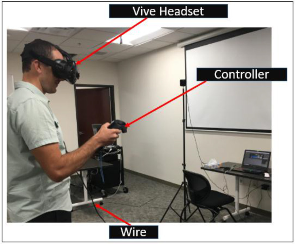
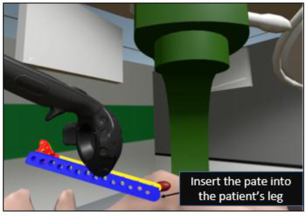
An innovative Immersive VR (IVR) simulator has been developed using the HTC Vive system for both LISS plating and Condylar plating surgical procedures [7]. This Vive based simulator system consists of a fully immersive headset, pair of wireless handheld controllers and two tracking cameras (figure 3). Cameras are used to track the position of the user in the virtual environment. The Vive headset has a field of view of 110˚ which increases the level of immersion allowing users to interact with the environment more naturally. The two wireless handheld controllers allow users to freely explore and interact with the virtual environment. In the context of LISS plating surgery (for example), users (or residents) can perform various surgical tasks such as assembly, screw insertion and tightening, drilling among others using the controllers. A view of a user interacting with the virtual environment wearing the Vive headset and using the controllers can be seen in figure 3. This IVR simulator was also developed using the Unity 3D game simulation engine C# and JavaScript were used to program the simulation environments. Steam VR toolkit and a third party VR toolkit were used to build this simulator. The major modules of the IVR simulator include a VR manager, Simulation Training manager and User Interface Manager. The Simulation Training manager coordinates all the interactions between the simulator and the users. The User Interface manager coordinates the input from the users and transmits it to the required training environment. The VR manager module serves as a bridge between Vive plugins and the Unity based simulation (graphical) environment. In figure 4, a view of the Condylar plate assembly environment for the IVR simulator is shown.

SRR 2018-101 - J. Cecil USA_F3

Figure 3. A user interacting with the IVR simulator

SRR 2018-101 - J. Cecil USA_F4

Figure 4. A view of Plate Insertion Training Environment (one of the six training environments developed for IVR condylar plating simulator)

Mixed Reality based Platforms

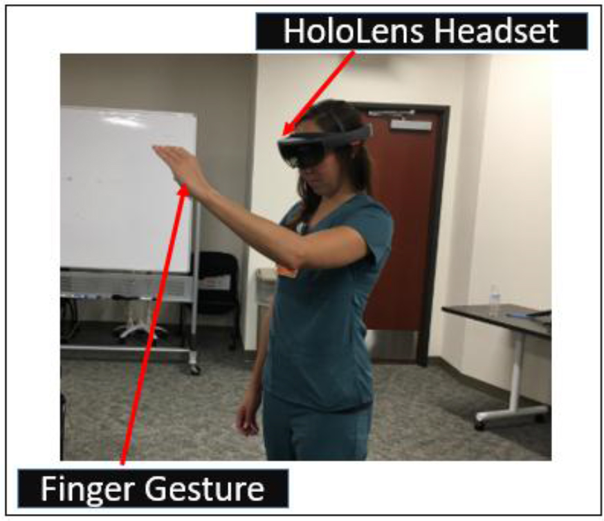
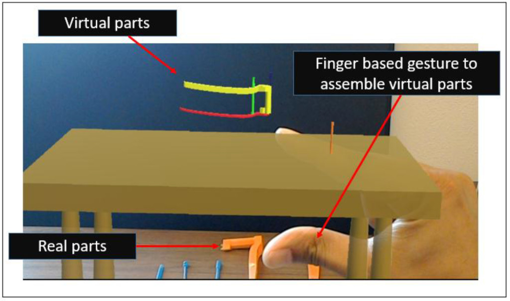
The Mixed Reality (MR) platform typically allows users to interact with both the real and virtual worlds at the same time; the primary advantage of such an MR platform is that a user can be guided by a simulation scenario to practice specific surgical steps using components in a physical environment; this allows a more integrated training scenario where residents and budding surgeons can practice various steps in a procedure with appropriate guidance from the virtual environment (running on the MR headset). A MR based simulator has been built using the Microsoft HoloLensTM platform and the Unity 3D engine [7, 31] to support training activities in both LISS plating and Condylar plating procedures and developed using the Unity 3D engine. The HoloLens is an untethered and portable MR based device in which user can interact with the virtual environment immersively without losing the sense of the real world environment. In the context of surgical training, the user can interact with virtual surgical objects using the finger based gesture supported by HoloLens and practice the same steps with corresponding physical surgical instruments, plates and other components. A programming toolkit known as the Windows 10 SDK was used during the building of the simulator. A user can be seen interacting with the HoloLens using the finger gesture in the figure 5. In figure 6, a view of the user assembling the LISS plating components using finger gestures can be seen.

SRR 2018-101 - J. Cecil USA_F5

Figure 5. A medical resident interacting with the HoloLens using finger gestures

SRR 2018-101 - J. Cecil USA_F6

Figure 6. A view of one of the HoloLens’ based training scenarios (to assemble the LISS plate)

Distributed/Web based Platforms

Information centric approaches have gained prominence recently due to the emergence of smart technologies that support web based interactions. However, the need to create such information centric approaches has been underscored by earlier research in manufacturing and engineering; one of the earliest initiatives involved creating an information centric function model to understand the complexities of designing fixtures for use in computer aided manufacturing contexts [33]; other more recent efforts have focused on creating information centric process models to understand the complexities of surgical activities prior to designing and building simulation based training environments in orthopedic surgery [6, 9, 34–38].

Another emerging area revolves around the term ‘Internet of Things (IoT)’, which can play an important role in the development of distributed/web based collaborative framework in engineering, medical and other application contexts. IoT, in general, can be described as a network of physical objects or ‘things’ enhanced with electronics, software and sensors [20]; the ‘things’ refer to sensors and other data exchange or processing devices which are part of a connected network linking cyber and physical resources in various contexts.  In the healthcare and medical context, the adoption of IoT based approaches can be useful in reducing costs and improving the health care quality while facilitating use of distributed (and remote) software and physical resources. The term ‘Internet of Medical Things’ (IoMT) has been proposed by the researchers to refer to IoT for medical applications [21, 22]. However, the term IoMT can lead to misinterpretation as this term is also being used by manufacturing researchers to refer to ‘Internet of Manufacturing Things’ [23, 24]. We propose to use the acronym ‘IoMedT’ to refer to the Internet of Medical Things, which can be defined as an IoT network which links medical devices, software, sensors and other cyber/physical resources through a network to support various activities including tele-medicine, web based surgical training, remote patient monitoring, among others. In the surgical training context, IoMedT based approaches can provide collaborative training to the medial residents and students along with proving crucial patient data online to improve their surgical capabilities. The role of collaborative learning has been underscored by only few researchers in medical education; for such cyber training activities, users and students can interact from different locations through the Internet [25–29]. One such environment is discussed in [30] which outlines a collaborative VR based environment for temporal bone surgery. They used a private gigabit Intranet to send only the modifications in drill positions/forces through the network whereas the model was made available in both locations.  Another approach involving collaborative haptic surgical system is described in [25] where users can simulate surgical processes using virtual tools independently in two different locations. The literature shows an evident lack of cloud based training simulators through which medical residents can practice surgical steps remotely and collaboratively.

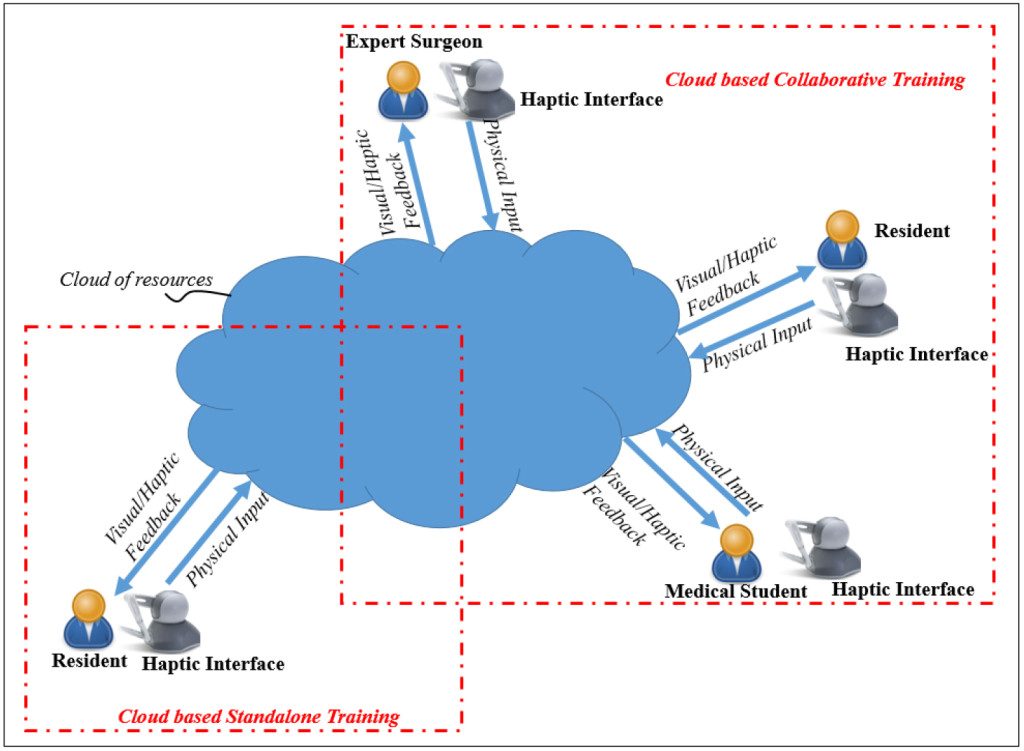
The use of cloud based approaches along with Virtual Reality (VR) based technologies can transform the way medical training is performed. Software Defined Networking (SDN) principles can be used to support such cloud based training approaches which not only reduce the complexity seen in today’s networks, but also help Cloud service providers host millions of virtual networks without the need for common separation/isolation methods [9, 15, 32]. In [15], a cloud based training framework to support haptic interface based training activities in LISS plating surgery has been discussed. Using such framework (figure 7), residents from different locations can interact with an expert surgeon who can guide them or supervise their virtual training activities in a collaborative manner. Initial results on the performance of the network as well as assessment of learning/training [8, 14, 39, 40, 43] have underscored the potential of such distributed approaches based on IoMedT principles.

SRR 2018-101 - J. Cecil USA_F7

Figure 7. A Cloud based IoMedT Framework for Surgical Training

Discussion

An important aspect to emphasize is that during the creation of these four VR based simulation platforms, the role of the expert surgeons was important. They were involved with the design, planning and development of the content of the training modules from the beginning; they provided reading materials and surgery videos along with discussing the surgical procedures in detail to ensure the software engineers gained a better understanding of the details involved. They also provided suggestions on improving the user interface and making it user friendly for the students and residents.

The impact of the four platforms supporting VR based training have been assessed through interactions with surgeons and medical residents at Texas Tech University Health Sciences Center, El Paso, Texas [7–9, 15, 39] and the Burrell College of Osteopathic Medicine (BCOM), Las Cruces, Mexico.

Survey based comparisons [6, 7, 14, 40] was conducted involving haptic based, IVR and MR based simulators; the criteria considered included:

Head movement/Comfort: did the users feel comfortable interacting with the training environments using the VR headsets? (Vive and HoloLens)

Field of view: how much of the training scenarios within the simulation environments could be seen when wearing when wearing the HoloLens or Vive device?

Navigation: how easy and intuitive were these platforms to allow users to explore a target training scenario including walking around, changing direction, zooming in on a specific area, etc. using a haptic pen (shown in figure 1), finger gestures in a HoloLens platform (figure 5) or a controller in a Vive platform (figure 3).

Ease of interaction: how easy was it for a user to interact with the training environment using the available user interface options?

Mobility during training: how much mobility did users have while interacting with the training simulator? (could they move freely or was their mobility restricted to a specific position during training)

The IVR simulator platform received a higher score on the first criterion. In terms of the Field of View, both IVR and MR simulators received comparable scores as both allowed a user to turn their heads and interact with the environment. In terms of Navigation, the three platforms (haptic, IVR and MR simulator) received similar scores. For criterion 4 (ease of user interaction), the controllers provided with the IVR simulator and the haptic simulators received a higher score than the MR based simulator (some users had difficulty using the finger gestures to interact with the user interfaces). For criterion 5 (Mobility during user interaction), the MR based simulator received the higher score as it allowed users unhindered movement during training. The IVR simulator had wires connected to the Headset which could potentially hinder or trip users when they were moving and navigating during a training session (the wires can be seen figure 3). The cost of these platforms devices were comparable. In general, the emergence of both the HoloLens and Vive based platforms provide a low cost alternative to the more traditional expensive VR environments such as the VR CAVE or Powerwall.

Conclusion

In this paper, a discussion of four simulator platforms for training medical residents in orthopedic surgery has been discussed. The four platforms discussed were haptic based, immersive, mixed reality based and distributed/web based platforms. These simulators were built to provide residents training in two orthopedic surgical procedures (LISS plating and condylar plating) to address fractures of the femur bone. A comparison of their interactive capabilities along with results from studies assessing their impact on education and training was also provided. The results from the learning interactions were predominantly positive and the comparison studies revealed that the low cost emerging platforms have the potential to be incorporated into medical training activities.

Acknowledgment

We would like to express our thanks to the surgeons, residents and students at the Paul L. Foster School of in El Paso (Texas) and Burrell College of Osteopathic Medicine (BCOM), Las Cruces, Mexico for participating in some of these project activities.

Competing interests

The author(s) declare(s) that they have no competing interests.

Funding Information

This material is based upon work supported by the National Science Foundation [under grant number CNS 1257803].

References

  1. Sørensen, T. S., Therkildsen, S. V., Makowski, P., Knudsen, J. L., & Pedersen, E. M. (2001). A new virtual reality approach for planning of cardiac interventions. Artificial Intelligence in Medicine, 22(3), 193–214.
  2. M. Peters, C. A. Linte, J. More, D. Bainbridge, D. L. Jones, G. M. Guiraudon (2008). Towards a medical virtual reality environment for minimally invasive cardiac surgery. In Miar 2008, Lncs 5128, pp. 1–11
  3. Azzie, JT. Gerstle, A. Nasr, D. Lasko, J. Green, O. Henao, M. Farcas, A. Okrainec (2011) Development and validation of a pediatric laparoscopic surgery simulator. In Journal of Pediatric Surgery, vol. 46, pp. 897–903 [Crossref]
  4. Makiyama, M. Nagasaka, T. Inuiya, K. Takanami, M. Ogata, Y. Kubota (2012) Development of a patient-specific simulator for laparoscopic renal surgery. In International Journal of Urology 19, pp. 829–835 [Crossref]
  5. Pettersson, J., Palmerius, K. L., Knutsson, H., Wahlstrom, O., Tillander, B., & Borga, M. (2008). Simulation of patient specific cervical hip fracture surgery with a volume haptic interface. IEEE Transactions on Biomedical Engineering, 55(4), 1255–1265.
  6. Cecil, J., Gupta, A., & Pirela-Cruz, M. (2018). An advanced simulator for orthopedic surgical training. International journal of computer assisted radiology and surgery, 13(2), 305–319. [Crossref]
  7. Gupta, A., Cecil, J., & Pirela-Cruz, M. (2018, August). A Virtual Reality enhanced Cyber Physical Framework to support Simulation based Training of Orthopedic Surgical Procedures. In 2018 14th IEEE Conference on Automation Science and Engineering (CASE). IEEE.
  8. Cecil, J., Gupta, A., Pirela-Cruz, M., & Ramanathan, P. (2017). A cyber training framework for orthopedic surgery. Cogent Medicine, 4(1), 1419792.
  9. Cecil, J., Kumar, M. B. R., Gupta, A., Pirela-Cruz, M., Chan-Tin, E., & Yu, J. (2016, October). Development of a virtual reality based simulation environment for orthopedic surgical training. In OTM Confederated International Conferences” On the Move to Meaningful Internet Systems” (pp. 206–214). Springer, Cham.
  10. Ming-Dar Tsai, Ming-Shium Hsieh, Chiung-Hsin Tsai (2007) Bone drilling haptic interaction for orthopedic surgical simulator. In Computers in Biology and Medicine, vol. 37, pp. 1709 – 1718 [Crossref]
  11. Lin, Y., Wang, X., Wu, F., Chen, X., Wang, C., & Shen, G. (2014). Development and validation of a surgical training simulator with haptic feedback for learning bone-sawing skill. Journal of biomedical informatics, 48, 122–129. [Crossref]
  12. Ming-Dar Tsai, Ming-Shium Hsieh, Shyan-Bin Jou (2001) Virtual reality orthopedic surgery simulator. In Computers in Biology and Medicine, vol. 31, pp. 333–351 [Crossref]
  13. J.W. Park, J. Choi, Y. Park, K. Sun (2011) Haptic Virtual Fixture for Robotic Cardiac Catheter Navigation. In Artificial Organs, ISSN 0160-564X, Vol. 35, Issue 11, pp. 1127–1131 [Crossref]
  14. Cecil, J., Gupta, A., Pirela-Cruz, M., & Ramanathan, P. (2018). A Network-Based Virtual Reality Simulation Training Approach for Orthopedic Surgery. ACM Transactions on Multimedia Computing, Communications, and Applications (TOMM), 14(3), 77.
  15. Cecil, J., Gupta, A., Ramanathan, P., & Pirela-Cruz, M. (2017, April). A distributed collaborative simulation environment for orthopedic surgical training. In 2017 Annual IEEE International Systems Conference (SysCon) (pp. 1–8). IEEE.
  16. Suresh, P. and Schulze, J.P., 2017. Oculus Rift with Stereo Camera for Augmented Reality Medical Intubation Training. Electronic Imaging, 2017(3), pp.5–10.
  17. Egger, J., Gall, M., Wallner, J., Boechat, P., Hann, A., Li, X., Chen, X. and Schmalstieg, D., 2017, March. Integration of the HTC Vive into the medical platform MeVisLab. In SPIE Medical Imaging (pp.
  18. Huber, T., Paschold, M., Hansen, C., Wunderling, T., Lang, H. and Kneist, W., 2017. New dimensions in surgical training: immersive virtual reality laparoscopic simulation exhilarates surgical staff. Surgical Endoscopy, pp.1–6. [Crossref]
  19. Harrington, C. M., Kavanagh, D. O., Quinlan, J. F., Ryan, D., Dicker, P., O’Keeffe, D., & Tierney, S. (2018). Development and evaluation of a trauma decision-making simulator in Oculus virtual reality. The American Journal of Surgery, 215(1), 42–47.  [Crossref]
  20. http://www.techopedia.com/definition/28247/internet-of-things-iot
  21. https://www.healthcaretechnologies.com/the-internet-of-medical-things-internet-of-things-health
  22. Jha, N. K. (2017, May). Internet-of-medical-things. In Proceedings of the on Great Lakes Symposium on VLSI 2017 (pp. 7–7). ACM.
  23. Zhang, Y., Zhang, G., Wang, J., Sun, S., Si, S., & Yang, T. (2015). Real-time information capturing and integration framework of the internet of manufacturing things. International Journal of Computer Integrated Manufacturing, 28(8), 811–822.
  24. Cutler, T. R. (2014). The internet of manufacturing things. Ind. Eng, 46(8), 37–41.
  25. Chebbi, B., Lazaroff, D., Bogsany, F., Liu, P. X., Ni, L., & Rossi, M. (2005, July). Design and implementation of a collaborative virtual haptic surgical training system. In IEEE International Conference Mechatronics and Automation, 2005 (Vol. 1, pp. 315–320). IEEE.
  26. Paiva, P. V., Machado, L. D. S., Valença, A. M. G., De Moraes, R. M., & Batista, T. V. (2016, June). Enhancing Collaboration on a Cloud-Based CVE for Supporting Surgical Education. In 2016 XVIII Symposium on Virtual and Augmented Reality (SVR) (pp. 29–36). IEEE.
  27. Tang, S. W., Chong, K. L., Qin, J., Chui, Y. P., Ho, S. S. M., & Heng, P. A. (2007, March). ECiSS: A middleware based development framework for enhancing collaboration in surgical simulation. In Integration Technology, 2007. ICIT’07. IEEE International Conference on (pp. 15–20). IEEE.
  28. Jay, C., Glencross, M., & Hubbold, R. (2007). Modeling the effects of delayed haptic and visual feedback in a collaborative virtual environment. ACM Transactions on Computer-Human Interaction (TOCHI), 14(2), 8.
  29. Acosta, E., & Liu, A. (2007, March). Real-time interactions and synchronization of voxel-based collaborative virtual environments. In 2007 IEEE Symposium on 3D User Interfaces. IEEE.
  30. Morris, D., Sewell, C., Blevins, N., Barbagli, F., & Salisbury, K. (2004, September). A collaborative virtual environment for the simulation of temporal bone surgery. In International Conference on Medical Image Computing and Computer-Assisted Intervention (pp. 319–327). Springer Berlin Heidelberg.
  31. Cecil, J., Gupta, A., & Pirela-Cruz, M. (2018, April). Design of VR based orthopedic simulation environments using emerging technologies. In 2018 Annual IEEE International Systems Conference (SysCon) (pp. 1–7). IEEE.
  32. Cecil, J., Cecil-Xavier, A., & Gupta, A. (2017, August). Foundational elements of next generation cyber physical and IoT frameworks for distributed collaboration. In 2017 13th IEEE Conference on Automation Science and Engineering (CASE) (pp. 789–794). IEEE.
  33. Cecil, J., (2002) A Functional Model of Fixture Design to aid in the design and development of automated Fixture Design systems’, Journal of Manufacturing Systems, Vol. 21, No.1, pp.58–72
  34. Cecil, J. and Cruz, M., (2015) An Information Centric framework for creating virtual environments to support Micro Surgery, International Journal of Virtual Reality, Vol. 15, No. 2, pp. 3–18.
  35. Cecil, J., and Cruz, M., (2011) An Information Model based framework for Virtual Surgery, International Journal of Virtual Reality, International Journal of Virtual Reality, Vol 10 (2), pp. 17–31
  36. Cecil, J., Pirela-Cruz, M., (2013) An information model for designing virtual environments for orthopedic surgery, Proceedings of the 2013 EI2N workshop, OTM Workshops, Graz, Austria. Vol 8186, Lecture Notes in Computer Science, page 218–227. Springer,
  37. Cecil, J., Pirela-Cruz, M., Information Model for a Virtual Surgical Environment, presentation at the 2010 Industrial Engineering Research Conference, Cancun, Mexico, June 5 -9, 2010
  38. Cecil, J., Pirela-Cruz, M., Development of an Information Model for Virtual Surgical Environments, Proceedings of the 2010 Tools and Methods in Competitive Engineering, Ancona, Italy, April 12–15, 2010.
  39. Cecil, J., Gupta, A., Pirela-Cruz, M., & Ramanathan, P. (2018). An IoMT-based cyber training framework for orthopedic surgery using next generation internet technologies. Informatics in Medicine Unlocked.
  40. Cecil, J., Gupta, A., Pirela-Cruz, M., The Design of IoT based smart simulation environments for orthopedic surgical training, accepted for presentation and publication in the proceedings of the Twelfth International Symposium on Tools and Methods of Competitive Engineering (TMCE 2018),Las Palmas de Gran Canaria, Gran Canaria, Spain, May 7 – 11 2018.
  41. Cecil, J., Gupta, A., Pirela-Cruz, M., Design of a Virtual Reality Based Simulation Environment and its Impact on Surgical Education, 2016 International Conference of Education, Research and Innovation (ICERI), Seville, Spain, Nov 14–16, 2016.
  42. Cecil, J., P. Ramanathan. M. Cruz, and Raj Kumar, M.B., A Virtual Reality environment for orthopedic surgery, Proceedings of the 2014 EI2N workshop, Amantea, Calabria, Italy, Oct 29–30.
  43. Cecil, J., Ramanathan, P., Prakash, A., Pirela-Cruz, M., et al., Collaborative Virtual Environments for Orthopedic Surgery, Proceedings of the 9th annual IEEE International Conference on Automation Science and Engineering (IEEE CASE 2013), August 17 to 21, 2013, Madison, WI.

Don’t Break My Heart: A Case Series Report of Acute Traumatic injury in Left Ventricular Assist Device (LVAD) Patients

DOI: 10.31038/SRR.2018112

Abstract

Purpose

The left ventricular assist device (LVAD) population is rapidly expanding. Unique characteristics, including lack of knowledge concerning LVADs and necessary anticoagulation, complicate acute traumatic injury.

Methods

A descriptive retrospective case series was created by cross-referencing our LVAD and trauma databases at our academic, Level 1 trauma center from 2004 – Present. A chart review was performed to obtain demographics, admission data, traumatic work-up and management, LVAD data, and outcomes.

Results

Three patients sustained traumatic injuries with a LVAD in place. None were admitted by the Trauma service; 2 had routine consultations, delaying imaging and injury diagnoses. Arrival vitals were not predictive of life threatening injuries; no palpable pulse with no recorded blood pressure. No mechanical pump complications resulted; 1 patient experienced a pump flow pulsatility/suction event.

Conclusion

Trauma providers must understand LVAD function to effectively evaluate these patients. Initial work-up should involve the trauma service to prevent delays in diagnosis and cardiothoracic surgery to evaluate the LVAD.

Keywords

Left ventricular assist device (LVAD); Trauma; Complications; Acute injury; Presentation

Introduction

Heart disease continues to plague Americans. It remains the leading cause of death for both men and women, and 1 out of every 4 deaths in America is due to heart disease [1]. For those with advanced or end stage heart failure aside from medical management, left ventricular assist devices (LVADs) are the most common therapy. Between 2,500 and 3,000 LVADs are implanted annually in the United States at nearly 200 facilities [2]. LVADs can serve as a bridge to heart transplantation (BTT) or as destination therapy (DT) for patients not meeting transplant eligibility which has exponentially increased the number of LVAD implantations in recent years [3]. Interestingly, one and two year survival rates of patients with LVADs is equivalent to those undergoing heart transplantation [4].

Despite this growing population of patients, some are transplanted and others destined never to be, few centers have LVAD programs, and even fewer providers are familiar with how LVADs function. Differing LVAD models and patient physiology will affect the presence of a pulse and requirements for anticoagulation. Emergency department providers in hospitals with LVAD programs may be primed to recognize common adverse events such as gastrointestinal bleeding, device thrombosis, and strokes but this is widely variable. Providers intimately involved with LVAD programs recognize aortic insufficiency, right ventricular failure, and driveline infections as serious complications which can lead to mortality, but unfortunately, literature about LVADs is primarily confined to cardiology and cardiothoracic surgery.

Following implantation, LVAD patients have an important improvement in their quality of life. Nearly 80% are satisfied with their decision to undergo implantation [2]. They are able to regain mobility and usual activities [5]. These patients are no longer house bound, return to driving, traveling, and partaking in risky behaviors such riding motorcycles. Therefore, they are at risk for traumatic injury and may present to facilities without LVAD programs receiving care from emergency department physicians, traumatologists, and intensivists – who may be unfamiliar with the presentation of a traumatically injured patient with an LVAD. The goal of this study was to review our cases of acute traumatic injuries in LVAD patients to better understand this patient population and approach to treatment.

Materials and Methods

Following IRB approval, we cross-referenced our LVAD and trauma registries at our academic, Level 1 trauma center from 2004 – 2017. We then created a descriptive, retrospective case series of patients appearing in both databases. A chart review was performed to obtain demographics, admission data, traumatic work-up and management, LVAD data, and outcomes.

Results

A total of 302 patients received LVADs at our Level 1 trauma center from 2004–2017. After the cross-reference process, 3 patients were identified as having sustained acute traumatic injuries with an LVAD in place. Following is a summary of each of their cases.

Case #1

A 74 year old male with a history of ischemic cardiomyopathy who had an Heartmate II LVAD in place for 4 years, 2 months, and 9 days as destination therapy at the time of his acute traumatic injury. He was not a candidate for transplant due to his high operative risk. His co-morbidities included renal insufficiency, chronic back pain, COPD, atrial fibrillation, hypothyroidism, and a 30-year pack year tobacco use. The patient was a restrained driver in a motor vehicle accident. He was traveling at 35 mph, got distracted and struck the back of another vehicle. Airbags were deployed. He denied loss of consciousness. He sustained bruising of his abdomen. He presented later that afternoon to his local infusion center for a transfusion and upon hearing the history, was sent to the nearest emergency department to be evaluated. Given the complex nature of his health, he was transferred to our tertiary center, arriving 7 hours post injury complaining of pain across his upper abdomen in a band-like distribution.

His vitals at the initial hospital and during transport demonstrated a heart rate of 70–100 with blood pressures of 90/palpable. At our facility, MAPs were recorded upon arrival until an arterial line was placed. The trauma service was the first service to document 2+ pulses symmetry throughout all extremities followed by the intensivists who documented bilateral radial and dorsalis pedis signals by Doppler, otherwise he was non-pulsatile.

Prior to transfer, the patient received a chest x-ray and CT head demonstrating a small acute tentorial and falcine subdural hematoma. He was directly admitted to our heart failure service who consulted our neurosurgical, cardiothoracic surgery and trauma services approximately 13 hours later. The trauma service ordered maxillofacial, chest, abdomen, and pelvis, cervical, thoracic, and lumbar spine CTs to complete his work-up. Injuries identified included a subdural hematoma, splenic laceration with associated hemoperitoneum, left 4–8 rib fractures, and right 4–7 rib fractures. The splenic laceration could not be graded and a liver laceration was suspected, however, the CTs were non-contrast due to the patients underlying renal insufficiency (Cr 1.5). His injury severity score was 29.

The patient was initially admitted to the floor, but given the patient’s subdural hematoma and therapeutic anticoagulation with warfarin, he was transferred to the ICU for frequent neurochecks. The patient had an INR of 2.48 at presentation with a lactic acid of 2.1, no leukocytosis, and a H/H of 7.6 and 26.8 respectively. Platelet count was 118. He initially received a transfusion of two units pRBCs due to a decrease in Hg to 6.4 on serial check. The patient’s warfarin was held at admission, and cardiothoracic surgery preferred not to give any reversal agents and restart the warfarin when all services were amenable. By hospital day (HD) 3, neurosurgery agreed to restarting warfarin and trauma was agreeable to restarting it on HD 4, so the first dose was given HD 4. Unfortunately on HD 5, the patient’s INR jumped to 3.7 and prothrombin complex concentrate 2500 units was given. Additionally, he was transfused 2 more units of pRBCs for worsening anemia and a stat CT angiogram of the abdomen was performed in anticipation of splenic embolization. During this time, he was transferred back to the ICU for close monitoring. Ultimately, the CT demonstrated no change and no further interventions or transfusions were necessary. He spent 6 of his 12-day hospitalization in the ICU and was discharged to a skilled nursing facility.

With regard to the LVAD, this patient did not have an EKG performed during this admission. An echocardiogram was performed hospital day 3 and a normal troponin was drawn at the transferring hospital with no repeats. His LVAD was interrogated during his hospital admission demonstrating low pulsatility indices (PIs) down to 1.7 post-accident, but no low PIs at the time of the accident. There was no evidence of device damage as a result of the motor vehicle crash. The patient did not receive any intervention for his traumatic injuries.

Case #2

This was a 72 year old female with a history of non-ischemic cardiomyopathy. At the time of her acute traumatic injury, her Heartmate II LVAD had been in place for 1 year, 5 months, and 22 days as destination therapy. Her co-morbidities included hypertension, chronic kidney disease, and diabetes. After standing up, the patient became unsteady, fell and hit her head. She did not recall any preceding symptoms of dizziness, lightheadedness, or palpations. She described the event as losing her balance and not being able to catch herself. She presented to the emergency department as a walk-in, complaining mainly of a headache.

Initially, heart rates in the 70s and no blood pressures were documented. The first documented blood pressure was 0/0 and a MAP of 110 five hours after her arrival. MAPs continued to be recorded without blood pressures. Pulses were not documented by emergency medicine. However, they were document as 2+ and symmetric by cardiology and nephrology, but 0/4 in all extremities bilaterally by cardiothoracic surgery.

The patient underwent a maxillofacial, head and cervical spine CT that were all negative. On physical exam, she had a 2 centimeter scalp laceration repaired by the emergency medicine providers
(ISS = 1). She, too, was admitted to our heart failure service to the general floor and cardiothoracic surgery and nephrology were consulted. She was hospitalized for 3 days of observation and discharged home with home health care.

On admission, her INR was 2.6 due to warfarin. Neither a lactic acid, nor troponin were checked. She had no leukocytosis, and her H/H was 10.1/33.7 with a platelet count of 339. She had an EKG performed on arrival, but no ECHO during her hospitalization. Her LVAD was interrogated and demonstrated normal values without any PI events. There was no evidence of device damage.

Case #3

A 51 year old male with a history of ischemic cardiomyopathy who had a HeartWare LVAD in place for 7 months and 25 days presented to our emergency department reporting two episodes of syncope the previous night. He initially had been listed for transplant but was delisted 2 months prior for drug use. His co-morbidities included hypertension, cardiorenal syndrome, pulmonary hypertension, chronic hyponatremia, tobacco abuse (quit 3 years prior), and drug abuse (methamphetamines). At presentation, he had an abrasion to his forehead and stated in the past week he had experienced worsening dizziness with falls. He could not recall all the events with accuracy. He reported 2 months of sobriety.

Initial blood pressures were recorded as 102/0 a little over two hours after arrival, followed by serial MAPs beginning 4 hours after arrival. Pulses were not documented until HD 2 by the critical care team as bilateral radial and pedal Doppler signals. Additionally, neither the trauma service nor emergency medicine documented a pulse or Doppler exam.

A CT head was obtained which demonstrated a 1.4 cm left cerebellar hyperattenuating lesion which appeared hemorrhagic and a 1.1 cm left temporal hypoattenuating lesion which was concerning for embolic cerebrovascular accident, intracranial abscess, or malignancy. Unfortunately, an MRI could not be performed due to the LVAD. The patient was admitted to the ICU by medical intensivists who consulted neurosurgery, cardiothoracic surgery, and nephrology. On admission, his WBC was 21 and blood cultures were sent that grew gram-positive cocci in pairs/chains within 12 hours. A lactic acid was not drawn and his H/H was 9.8/30.6 with a platelet count of 317.

This patient was anticoagulated with warfarin, and his INR on admission was 2.8. Due to his LVAD device, he was an elevated risk of embolic events. Thus, unless his head bleed worsened or he needed operative intervention, cardiothoracic surgery recommended keeping him his INR therapeutic. On HD 2, however, the patient had a 2 gram decrease in hemoglobin. Combined with the concern for septic emboli and malignancy, a CT chest, abdomen and pelvis were performed. A grade 3 splenic laceration (4 cm) with subcapsular hematoma with active bleeding and moderate hemoperitoneum was identified (ISS = 9). At this point 26 hours after admission, the trauma service was consulted. Fresh frozen plasma was transfused with a goal INR of 2.0. Serial hemoglobins were checked with the plan for embolization if the patient demonstrated continued transfusion requirements. He received FFP and 5 units of pRBCS within 24 hours. A repeat head CT was obtained and remained unchanged. Review of the first CT abdomen and pelvis was concerning for splenic pseudoaneurysms. Therefore, on HD 3, a repeat CT angiogram of the abdomen was performed which confirmed multifocal pseudoaneurysms in the spleen. The patient underwent successful selective embolization of these pseudoaneurysms and feeding arteries. He required no further transfusions, but did receive antibiotics for 1 month for strep viridans bacteremia. He was bridged back to warfarin with heparin beginning HD 6 after much deliberation amongst the teams. His brain lesions were felt to be hemorrhagic as his septic emboli work-up was negative except for bacteremia. He was discharged home after 11 days, spending 4 of those days in the ICU.

This patient had an EKG on HD 1, a transthoracic echocardiogram on HD 1, and a transesophageal echocardiogram on HD 3 to evaluate his LVAD and valves for vegetation. No troponins were checked. LVAD interrogation revealed stable parameters without any worrisome events. There was no evidence of device damage as a result of the MVC.

Discussion

A review of the literature demonstrates only a single other case series of trauma in LVAD patients. In 2013, Sarsam et al. described 4 patients who sustained external trauma; 3 falls and one blow to the chest [6]. Two of these patients from this series presented acutely, either immediately after the traumatic injury or at 48 hours, while one patient waited 3 weeks and another 14 months [6]. All readmissions, however, were due to device complication. Unfortunately, their report focused on damage to the LVAD, presentation of symptoms related to the LVAD and the operative findings of the damage [6]. To our knowledge, this case series is the first report of traumatic injury in LVAD patients and the findings and considerations at the time of presentation.

There is a paucity of literature about the presentation of these patients and a lack of general understanding about initial management [6,7]. This is evidenced by the varying documentation of the pulse exam and blood pressures in our cases. Providers unfamiliar with LVADs may not understand the continuous blood flow from the LVAD and that pulses may be thready or absent, therefore must be listened to with a Doppler. Additionally, systolic and diastolic blood pressures cannot be technically obtained, but mean arterial pressures are trended even by arterial line if needed. This poses a problem to initial providers who are taught to recognize hemorrhagic shock in traumatically injured patients by tachycardia, a narrowed pulse pressure, and hypotension. Kenyhercz et al. describes this distraction in emergency medicine simulation where learners were disoriented by the LVAD, had little to no knowledge regarding the mechanics or physiology of an LVAD, and therefore, failed to implement Advanced Trauma Life Support (ATLS) resulting in the failure to diagnose life threatening injuries resulting in the death of the simulated patient [8]. This was echoed in our third case with the splenic laceration. In fact, the providers involved considered the acute anemia to be a result of hydration and did not consider a hemorrhagic traumatic injury. The lack of consideration of traumatic injuries is echoed numerous times in the INTERMACS reports [2,4], and even the article “How to Manage the Patient in the Emergency Department With a Left Ventricular Assist Device” only mentions fall with hematoma [9]. Emergency department presentation focuses solely on feared complications of the LVAD and anticoagulation that lead to death, gastrointestinal bleeding, device thrombosis, and strokes, and not the potential for life threatening injuries [9]. Sen et al. does state ATLS protocols should be followed and device malfunction needs to be assessed immediately [10]. While ensuring the VAD coordinator and cardiothoracic surgery team are consulted, a failure echoed in the simulations, Sen et al. does not mention involvement of the trauma service or a trauma surgeon who knows the indicated imaging, concerns, and management of common injury patterns [8].

In our cases, the involvement of the trauma service earlier may not have changed the outcomes, but may have identified injuries earlier changing the plan of anticoagulation reversal. This decision certainly must be made in a multidisciplinary fashion dependent on the patient, type of LVAD, and injuries present with risks and benefits weighed. Unfortunately, at this time there is too little data for any generalized recommendations.

Conclusion

To our knowledge, this is the first report of acute traumatic injury in LVAD patients. Trauma providers (emergency medicine or trauma surgeons) must have an understanding of LVAD function to effectively evaluate these patients, especially if practicing in an area with a robust LVAD and heart transplant program. This includes an understanding of vital sign interpretation, physical exam findings, and need for anticoagulation. Admission may be to a trauma service or cardiology, heart failure, heart transplant, or critical care team. Ultimately, the involvement of a trauma surgeon can prevent delays in diagnosis. The device will need interrogated for pump flow events and to ensure no damage has occurred. However, the decision to reverse anticoagulation and method by which this should be completed must be a collaborative decision.

Acknowledgements

Candice M. Thompson, MSN, RN, CEN

Timothy R. Ryan, APRN-NP

Conflict of interest

The authors, Lisa Schlitzkus, Brett Waibel, John Um and Zachary Bauman, have no conflicts of interest.

Human and animal studies

The study was conducted in accordance with all institutional and national guidelines for the care and use of laboratory animals.

References

  1. Centers for Disease Control and Prevention. Heart Disease Facts.
    https://www.cdc.gov/heartdisease/facts.htm
  2. Kirklin JK, Pagani FD, Kormos RL, et al. (2017). Eighth annual INTERMACS report: Special focus on framing the impact of adverse events. J Heart Lung Transplant 36: 1080–1086. [Crossref]
  3. Prinzing A, Herold U, Berkefeld A, et al. (2016). Left ventricular assist devices – current state and perspectives. J Thorac Dis. 8(8): E660-E666. [Crossref]
  4. Kirklin JK, Naftel DC, Pagani FD, et al. (2015). Seventh INTERMACS annual report: 15,000 patients and counting. J Heart Lung Transplant. 34(12): 1495–504. [Crossref]
  5. Stehlik J, Estep JD, Selzman CH, et al. (2017). Patient-Reported Health-Related Quality of Life Is a Predictor of Outcomes in Ambulatory Heart Failure Patients Treated With Left Ventricular Assist Device Compared with Medical Management. Circ Heart Fail. 10: e003910. [Crossref]
  6. Sarsam SH, Meyers DE, Civitello AB, et al. (2013). Trauma in Patients With Continuous-Flow Left Ventricular Assist Devices. Am J Cardiol 112: 1520–1522. [Crossref]
  7. Gogas BD, Parissis JT, Filippatos GS, et al. (2009). Severe anemia and sub capital femur fracture in a patient with Left Ventricular Assist Device Heart Mate II: the cardiologist’s management of this rare patient. Eur J Heart Fail 11: 806–808. [Crossref]
  8. Kenyhercz WE, Perez JL, Wolfe AN, et al. (2017). Trauma Resuscitation in a Left Ventricular Assist Device Patient: An Emergency Medicine Simulation Scenario. Cureus 9(10): 31773. DOI 10.7759/cureus. 1773 [Crossref]
  9. Kroekel PA, George L, Eltoukhy N. (2013). How to Manage the Patient in the Emergency Department With a Left Ventricular Assist Device. J Emerg Nurs 39: 447–453.
  10. Sen A, Larson JS, Kashani KB, et al. (2016). Mechanical circulatory assist devices: a primer for critical care and emergency physicians. Critical Care 20: 153–173. [Crossref]
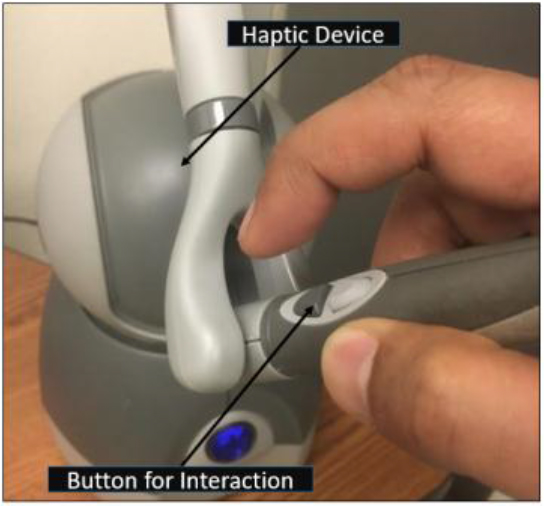
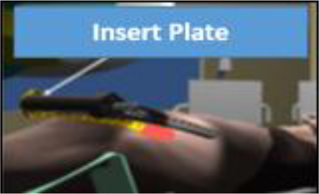
Safty and Efficacy of Collagen Tissudura in Repair of Underlying Dural Defects after Craniotomy

DOI: 10.31038/SRR.2018111

Abstract

Reconstruction and restoration of dura is an important issue in many cases of craniotomy. In cases that must be replaced large defects of dura, the usage of artificial dura is increasing.

During the period between 2013 till 2017, a total of 94 patients who underwent craniotomy for various reasons and collagen Dural plates were applied for Dural defects repairing were recruited to the study. During the follow-up of these patients, 10 patients were excluded from the study, which from the total of the remaining 84 patients, 11 cases of infection “13%” were observed. Eight cases of infection occurred as subdural empyema which were cured with reoperation treatment and evacuation of pus and dural repair. Cerebrospinal fluid leakage was seen 15.4% in 13 patients that all of them were improved with non-surgical supportive care.

Conclusion

With regards to the ease of using and least complications associated with the use of these artificial dural plates, it can be recommended to use it to dural restoration in the various craniotomy surgeries

Introduction

Proper Dural repair should be done to prevent cerebrospinal fluid leakage occurrence in craniotomy with any reason. In the cases that the primary defect of dura is too large that cannot be restored by the primary Dural tissue of the brain, we have to use alternatives to repair the underlying Dura. It is recommended to use a various type of dural alternatives that include the use of fasciae latae or peri-cranial tissue or artificial synthetic dura or ultimately tissues derived from allografts (cadaveric Dura) that each one has its own advantages and disadvantages [1–5].The usage of fasciae latae auto grafts don’t have much attractive due to the need for cutting and additional procedures with possible complications and pain caused by the removal of grafts ‘site or in cases where there are some reports from transmission of Creutzfeldtjakob caused by the use of allograft cases[6–8]. Synthetic collagen dural sheets are also safe alternatives to use as dural repair; because they are both easy to use and their reports of granulomatous reaction occurrence are rare[5, 9–12].In this study, we have discussed about results of the use of “ Aesculap onlay dura mater “ which exist as an artificial sticky sheets of Dura.

Materials and method

During the period between2013 till 2017, a total of 94 patients who underwent craniotomy for various reasons and collagen Dural plates were applied for Dural defects repairing were recruited prospectively to the study. The reasons that Patients were undergoing craniotomy include: tumors, brain hemorrhages, cerebral infarction and cerebro-vascular disease. In the process of dural restoration, we have used absorbable collagen plates of Dura “Aesculap onlay dura mater” in the size of 10cm × 10cm for each patient based on the underlying dural defects. These plates have been made of the underlying collagen scaffolds [10–12]. Sheets application is without the need for suture them to underlying dura. It will be embedded with the cover margin of at least 1cm from surrounding healthy Dura on the defect site (figure 1). Patients were followed up for three months after surgery. During the follow-up period, 10 patients died in a short period after surgery due to underlying medical conditions and were excluded from study. Of the remaining 84 patients, 25 patients “29.7%” had been subjected to surgery because of trauma, and 11 patients “% 13” had undergone craniotomy for brain infarction, 15 patients (17.8%) due to cerebral hemorrhage, and 12 patients “14.2%” for brain tumor and 21 patients “% 25” followed by cerebro-vascular lesions. (Table 1)

Table 1. Collagen dura usage results in patients

All cases

Sub-galeal space Infections

Sub-dural empyema

Leak of cerebrospinal fluid

trauma

25

2

4

4

Cerebral infarctions

11

0

0

0

Cerebral hemorrhage

15

1

1

1

Tumor

12

0

1

2

Vascular lesions

21

0

2

6

total

84

3

8

13

SRR 2018-103 - Safari H Iran_F1

Figure 1. Collagen tissue dura sheets application

Underlying dural defects in traumatic bifrontal craniotomy , without the need for suture collagen tissue dura sheet is embedded with the cover margin of at least 1cm from surrounding healthy Dura on the defect site.

Results

Totally, infection was observed in 11 patients (13%) during follow-up period (Table1). According to the clinical and MRI findings, eight cases “% 9.5” were diagnosed as subdural empyema infection. sub-galeal space infection in three cases (3.5%). Diagnosis of sub-galeal space infection was obtained by pus discharge at the incision site and MRI findings. The cerebrospinal fluid leakage was observed in a total of 13 patients (15.4%) as a discharge of cerebrospinal clear fluid secretions from the incision site during the follow-up period. Of the total cases suffered from leakage, six cases of them were underwent surgery for cerebro-vascular lesions (46%) and four cases underwent surgery for traumatic brain injury (% 31); tumor and brain hemorrhage were constituted two patients (15%) and one patient (8%) respectively (Chart 1). All of CSF leakages were treated by conservative measures; however, there were three cases from four patients with cerebrospinal fluid leaks among patients undergoing surgery due to traumatic events experiencing subdural empyema. Two cases of cerebrospinal fluid leakage were among patients that undergoing surgery due to cerebro-vascular lesions who were experiencing subdural empyema. Cerebrospinal fluid leak in patients with cerebral hemorrhage and brain tumors has been improved by the conservative measures generally and none of the cases led to subdural empyema, or infection.

SRR 2018-103 - Safari H Iran_F2

Chart 1. Rate of CSF leakage in different causes of craniotomy

Epidural accumulation of pus was occurred in all the three patients that two of them were observed in traumatic patients and one case occurred in a patient who was undergoing surgery due to brain hemorrhage. Subdural empyema patients were undergoing reoperation and the products of previous synthetic dura and pus collection were irrigated and evacuated completely, so that it was used from fasciaelatae autograft tissue to repair dural defect. After drainage, washing and continuing antibiotic treatment, six patients of a total of eight patients with subdural empyema were improved completely and pus accumulation in the subdural space was resolved in subsequent imaging and did not recur. These two patients were again subjected to surgery and re-drainage of the pus and subdural space washing due to the formation of pus in the subdural space that full recovery was observed with surgical repetition in these patients.

Of the three patients with sub-galeal accumulation, all of them were initially subjected to reoperation and pus discharge from the space of sub-galeal. One patient had fully recovered by this action and re-accumulation of pus was not found, but two other patients were undergoing surgical removal of previous synthetic dural tissues and replacing it with autograft Fasciae latae tissue due to re-accumulation of pus in follow-up MRIs which finally resulted in complete remission in these patients.

Discussion

Overall, infections in our patients was ultimately 13% during follow-up period. Infection rate of traumatic patients was totally more than the other groups that were subjected to surgery [54% of overall infection cases]. The least infections were among patients with cerebral infarction (no patient).

According to the Setsuko et al. (2003) which finally completed on 56 patients[13].The overall infection rates(14.3%) were reported in the use of artificial dural plates which was slightly higher than our study of collagen plates in the repair process. The percentage of sub-dural empyema incidence was reported 12.5% after the use of Dura plates here. While it has been observed 9.5% in our research.

Generally, the incidence of CSF leakage has been reported between4 -17% in other studies [14, 15], that is defined as sub-galeal accumulations of cerebrospinal fluid and its outflow through the sutures or its surrounding areas as the rhinorrhea or othorrhea . Totally, cerebrospinal fluid leakage was 15.4% in all of our studied patients. Highest amount of observed leakage was happened among the patients undergoing surgery for cerebro-vascular lesions “46%”, and the lowest amount was in patients undergoing surgery for cerebral infarctions that no case of leakage was found among them. The CSF leakage in patients who subjected to cerebro-vascular lesions surgery may be due to the need for a wider dissection in the sub-arachnoid space during surgery to create a wider space and visibility.

In the research of Than ko (2008), the amount of CSF leakage in the patients who had been used artificial dura to repair dural defects, was reported10%[14]. Huttera et al (2014) did not report any advantage in the reduction of CSF leakage by this method on patients using artificial dura as a normal suture compared to the use of artificial dura as a strengthening method; but in general, announced their two groups of patients CSF leakage incidence as 13.5% [15].

In general, it has been used artificial collagen plates in the studies and have shown good results, also the granulomatous reactions are relatively little to it. According to the present studies, fibroblastic cells begin to multiply after about a month in underlying tissue of artificial dura and coverage of fibroblastic tissue will be created along with surrounding dura [16, 17].

In our study, all the patients with subdural or sub-galeal infections were eventually recovered completely by reoperation, washing and removing the remnants of artificial dura and using fasciae latae tissue to repair underlying dura.

By comparing the results of similar studies in the use of artificial dural plates and the results of our study, the acceptable complications rate and control power to prevent the cerebrospinal fluid leakage incidence was perfect in the use of artificial dural plates, although using this material could be imagine a proper alternative to dural repair and prevention of a cerebro-spinal fluid leakage considering to the easiness of these artificial dural plates and no need to remove autograft tissue to repairing dura, which has its own complications.

Conclusion

With regards to the ease of using and least complications associated with the use of these artificial dural plates, it can be recommended to use it to dural restoration in the various craniotomy surgeries.

References

  1. Costa BS, Cavalcanti-Mendes Gde A, de Abreu MS, de Sousa AA. (2010) Clinical experience with a novel bovine collagen dura mater substitute. Asian J Neurosurg. 5(2): 31–4. [Crossref]
  2. Danish SF, Samdani A, Hanna A, Storm P, Sutton L. (2006) Experience with acellular humandura and bovine collagen matrix for duraplasty after posterior fossa decompression for Chiari malformations. Journal of neurosurgery. 104(1 Suppl): 16–20. [Crossref]
  3. Knopp U, Christmann F, Reusche E, Sepehrnia A. (2005) A new collagen biomatrix of equine origin versus a cadaveric dura graft for the repair of dural defects–a comparative animal experimental study. Acta neurochirurgica. 147(8): 877–87. [Crossref]
  4. Vakis A, Koutentakis D, Karabetsos D, Kalostos G. (2006) Use of polytetrafluoroethylene dural substitute as adhesionpreventive material during craniectomies. Clinical neurology and neurosurgery. 108(8): 798–802. [Crossref]
  5. Zahrai A, Shah J, Narotam P, M. G. (2005) A PROSPECTIVE CLINICAL STUDY OF THE USE OF COLLAGEN MATRIX AS A DURAL GRAFT IN SPINAL SURGERY. J Bone Joint Surg. 87: 295. [Crossref]
  6. Fushimi M, Sato K, Shimizu T, Hadeishi H. (2002) PLEDs in Creutzfeldt-Jakob disease following a cadaveric dural graft. Clinical neurophysiology: official journal of the International Federation of Clinical Neurophysiology. 113(7): 1030–5. [Crossref]
  7. Heath CA, Barker RA, Esmonde TF, Harvey P, Roberts R, Trend P, et al. (2006) Dura mater-associated Creutzfeldt-Jakob disease: experience from surveillance in the UK. Journal of neurology, neurosurgery, and psychiatry. 77(7): 880–2. [Crossref]
  8. Martinez-LageJF, Rabano A, Bermejo J, Martinez Perez M, Guerrero MC, Contreras MA, et al. (2005) Creutzfeldt-Jakob disease acquired via a dural graft: failure of therapy with quinacrine and chlorpromazine. Surgical neurology. 64(6): 542–5, discussion 5. [Crossref]
  9. Dongmei He, David G. Genecov, Morley Herbert, Raul Barcelo, Mohammed E. Elsalanty, Bradley E. Weprin, et al. (2010) Effect of recombinant human bone morphogenetic protein–2 on bone regeneration in large defects of the growing canine skull after dura mater replacement with a dura mater substitute. Journal of neurosurgery. 112(2): 319–28. [Crossref]
  10. Parlato C, di Nuzzo G, Luongo M, Parlato RS, Accardo M, Cuccurullo L, et al. (2011) Use of a collagen biomatrix (TissuDura) for dura repair: a long-term neuroradiological and neuropathological evaluation. Acta neurochirurgica. 153(1): 142–7. [Crossref]
  11. Ruediger Stendel, Marco Danne, Ingo Fiss, Ilse Klein, Andreas Schilling, Stefanie Hammersen, et al. (2008) Efficacy and safety of a collagen matrix for cranial and spinal dural reconstruction using different fixation techniques. Journal of neurosurgery. 109(2): 215–21. [Crossref]
  12. Narotam PK, Qiao F, Nathoo N. (2009) Collagen matrix duraplasty for posterior fossa surgery: evaluation of surgical technique in 52 adult patients. Clinical article. Journal of neurosurgery. 111(2): 380–6. [Crossref]
  13. Nakagawa S1, T. H. (2003) Postoperative infection after duraplasty with expanded polytetrafluoroethylene sheet. Neurol Med Chir (Tokyo). 43(3): 120–4. [Crossref]
  14. Than KD, Baird CJ, Olivi A. (2008) Polyethylene glycol hydrogel dural sealant may reduce incisional cerebrospinal fluid leak after posterior fossa surgery. Neurosurgery. 63(1 Suppl 1): ONS182–6; discussion ONS6-7. [Crossref]
  15. Hutter G, von Felten S, Sailer MH, Schulz M, Mariani L. (2014) Risk factors for postoperative CSF leakage after elective craniotomy and the efficacy of fleece-bound tissue sealing against dural suturing alone: a randomized controlled trial. Journal of neurosurgery. 121(3): 735–44. [Crossref]
  16. Neulen A, Gutenberg A, Takacs I, Weber G, Wegmann J, Schulz-Schaeffer W, et al. (2011) Evaluation of efficacy and biocompatibility of a novel semisynthetic collagen matrix as a dural onlay graft in a large animal model. Acta neurochirurgica. 153(11): 2241–50. [Crossref]
  17. Matsumoto Y, Aikawa H, Tsutsumi M, Narita S, Yoshida H, Etou H, et al. (2013) Histological examination of expanded polytetrafluoroethylene artificial dura mater at 14 years after craniotomy: case report. Neurol Med Chir (Tokyo). 53(1): 43–6. [Crossref]

A Component of the Puzzle, When Attempting to Understand Antipsychotics: A Theoretical Study of Chemical Reactivity Indexes

DOI: 10.31038/JPPR.2018115

Abstract

Schizophrenia is a human condition that has attracted the attention of researchers. There is no cure for schizophrenia but several treatments can help control symptoms (hallucinations and delusions). Dopamine D2 receptor antagonists are mainly effective for the treatment of psychotic symptoms, hence the name “antipsychotic”. This study principally aims to conduct a quantum chemical analysis of one family of antipsychotics. New physical insights have attempted to explain the pharmacological action mechanism of these drugs. Two questions for which we found a possible answer are: why the Defined Daily Dose (DDD) of these drugs varies and what do we have to consider when attempting to improve the effectiveness of medications? Although DDD is a complex concept related with pharmacokinetics, in this investigation we report some insights concerning the chemical reactivity that could be useful. These drugs are antagonists of dopamine. This means that they occupy the same receptor, but refrain from activating it. We found that the more they differ from those found in dopamine, the lower DDD is required. Chemical reactivity indexes of antipsychotics should to be different from those of dopamine. These ideas represent an aspect of the complex puzzle that contributes to define the pharmacological action of antipsychotics.

Keywords

Antioxidant, DAM, Density Functional Theory, Lagartil®, Schizophrenia

Note: *On sabbatical leave at Departamento de Química, División de Ciencias Básicas e Ingeniería, Universidad Autónoma Metropolitana-Iztapalapa, CDMX.

Introduction

Psychosis comprises a group of symptoms. An episode of psychosis is recognized when people “break” with reality. One type of psychosis is known as schizophrenia. Schizophrenia is a chronic brain disorder, affecting one percent of the global population [1–12]. The symptoms of this disorder include delusions and hallucinations. When these symptoms are treated, most people with schizophrenia improve their social function and can be re-integrated into their family and workplace. Specific treatment can help people with schizophrenia to become highly productive and develop social skills, which allow them to become adapted to their social environment.

Schizophrenia is a human condition that has attracted the attention of researchers. The etiology is complex and unknown. Moreover, there is no cure for schizophrenia, although several remedies can control symptoms [5–8]. Until the 1950s, a number of medicines manifesting restricted clinical effectiveness were used to treat the symptoms of psychosis. Electroconvulsive therapy or treatments containing a number of unspecified pharmacological agents, such as opium, morphine and cocaine, were administered with limited success [9]. The most important advance occurred in 1952 with the serendipitous observations by Laborit, which described the effect of chlorpromazine on patients suffering an episode of psychosis [10,11]. This drug has mainly proved effective for the treatment of psychotic symptoms (hallucinations and delusions), hence the name “antipsychotic”. Chlorpromazine is considered as the prime antipsychotic drug and works as a dopamine D2 receptor antagonist. Since the initial discovery of chlorpromazine, many investigations have focused on the development of new and safer treatments [12–19].

The introduction of chlorpromazine represented the first selective and effective approach to the treatment of schizophrenia, initiating the psychopharmacological era [10–19]. Ever since this finding, many investigations were undertaken, focusing on the synthesis of numerous antipsychotic drugs [1]. Those that function as a dopamine D2 receptor antagonist are considered as the first generation of antipsychotics and include different chemical compounds, such as derivatives of phenothiazine. They can be classified according to their clinical potency that correlates with dopamine D2 receptor affinity [19].

Phenothiazine is an organic compound, the molecular formula for which is presented in Figure 1. Derivatives of phenothiazine are substances that present antiemetic, antipsychotic, antihistaminic and anticholinergic activities [20–23]. The phenothiazine group of drugs is used when patients do not respond to other antipsychotics and they were one of the most widely prescribed psychotropic drugs in the world [22]. Information relating to the toxic and beneficial effects of these drugs has been discussed [21] and the value of gaining a comprehensive knowledge about their mechanism of action in order to ensure appropriate clinical application of phenothiazines [22] is recognized. Other reports suggest that the level of substitutions [23] is a factor defining the efficaciousness of this group of drugs and it has been proposed that the involvement of different molecular orbitals is related to the expression of distinct biological activities. Previous results reported that pharmacological action is influenced by the nature of substitutions, which modify receptor specificity [24, 25].

Although many studies have investigated the pharmacological action of these drugs, the exact antagonistic mechanism of the dopamine D2-receptor is still unknown. In this investigation, we aim to undertake a quantum chemical analysis of antipsychotics derived from phenothiazine. Principally, our strategy is to apply simple quantum chemical models in order to reveal the complex operation of these molecules. We applied chemical reactivity indexes and electron transfer models, previously used to successfully explain a number of reactions [26–29], to provide new explanations for the pharmacological action mechanism of these drugs. We found a possible answer to the following questions: why the Defined Daily Dose (DDD) of these antipsychotics varies, and which future strategies appear competent for improving the effectiveness of medications? Any possible solutions, not only relate to geometry, but also to electron transfer ability. Results from this research indicate that different doses of medication, reported previously, relate to electron donor-acceptor capacity. These drugs are antagonists of dopamine, because they occupy the same receptor without activating it. This leads us to the conclusion that an effective antagonist of dopamine, in the form of antipsychotic drugs, must have different electron donor acceptor properties and moreover, the more they vary from dopamine, the more efficient they will be. The outcomes reported here help to elucidate the complicated action mechanism manifested by drugs used in the treatment of schizophrenia (Figure 1).

JPPR-18-105-Ana Martinez_Mexico_F1

Figure 1. Molecular formula of different phenothiazine derivatives. R1 is equal to H for phenothiazine.

Computational Details

Gaussian09 was used for all electronic calculations [30]. Geometry optimizations without symmetry constraints were implemented at M06/6-311+G(2d, p) level of theory [31–35], while applying the continuum solvation model density (SMD) with water, in order to mimic a polar environment [36]. Harmonic analyses were calculated to verify local minima (zero imaginary frequencies). Initial geometries were obtained from PubChem database, but different conformers of each molecule were also optimized. The ground states are those that come from PubChem [37].

In order to analyze electron-donor acceptor properties, vertical ionization energy (I) and vertical electron affinity (A) were obtained from single point calculations of the corresponding cationic and anionic molecules, using the optimized structure of the neutrals. The same level of theory was used for all computations. Electrodonating (ω) and electroaccepting (ω+) power was previously reported by Gázquez et al [38, 39]. These authors defined the propensity to donate charge or ω as follows:

JPPR-18-105-Ana Martinez_Mexico_Eq1 (1)

Whereas the propensity to accept charge or ω+ is defined as

JPPR-18-105-Ana Martinez_Mexico_Eq2 (2)

Lower values of ω imply greater capacity for donating charge. Higher values of ω+ imply greater capacity for accepting charge. In contrast to I and A, ω and ω+ refer to fractional charge transfer. This definition is based on a simple charge transfer model expressed in terms of chemical potential and hardness. The Donor-Acceptor Map defined previously [40, 41] is a useful graphic tool. We have plotted ω and ω+ (Figure 2) on this map, enabling us to classify substances as either electron donors or acceptors. Electrons are transferred from good donor systems (down to the left of the map) to good electron acceptor systems (up to the right of the map) (Figure 2).

JPPR-18-105-Ana Martinez_Mexico_F2

Figure 2. Donor-Acceptor Map (DAM).

Results and Discussion

Figure 3 presents a schematic representation of the compounds that are analyzed in this investigation. There are phenothiazine-derivatives with different substituents. Promethazine is not an antipsychotic drug and is included for comparison purposes. Defined Daily Doses (DDDs) are also cited, as this parameter relates to the efficacy and potency of the compounds. Certain reports correlate the action at the receptor levels of these drugs with required doses. Those that require smaller doses are more effective and usually present fewer side effects [42 – 44]. In this investigation, we correlate DDD with different structural parameters and distinct electronic properties.

JPPR-18-105-Ana Martinez_Mexico_F3

Figure 3. Schematic representation of the molecular formula of phenothiazine derivatives investigated in this work. Defined Daily Doses (DDDs) are included (as mg/day) as is the NCCC dihedral angles (in degrees) for the optimized structures obtained in this investigation.

All antipsychotic drugs in Figure 3 are molecules that include R1 as their substituent with the following chain; -CH2-CH2-CH2-N-. Promethazine contains a substituent that is -C-C-N-, and it is not an antipsychotic drug. The first three antipsychotic molecules presented in Figure 3 have similar structures and the same DDD. Apparently, the presence of -Cl, -O-CH3, or -CH3 groups does not modify their efficacy. Molecules with lower DDDs are those compounds with a six-member ring instead of methyl groups in R1. In order to increase their efficacy and therefore decrease DDD, it is important to replace the methyl groups with a six-member ring. Comparing the NCCC dihedral angles, also depicted in Figure 3, there are two types of compounds: those for which NCCC < 100º and those for which NCCC > 150º. DDD is less for compounds with larger dihedral angles than for those antipsychotic molecules with smaller dihedral angles; the only exception is trifluoperazine. These observations show that as expected, geometrical structure and chain composition relate to DDD. All these molecules bind to D2 receptors in the brain and act as dopamine D2 receptor antagonists. Therefore geometry is important.

Even when most R2 in the antipsychotic molecules represent electron-withdrawing substituents, different effects are evident. Comparing the molecular formula of perphenazine with that of fluphenazine, the difference is the R2 substituent (Cl and CF3, respectively) and the DDD of fluphenazine is one third that of perphenazine. When we compare trifluoperazine and thioproperazine, results are the same. Molecular structures are similar with different R2 substituents. Trifluoperazine contains CF3 substituent, whereas thioproperazine does not; DDD of trifluoperazine is also almost one third of thioproperazine. It appears that the presence of CF3 in the molecule decreases the required doses, i.e. efficacy increases (Figure 3).

Likewise, comparisons indicate that thioproperazine and pipotiazine have the same R2 but different R1 (with -NCH3 and -CH-CH2-CH2-OH, respectively). The last of these is more effective than the first. We can assume that the presence of OH in the chain increases efficiency. The last compound in Figure 3 has CN as R2 and OH in R1. The required dose is not one of the smallest (50). It seems that potency is not associated with the presence of CN. With all these evaluations, it is apparent that chemical structure does not completely explain the variation in required doses.

An association can be found between DDD and electron transfer capacity using ω+ and ω−. For this purpose, figure 4 reports the DAM of the antipsychotic drugs. Phenothiazine and dopamine are also included for comparison. All compounds that need greater DDD are located down to the left (good electron donors), whereas compounds that required lower DDD are situated up to the left (good electron acceptors). The antipsychotics promazine and levopromazine represent the best electron donors, followed by chlorpromazine. Periciazine, pipotiazine and thioproperazine are the best electron acceptors. Compounds containing DDD equal to 300 mg/day constitute better electron donors than those with DDD of less than 100 mg/day. Moreover, antipsychotics that require a dose of less than 100 mg/day are better electron acceptors (ω+ > 1.5). The only exception is perphenazine, as ω+ is less than 1.5 but its DDD is 30 mg/day (Figure 4).

JPPR-18-105-Ana Martinez_Mexico_F4

Figure 4. DAM of the antipsychotic drugs studied in this investigation. Other compounds are included for comparison.

These antipsychotics are dopamine D2 receptor antagonists. Therefore, it is interesting to compare these with dopamine’s electron donor acceptor capacity. The optimized structure of dopamine is reported in Figure 5. The ω− and ω+ values are smaller for dopamine than for other compounds, meaning that it represents the best electron donor and also the worst electron acceptor. More efficient compounds (lower DDD) are those that differ most from dopamine. Apparently, it is not necessary to manifest an electron donor acceptor capacity similar to dopamine in order to be an effective antipsychotic. On the contrary, these differences appear to be necessary. One way of understanding these results is to bear in mind that these drugs are antagonists of dopamine. They occupy the same receptor but do not activate it. This activation may partly relate to the transfer of electrons. Concurring with this idea, if the dopamine D2 receptor is blocked without activating it, the capacity to transfer electrons should be different. This explains why those antipsychotics that require lower DDD are those that differ most from dopamine (Figure 5).

JPPR-18-105-Ana Martinez_Mexico_F5

Figure 5. Optimized structure of dopamine.

Other chemical indexes are valuable in this analysis, as they provide further physical insights that will enable us to answer the principal questions. These include first excitation energy and molecular hardness (η). The gap of the frontier orbitals (HOMO-LUMO gap) in the Kohn-Sham context is an approximation to the first excitation energy [45, 46]. An approximation of η is obtained with the following equation [38, 39, 47].

η = I – A (3)

The absolute values for eigenvalues of the Highest Occupied Molecular Orbitals (HOMO) and the Lowest Unoccupied Molecular Orbitals (LUMO) are reported in Figure 6. The HOMO-LUMO gap is also included. HOMO eigenvalues are similar (5.51–5.68 eV) but LUMO values show more variation (0.68–1.54 eV). The largest HOMO-LUMO gap is for dopamine (5.94 eV) and the smallest for periziacine (4.14 eV). There seems to be a correlation between HOMO-LUMO gap and efficacy, as the smallest values correspond to antipsychotics drugs that require lower doses. According to Figure 6, lower excitation energies correlate with lower DDDs. This means that they are more efficient as antipsychotic drugs. As antagonists, these molecules also differ from dopamine in terms of this property. The excitation energy may also relate to the activation of the D2 receptor. In order to be an antagonist, excitation energy should be less than that of dopamine. Accordingly, the antagonist will not activate the receptor (Figure 6).

JPPR-18-105-Ana Martinez_Mexico_F6

Figure 6. Eigenvalues (absolute values in eV) of HOMO and LUMO of the molecules studied here. HOMO-LUMO gap is also reported.

Figure 7 presents the results for η. In conformity with ideas from Parr et al [47], systems are more reactive when η is small. In our systems, lower values of η are associated with lower DDD values and therefore, greater efficiency. For example, the η of periciazine is 0.8 eV smaller than the corresponding value for chlorpromazine, and for periciazine it is 1.8 eV smaller than the corresponding value for dopamine. Compounds that require lower DDD manifest the greatest variation, when compared to dopamine. To be an effective antagonist, η has to be smaller than the corresponding value for dopamine. This could be related with the receptor binding affinity of antipsychotics. Investigations concerning the receptor and the interactions with these drugs are in process. However, these results could explain why there is a need for varying DDD and they also provide inspiration concerning possible strategies to improve the effectiveness of medications (Figure 7).

JPPR-18-105-Ana Martinez_Mexico_F7

Figure 7. Molecular hardness (η = Ι−Α) of antipsychotics. Dopamine value is included for comparison. Values in eV

In summary, electron donor acceptor capacity, excitation energies and molecular hardness are global chemical indexes that could help us to explain different DDD values. In order to investigate local properties, we analyzed frontier molecular orbitals and Model Electrostatic Potential (MEP). It was previously reported that different molecular orbitals might relate to distinct biological activities. Figure 8 reports HOMO and LUMO of the compounds under study. In all cases, HOMOs and LUMOs are π bonding orbitals located in the phenothiazine fragment. The exceptions are for pipotiazine and periciazine, as LUMOs are antibonding π orbitals. No system has the participation of halogens, sulfur or CN in the frontier molecular orbitals. Because there are no differences, it is not possible to use molecular orbitals in order to explain dissimilarities in the efficacy and potency of these drugs. According to these results, the involvement of different molecular orbitals is not related to the expression of distinct activities (Figure 8).

JPPR-18-105-Ana Martinez_Mexico_F8

Figure 8. Frontier molecular orbitals of the systems being studied.

Figure 9 reports MEP for all compounds under study. Red zones are negative regions, whereas blue sections are positive. The biggest difference is the presence of negative sections in those compounds that require lower DDD. Dopamine does not present these red zones, possibly indicating that local properties need to differ from those of dopamine, in order to increase efficiency (Figure 9).

JPPR-18-105-Ana Martinez_Mexico_F9

Figure 9. Molecular Electrostatic Potential (MEP) of the systems under study.

Conclusions

All results reported in this investigation focus on the properties of one family of antipsychotic drugs. It is apparent that the geometry and nature of substituents are important for increasing efficiency. This is a logical finding, as all these molecules bind to D2 receptors in the brain, acting as dopamine D2 receptor antagonists. Structure should be an important factor, in terms of occupying the receptor site. Nevertheless, geometrical comparisons are not sufficient to explain differences in DDD as no differences were found. Neither is possible to use frontier molecular orbitals to explain dissimilarities in the efficacy and potency of these drugs.

Electronic properties allow us to classify the best antipsychotic drugs as good electron acceptors and also as molecules that have negative sections in the MEP. Lesser hardness and lower excitation energies are also associated with lower DDD values. Dopamine is the best electron donor and the worse electron acceptor, whereas one of the most efficient antipsychotic drugs (pipotiazine) represents one of the best electron acceptors and one of the worst electron donors. Concerning molecular hardness, dopamine presents the greatest hardness, whereas the best antipsychotics present the least. Similar results are observed when comparing excitation energies. In summary, compounds with greater efficiency (lower DDD) constitute those that differ most from dopamine. It appears that it is not necessary to have similar capacities to those manifested by dopamine. Contrarily, given that these antipsychotics act as antagonists that occupy the receptor but do not activate it, their properties should vary from those of dopamine. Although the DDD concept is complicated and it is related with the pharmacokinetics, these results allow us to give some insights concerning the activity of these drugs. The results presented here represent a component in the complex puzzle that defines the action mechanism of antipsychotics.

Acknowledgment

This study was funded by DGAPA-PAPIIT, Consejo Nacional de Ciencia y Tecnología (CONACyT), and resources provided by the Instituto de Investigaciones en Materiales (IIM). This work was carried out using a NES super computer, provided by Dirección General de Cómputo y Tecnologías de Información y Comunicación (DGTIC), Universidad Nacional Autónoma de México (UNAM). We would like to thank the DGTIC of UNAM for their excellent and free supercomputing. We also thank the Laboratorio de Supercómputo y Visualización en Paralelo at the Universidad Autónoma Metropolitana- Iztapalapa for the access to its computer facilities. Authors would like to acknowledge Oralia L Jiménez A., María Teresa Vázquez and Caín González for their technical support.

References

  1. Li P, Snyder GL, Vanover KE (2016) Dopamine Targeting Drugs for the Treatment of Schizophrenia: Past, Present and Future. Curr Top Med Chem 16: 3385–3403. [crossref]
  2. Wickelgren I (1998) A new route to treating schizophrenia? Science 281: 1264–1265. [crossref]
  3. Marino MJ, Knutsen LJ, Williams M (2008) Emerging opportunities for antipsychotic drug discovery in the postgenomic era. J Med Chem 51: 1077–1107. [crossref]
  4. Forray C, Buller R (2017) Challenges and opportunities for the development of new antipsychotic drugs. Biochem Pharmacol 143: 10–24. [crossref]
  5. Hosák L, Hosakova J (2015) The complex etiology of schizophrenia general state of the art. Neuro. Endocrinol Lett 36: 631–637. [crossref]
  6. Rădulescu A (2009) A multi-etiology model of systemic degeneration in schizophrenia. J Theor Biol 259: 269–279. [crossref]
  7. Walker E, Kestler L, Bollini A, Hochman KM (2004) Schizophrenia: etiology and course. Annu Rev Psychol 55: 401–430. [crossref]
  8. Dean B (2012) Neurochemistry of schizophrenia: the contribution of neuroimaging postmortem pathology and neurochemistry in schizophrenia. Curr Top Med Chem 12: 2375–2392. [crossref]
  9. Delay J, Deniker P, Harl JM (1952) [Therapeutic method derived from hiberno-therapy in excitation and agitation states]. Ann Med Psychol (Paris) 110: 267–273. [crossref]
  10. Laborit H, Huguenard P, Alluaume R (1952) [A new vegetative stabilizer; 4560 R.P..]. Presse Med 60: 206–208. [crossref]
  11. López-Muñoz F, Alamo C, Cuenca E, Shen WW, Clervoy P, et al. (2005) History of the discovery and clinical introduction of chlorpromazine. Ann Clin Psychiatry 17: 113–135. [crossref]
  12. Ban TA (2007) Fifty years chlorpromazine: a historical perspective. Neuropsych Disease Treat 3: 495–500. [crossref]
  13. Kim DH, Maneen MJ, Stahl SM (2009) Building a better antipsychotic: receptor targets for the treatment of multiple symptom dimensions of schizophrenia. Neurotherapeutics 6: 78–85. [crossref]
  14. Mailman RB, Murthy V (2010) Third generation antipsychotic drugs: partial agonism or receptor functional selectivity?. Curr Pharm Des 16: 488–501. [crossref]
  15. CARLSSON A, LINDQVIST M (1963) EFFECT OF CHLORPROMAZINE OR HALOPERIDOL ON FORMATION OF 3METHOXYTYRAMINE AND NORMETANEPHRINE IN MOUSE BRAIN. Acta Pharmacol Toxicol (Copenh) 20: 140–144. [crossref]
  16. Creese I, Burt DR, Snyder SH (1976) Dopamine receptor binding predicts clinical and pharmacological potencies of antischizophrenic drugs. Science 192: 481–483. [crossref]
  17. Madras BK (2013) History of the discovery of the antipsychotic dopamine D2 receptor: a basis for the dopamine hypothesis of schizophrenia. J Hist Neurosci 22: 62–78. [crossref]
  18. Meltzer HY (2013) Update on typical and atypical antipsychotic drugs. Annu Rev Med 64: 393–406. [crossref]
  19. Samara MT, Cao H, Helfer B, Davis JM, Leucht S (2014) Chlorpromazine versus every other antipsychotic for schizophrenia: a systematic review and meta-analysis challenging the dogma of equal efficacy of antipsychotic drugs. Eur Neuropsychopharmacol 24: 1046–1055. [crossref]
  20. Jaszczyszyn A, GÄ…siorowski K, ÅšwiÄ…tek P, Malinka W, CieÅ›lik-Boczula K, et al. (2012) Chemical structure of phenothiazines and their biological activity. Pharmacol Rep 64: 16–23. [crossref]
  21. Sudeshna G, Parimal K (2010) Multiple non-psychiatric effects of phenothiazines: a review. Eur J Pharmacol 648: 6–14. [crossref]
  22. Fourrier A, Gasquet I, Allicar MP, Bouhassira M, Lépine JP, et al. (2000) Patterns of neuroleptic drug prescription: a national cross-sectional survey of a random sample of French psychiatrists. Br J Clin Pharmacol 49: 80–86. [crossref]
  23. Molnár J, Sakagami H, Motohashi N (1993) Diverse biological activities displayed by phenothiazines, benzo[a]phenothiazines and benz[c]acridins (review). Anticancer Res 13: 1019–1025. [crossref]
  24. Horn AS, Snyder SH (1971) Chlorpromazine and dopamine: conformational similarities that correlate with the antischizophrenic activity of phenothiazine drugs. Proc Natl Acad Sci U S A 68: 2325–2328. [crossref]
  25. Ford JM, Prozialeck WC, Hait WN (1989) Structural features determining activity of phenothiazines and related drugs for inhibition of cell growth and reversal of multidrug resistance, Mol Pharmacol 35: 105–115. [crossref]
  26. Reina M, Martínez A (2017) Silybin interacting with Cu4, Ag4 and Au4 clusters: do these constitute antioxidant materials. Comput Theor Chem 1112: 1–9.
  27. Reina M, Martínez A (2017) How the presence of metal atoms and clusters can modify the properties of silybin: a computational prediction. Comput Theor Chem 1099: 174–184.
  28. Pillegowda M, Periyasamy G (2018) DFT studies on interaction between bimetallic [Au2M] clusters and cellobiose, Comput Theor Chem 1129: 26–36.
  29. Martínez A, Vargas R, Galano A (2018) Citric acid: a promising copper scavenger. Comput Theor Chem 1133: 47–50.
  30. Frisch MJ, Trucks GW, Schlegel HB, Scuseria GE, et al. (2009) Gaussian 09, Revision A.08 Inc. Wallingford, CT.
  31. Zhao Y, Truhlar DG (2008) The M06 suite of density functionals for main group thermochemistry, thermochemical kinetics, noncovalent interactions, excited states, and transition elements: two new functionals and systematic testing of four M06-class functionals and 12 other functionals, Theor Chem Acc 120: 215–241.
  32. Petersson GA, Bennett A, Tensfeldt TG, Al-Laham MA, William A. Shirley (1988) A complete basis set model chemistry. I. The total energies of closed-shell atoms and hydrides of the first-row atoms, J Chem Phys 89: 2193.
  33. Petersson GA, Al-Laham MA (1991) A complete basis set model chemistry. II. Open-shell systems and the total energies of the first-row atoms, J Chem Phys 94: 6081.
  34. McLean AD, Chandler GS (1980) Contracted Gaussian-basis sets for molecular calculations. 1. 2nd row atoms, Z = 11–18. J Chem Phys 72: 5639.
  35. Raghavachari K, Binkley JS, Seeger R, Pople JA (1980) Self-Consistent Molecular Orbital Methods. 20. Basis set for correlated wave-functions. J Chem Phys 72: 650.
  36. Marenich AV, Cramer CJ, Truhlar DG (2009) Universal solvation model base on solute electron density and on a continuum model of the solvent defined by the bulk dielectric constant and atomic surface tensions. J Phys Chem. 113: 6378.
  37. Kim S, Thiessen PA, Bolton EE, Chen J, Fu G, et al. (2016) PubChem Substance and Compound databases. Nucleic Acids Res 44: D1202–1213. [crossref]
  38. Gázquez JL, Cedillo A, Vela A (2007) Electrodonating and electroaccepting powers. J Phys Chem A 111: 1966–1970. [crossref]
  39. Gázquez JL (2008) Perspectives on the density functional theory of Chemicals reactivity. J Mex Chem Soc 52: 3.
  40. Martínez A, Rodríguez-Gironés MA, Barbosa A, Costas M (2008) Donator acceptor map for carotenoids, melatonin and vitamins. J Phys Chem A 112: 9037–9042. [crossref]
  41. Martínez A (2009) Donator acceptor map of psittacofulvins and anthocyanins: are they good antioxidant substances? J Phys Chem B 113: 4915–4921. [crossref]
  42. Danivas V, Venkatasubramanian G (2013) Current perspectives on chlorpromazine equivalents: comparing apples and oranges! Indian J. Psych 55: 207–208.
  43. Leucht S, Samara M, Heres S, Davis JM (2016) Dose Equivalents for Antipsychotic Drugs: The DDD Method. Schizophr Bull 42: 90–94. [crossref]
  44. Andreasen NC, Pressler M, Nopoulos P, Miller D, Ho BC (2010) Antipsychotic dose equivalents and dose-years: a standardized method for comparing exposure to different drugs. Biol Psychiatry 67: 255–262. [crossref]
  45. Baerends EJ, Gritsenko OV, van Meer R (2013) The Kohn-Sham gap, the fundamental gap and the optical gap: the physical meaning of occupied and virtual Kohn-Sham orbital energies. Phys Chem Chem Phys 15: 16408–16425. [crossref]
  46. Vargas R, Garza J, Cedillo A (2005) Koopmans-like approximation in the Kohn-Sham method and the impact of the frozen core approximation on the computation of the reactivity parameters of the density functional theory. J Phys Chem A 109: 8880–8892. [crossref]
  47. Parr RG, Gázquez JL (1993) Hardness fuctional. J Phys Chem 97: 3939–3940.

How to Help Women When Providing Outreach Visits to Rural and Remote Areas of a Low Income Country (LIC)

DOI: 10.31038/AWHC.2018125


In Papua New Guinea (PNG), as in many LICs, there are areas of the country where people do not have access to health services – either because there are no health services or because their village does not have a road link to the nearest (or not so near) health facility. In PNG it is estimated that 20% of people live more than 4 hours walk (travel) from the nearest health facility. In addition, over the past 30 years more than 50% of rural health posts have closed because of health funding, lack of supervisory support, lack of community support and tribal conflict issues. This means that there are large swathes of PNG where nowadays women have no access to health care for their pregnancies.

The maternal mortality ratio (MMR) for women having a supervised birth in a provincial hospital is 50–100 (per 100,000 live births); in a rural health facility the figure is about 200. But if a woman delivers at home and there is little possibility of transfer to a health facility if some serious complication develops (as it does in about 2% of births), then the woman is in great danger of dying; for these reasons the MMR risk of home birth in PNG is about 800/100,000 live births. (In high income countries MMRs are typically less than 10). Therefore, one of the most cost effective ways of helping women not die from pregnancy complications is to help them not get pregnant when they are not wanting to do so; ie. provide them with effective contraception.

In PNG the total fertility rate is 4.4, and Demographic Health Surveys [1] consistently show that women (and men) generally have a desired family size of one child less than the total fertility rate; this indicates a huge unmet need for family planning services. This unmet need for family planning is also an issues in urban areas and rural areas with access to health services, but in remote rural areas where there are no health services women have virtually no choice at all with regards fertility regulation. Research shows that 40% of pregnancies in PNG are unplanned and 20% are unwanted as well [2]; this means that out of the 250,000 births each year in PNG 50,000 – 100,000 are unplanned/unwanted, and probably result in 200–400 additional maternal deaths pa [3].

To be effective strategies to assist women and families obtain and use family planning need to be tailored for individuals, communities and socio-demographic circumstances. Recently a number of hospitals in PNG started offering contraceptive Implants to women as a family planning option for insertion immediately postpartum. Women are counselled in antenatal clinics and indicate to the maternity carers before their due date if they would like to take up this option. Last year about 7000 women received Implants the day after their supervised birth in a health facility, and this provided about 25,000 couple years of protection (CYPs) [4]. However, only 40% of women in PNG currently access a supervised birth: this being the case then how can we assist women and families who live in remote rural areas where there is no opportunity or reasonable access for supervised birth or contraceptive services.

In colonial days (ie prior to independence in 1975) rural health workers would regularly walk to remote areas from their rural health facilities to provide outreach services like immunization and family planning. Very little outreach of this kind continues today. The Missionary Aviation Fellowship is proposing that outreach to remote areas with no health services could be conducted using their small planes by flying a health outreach team into a remote rural airstrip. The outreach team conducts health and medical work over 2–3 days and then is picked up again by another flight. The question is how can we provide something useful for women on these proposal ‘fly in- fly out’ health outreach trips?

When health outreach patrols take place sick people come, women bring their newborns for vaccination services and their babies for ‘a health check’, and pregnant women also come for a ‘check up’. In remote areas fertility control is not something that people are aware of or consider as a life option. Therefore, couples do not present for ‘family planning’ assistance. Women who bring small babies for checks and vaccination can be offered Implants and there may be good uptake for these women because few women are contemplating another pregnancy in the near future when they have a small breast feeding baby. However, who can the outreach team assist the pregnant women who attend the outreach mobile clinic?

One antenatal check (with no possibility of a supervised birth) does not actually provide much benefit for the woman, her pregnancy nor improve the chance that she will survive when it comes to lab or and delivery time. However, effective contraception for several years after the birth will at least ensure that, if she survives this birth, she does not die from another (probably unplanned pregnancy) in the near future. Fly in fly out outreach health patrols of this kind are not provided on a regular basis, and even if there will be another such patrol in the same area when the currently pregnant women have delivered there is no guarantee that they will present or have access to the clinic at that time. For this reason we are considering offering pregnant women a 5 year contraceptive Implant (Jadelle) insertion while they are in the current pregnancy in these exceptional circumstances where supervised birth is not possible and post partum contraception not available either. The World Health Organization (WHO) has declared that inadvertent insertion of progestergen based Implants have no negative impact on a pregnancy [5]. It is reasonable therefore to suggest that ‘advertent’ insertion of the Implant in pregnancy will likewise have no negative impact, and in these circumstances offer the very great benefit to the woman of about 4 years of postpartum contraception.

References

  1. Papua New Guinea Demographic Health surveys; 1996, 2006, 2016. National Statistics Office, Kumul Ave, Port Moresby, Papua New Guinea
  2. Sanga K, Mola G, Wattimena J, Justesen A, Black KI (2014) Unintended pregnancy amongst women attending antenatal clinics at the Port Moresby General Hospital. Aust N Z J Obstet Gynaecol 54: 360–365. [crossref]
  3. Mola G, Kirby B (2013) Discrepancies between national maternal mortality data and international estimates: the experience of Papua New Guinea. Reprod Health Matters, 23:191–202. [crossref]
  4. Annual Report of the Division of Obstetrics and Gynaecology of the Port Moresby General hospital and annual report of the Post Partum Implant program, Marie Stopes PNG 2017
  5. World Health Organization Family Planning Decision Making Tool, 2016

A Parallel Computation Approach to Detailed 3D Modelling of the Complete Oxygen Distribution in Large Tumours

DOI: 10.31038/CST.2018343

Abstract

Purpose

To develop a general course of action for oxygen distribution calculations, in macroscopic tumours, using Graphics Processing Units (GPU) for parallel computation.

Methods

A vessel tree structure and an associated macroscopic (about 100 g) tumour were generated, using a stochastic method and Bresenham’s line algorithm. The vessel dimensions were adjusted to correspond to measured values and each vessel voxel was assigned an oxygen value, based on its distance from an incoming large vessel. Diffusion and consumption were modelled using a Green’s function approach together with Michaelis-Menten kinetics. The tumour was inscribed in a matrix of 1012 elements. The computations were performed using a parallel method (CUDA), where the tumour was sectioned into about 18000 sub-matrices, overlapping to avoid edge effects, which were processed individually by three GPU: s. The result matrices were cropped to original size to enable concatenation.

Results

The entire process took approximately 48 hours, corresponding to 20 seconds per sub-matrix, which is more than fifty times faster when compared to the equivalent CPU calculation. Sample images illustrate the oxygen distribution of our poorly vascularised example tumour.

Conclusions

Regardless of the model accuracy and performance, the improvement in computation time using GPU calculations is highly advantageous. The preferred, parallel calculation method lowers the computation time by over 98% in this example, while maintaining full quality of performance. This is a remarkable improvement, which makes it possible to test and develop relevant models significantly faster. This computation approach does not depend on how the tumour model was created, nor is it limited to the type of model used here, but may be applied to a variety of problems, involving element-wise operations on large matrices.

Keywords

Parallel Computing, Modelling, Hypoxia, Radiosensitivity

Introduction

Numerical approaches to diffusion-consumption problems are frequently used for estimating tumour tissue oxygen transport [1–5]. When used in combination with oxygen consumption calculations, using for example the Michaelis-Menten model [6], they can provide valuable information on the characteristics of tumour oxygenation. However, there are limitations due to the extensive calculations and required computer memory. Provided sufficient accuracy, the computation times depend basically on volume and resolution. In practice, this limits decent computers to downsized experiments. This is unfortunate, since some oxygenation related effects need to be studied on clinically relevant tumour volumes in order to be properly evaluated. As we have established in previous studies, simplified calculations, e.g. through collapsing dimensions, and sub-sampling, rarely generate adequate results [5, 7].

In the previous studies we investigated the accuracy of two different approximative methods aiming to give a fast, detailed estimation of the oxygenation in a large tumour [7]. With the first method, the Individual Tree Method (ITM), we calculated the oxygen contribution from each microvessel tree in the tumour vasculature individually, down sampling the results to 100 µm cubical voxels and superposing them according to the entire vasculature. In the second method, the combined tree method, the distribution including all microvessel trees simultaneously in a 10 µm voxel model was calculated, but only for five randomly selected sample volumes of the entire tumour model. The conclusion, however, was that neither ITM nor CTM, applied to tumour samples, was sufficient to describe the oxygen status of the tumour, but that highly resolved calculations must be applied to the whole tumour. In this study, we attempt to find a way to perform such calculations and bypass the above limitations, performing parallel computations on graphical processing units (GPU) using CUDA®.

Methods

A macrovessel tree was generated, using the same method and parameter values as in Lagerlöf et al 2016. A total of 100 microvessel trees were generated accordingly and randomly assigned to each of the leaf nodes of the macrovessel tree. The combined vessel tree defines the irregularly shaped tumour, with a total volume of 100 cm3 used in these calculations. The microvessel voxels were assigned pO2-values, depending on distance from the origin of the microvessel tree, ranging from 100 to 40 mmHg [7].

The smallest cuboid volume span by the vessels measured 9 × 10 × 11 cm3, approximately 1012 voxels, side 10 µm. The calculations were made using a Gaussian diffusion kernel (equation 1) [8,9] for repeated convolution with the oxygen distribution matrix in time steps of
100 ms.

CST 2018_117_Eq1(1)

The Michaelis-Menten model (Equation 2) was used to model concentration dependent oxygen consumption at every time step.

CST 2018_117_Eq2(2)

In total, this required four single precision floating point number arrays (using 4 bytes of memory per element), holding vessel information, current oxygenation, oxygen consumption and the calculation result. For faster calculations, also the fourier transform of the oxygenation and of the diffusion kernel are needed, since convolution in the spatial domain is equivalent to multiplication in the frequency domain and the latter operation is faster for large matrices [8]. This would, in turn, require 6 · 4 · 1012 bytes (21.8 TB) of available memory, which corresponds to 700 well equipped workstations. Therefore, sectioning the tumour matrix into sub-matrices was inevitable.

A large number of fairly simple, independent, matrix operations like these are well suited for performing on the computers (GPU). For this purpose, we used a computer equipped with three CUDA-compatible Nvidia titan X (gaming) GPUs. For optimum performance on the GPU, the preferred matrix size was 512 × 512 × 512, corresponding to 512 MB of data. With the purpose of limiting the edge effects of the convolutions to below 1%, each matrix needed 70 voxels of padding in all directions. This was achieved by overlapping the sub-matrices in the sectioning, leaving 372 × 372 × 372 voxels of valid data per sub-matrix, requiring a total of 18720 sub-matrices, of which 10156 were outside the actual tumour, therefore containing no oxygen or vessel data and thus were omitted from the calculations, leaving 8564 tumour sub-matrices. The sub-matrices were indexed to preserve the geometrical information of the original matrix.

Each of the matrices was passed to the first available GPU, along with the fourier transform of the 100ms oxygen diffusion kernel. On the GPU, the current oxygen distribution (at first iteration equal to vessel oxygen content) was fourier transformed and multiplied with the fourier transformed kernel. The resulting matrix was inversely transformed, the voxel-wise oxygen consumption was calculated (according to equation 2) and subtracted; the vessel oxygen data was restored. The process was repeated 300 times and the resulting matrices were returned to the CPU, where the padded edges were trimmed, an oxygen histogram was calculated and the data was saved to file. For quality assurance, the result of an individual sub-matrix calculation was compared voxel-wise to the equivalent CPU-calculation.

The indexing of the sub-matrices allows for any slice or (sufficiently small) sub-volume of the tumour to be easily loaded into the computer memory, for further calculation, visualisation or analysis.

Results

The entire process took approximately 48 hours, corresponding to 20 seconds per sub-matrix. When performed on an Intel® Core™ i7 CPU @ 2,7 GHz (representing normal conditions), the corresponding calculations would endure approximately four months (based on slightly over 20 minutes per sub-matrix in the comparative test calculations), providing the exact same result.

By this new simulation strategy of running the computations in parallel on GPUs detailed information about the oxygen level can be visualised and quantified. Figure 1 a-c shows the oxygenation in a central slice of the tumour in the x, y and z direction respectively. Irregular oxygen distributions are observed, and the oxygen level range from approximately 0 to 80 mmHg. Figure 2 illustrates the oxygen distribution in a 400 megavoxel sample in the centre of the tumour. Demonstrating the large variation in oxygen pressure within the tumour centre. A high fraction of the cells in this region will have a low oxygen pressure, which is non-beneficial for treatment strategies as radiation therapy.

CST 2018_117_F1

Figure 1. Illustration of the simulated oxygen distribution in the tumour centre slices in x (A), y (B) and z (C) direction. The colour bar shows the oxygen level from 0 to 65 mmHg. The white length scale is 10 mm.

CST 2018_117_F2

Figure 2. The cumulative oxygen distribution in a 400 megavoxel sample in the tumour centre.

Discussion

Regardless of the model accuracy and performance, the improvement in computation time using GPU calculations is highly advantageous. The preferred, parallel calculation method lowers the computation time by over 98% in this example, while maintaining full quality of performance. This is a remarkable improvement, which makes it possible to test and develop relevant models significantly faster. This computation approach does not depend on how the tumour model was created, nor is it limited to the type of model used here, but may be applied to a variety of problems, involving element-wise operations on large matrices and it only requires one standard workstation with GPU:s.

There is room for further progress though. Using state of the art equipment (newest NVIDIA Tesla GPUs dedicated for computation) there is roughly a speed gain of 6 compared to the GPUs used in this study [10]. Increasing the number of GPU:s to the maximum (8) gives another 2.7, which means that the calculation times may be reduced by approximately 16 times compared to what is presented here, which theoretically gives a total computation time of about three hours, 99.9 % faster than the estimated 120 days in our CPU comparison.

In addition, these GPUs are able to perform two arithmetic operations at the same time (under certain conditions), on half precision floating-point numbers. Lowering the precision to 16 bit also allows for twice the amount of data to be passed to the GPU per unit time, which means that the efficiency would increase by up to 4 and the computation time ideally could become as short as 45 minutes, a reduction by 99.97 %. However, this could possibly introduce rounding errors (depending on the numerical values used in the model) due to lowered precision. The results therefore would have to be validated. At these rates, the over-head times (data transfer between CPU, GPU and data storage) could become an issue, which makes it hard to estimate the actual time gain. In the event of lost performance due to this, the CPU may be bypassed and data transferred directly to and from GPU memory [11].

Finally, the maximum data transfer rates (8564 · 512 · 512 · 512 · 16/8 Bytes per 45 minutes) would exceed even the theoretical limitations of Superspeed+ (USB 3.1 gen 2 of 0.6 GB/s) as well as of conventional hard drives. Therefore, for convenient writing and reading of data to and from file, using current technology, a solid state drive (SSD) and an interface with sufficient throughput (PCI express or Thunderbolt 3), or at least two SSD:s (if Superspeed+, SATA III or Serial attached SCSI interface is preferred), are advised [12–15].

However, performance is continually improving and within a near future we can expect computation times to further decrease considerably, making this field of tumour modelling research even more exciting. The major initial advantage of faster computations is that it accelerates the evolution of realistic models due to a notable decrease in development cycle time. By extension it enables more accurate calculations to be performed faster, perhaps essentially in real time in clinical settings, for instance diagnostic imaging.

References

  1. Harting C, Peschke P, Borkenstein K, Karger CP (2007) Single-cell-based computer simulation of the oxygen-dependent tumour response to irradiation. Phys Med Biol 52: 4775–4789. [crossref]
  2. Adhikarla V, Jeraj R (2012) An imaging-based stochastic model for simulation of tumour vasculature. Phys Med Biol 57: 6103–6124. [crossref]
  3. Dasu A, Toma-Dasu I, Karlsson M (2003) Theoretical simulation of tumour oxygenation and results from acute and chronic hypoxia. Physics in medicine and biology 48: 2829–2842.
  4. Lagerlöf JH, Kindblom J, Cortez E, Pietras K, Bernhardt P (2013) Image-based 3D modeling study of the influence of vessel density and blood hemoglobin concentration on tumor oxygenation and response to irradiation. Medical physics 40: 024101.
  5. Lagerlof JH, Kindblom J, Bernhardt P (2014) The impact of including spatially longitudinal heterogeneities of vessel oxygen content and vascular fraction in 3D tumor oxygenation models on predicted radiation sensitivity. Med Phys 41: 044101.
  6. Secomb TW, Hsu R, Dewhirst MW, Klitzman B, Gross JF (1993) Analysis of oxygen transport to tumor tissue by microvascular networks. International journal of radiation oncology, biology, physics 25: 481–489.
  7. Lagerlof JH, Bernhardt P (2016) Oxygen Distributions—Evaluation of Computational Methods, Using a Stochastic Model for Large Tumour Vasculature, to Elucidate the Importance of Considering a Complete Vascular Network. PloS one 11: 0166251.
  8. ter Haar Romeny BM (2003) The Gaussian kernel,” Front-End Vision and Multi-Scale Image Analysis: Multi-Scale Computer Vision Theory and Applications. written in Mathematics 37–51.
  9. Lagerlöf J, Diss Göteborg Göteborgs universitet (2014) Department of Radiation Physics, Institute of Clincial Sciences, The Sahlgrenska Academy, University of Gothenburg, Sweden.
  10. WHAT IS GPU-ACCELERATED COMPUTING?, http://www.nvidia.com/object/what-is-gpu-computing.html
  11. NVIDIA GPUDirect., https://developer.nvidia.com/gpudirect
  12. Superspeed USB., http://www.usb.org/developers/ssusb
  13. Thunderbolt 3 tehology brief.,
    https://thunderbolttechnology.net/sites/default/files/HBD16235_Thunderbolt_TB_r05.pdf
  14. SATA-IO Releases SATA Revision 3.0 Specification.,
    https://sata-io.org/sites/default/files/documents/SATA-Revision-3.0-Press-Release-FINAL-052609.pdf
  15. Serial Attached SCSI Technology Roadmap.,
    http://www.scsita.org/content/library/serial_attached_scsi_technology_roadmap/

The Effect of Health Insurance Expansion under the Affordable Care Act on Maternal Mortality Rates

DOI: 10.31038/AWHC.2018124

Abstract

Although the risk of death from complications of pregnancy in the last century has decreased dramatically, maternal mortality rates are rising in the United States. A significant proportion of these events are preventable with a timely access to medical care. This study estimates the effect of insurance access and Medicaid expansion under the Affordable Care Act on the variance in maternal mortality rates across states. We use maternal mortality data as estimated by the Centers for Disease Control and Prevention (CDC) and multivariate regression analysis to explain the wide variation in maternal mortality across states. Regression results indicate that insurance has a significant impact on state maternal mortality rates (p<0.05). Medicaid expansion does not affect mortality rates but is effective at decreasing maternal mortality in states with higher poverty rates (p<0.1). This study finds that access to insurance and early medical interventions as measured by a prenatal care visit before the third trimester can have a statistically significant effect on decreasing maternal mortality in the United States.

Introduction

The Word Health Organization defines maternal death as: “The death of a woman while pregnant or within 42 days of termination of pregnancy, irrespective of the duration and the site of the pregnancy, from any cause related to or aggravated by the pregnancy or its management, but not from accidental or incidental causes” [1]. Worldwide between 1990 and 2013 maternal mortality rates decreased in nearly every country, except the United States where it increased [2]. Data from the Centers for Disease Control and Prevention’s (CDC) Pregnancy Mortality Surveillance System show that maternal mortality increased from about 10 deaths per 100,000 live births in the early 1990s to 16 deaths per 100,000 live births by 2010 [3]. Overall, 40% of pregnancy-related deaths are potentially preventable with improved use of medical care being the most important factor in decreasing maternal mortality statistics [4]. Previous research shows that in the United States maternity rates and trends vary widely by state. While California is showing a declining MMR trend, Texas MMR is increasing over time, doubling between 2011 and 2012 [5]. Overall, MMR for 48 states and Washington DC is increasing and this trend places the United States far behind other developed nations [5]. This study attempts to isolate the role of health insurance in explaining the variance in MMR across states.

When the Affordable Care Act (ACA) was passed in 2010, it mandated the expansion of Medicaid (effective January 1, 2014), increasing eligibility for nearly all US residents with household incomes up to 138% of the federal poverty level. However, the US Supreme Court struck down the mandatory expansion of Medicaid in 2012 and ruled that each state could choose whether to expand this state program. In 2016 (the year of the data in this study), 30 states and Washington, DC, have elected to expand Medicaid, whereas 20 states have not. Yet evidence regarding the effects of Medicaid on health outcomes remains unclear. On one hand, randomized Medicaid trial in Oregon showed no significant effect of Medicaid expansion on mortality rates despite higher medical care utilization rates [6]. On the other hand, Sommers et al. [7] find that state Medicaid expansions were associated with a significant reduction in adjusted all-cause mortality that benefited older adults, minorities and the poor the most. Since 40 percent of maternity-related deaths are potentially preventable with improved quality of medical care [4] we hypothesize that insurance will play a greater role in decreasing MMR than overall mortality rates.

Empirical Model

This study attempts to explain the variance in MMR rates across states in the United States. Empirical model below shows the estimating equation:

MMR = α + β0 (Medicaid Expansion) + β2 (Uninsured) + β3 (Poverty) + β4 (Poverty*Medicaid Expansion) + β5 (Median Income) + β6 (Food Stamps) + β7 (Teen Births)+ β8 (Dedicated Health Provider) + β9(Physical Inactivity) + β10 (Obesity) + β11 (Smoking) + β12 (Prenatal Visit)

Our dependent variable (MMR) measures maternal mortality rates across states. Our independent variables of interest are percent uninsured, Medicaid expansion and interaction variable between Medicaid expansion and poverty rates. Medicaid expansion equals 1 for all states that expanded Medicaid under the Affordable Care Act and 0 for states that did not expand. Since Medicaid expansion benefits states with higher poverty rates the most, interaction between Medicaid expansion and Poverty rate will capture this effect. We hypothesize that Medicaid expansion may not affect all states but rather states with higher proportion of lower income households. We run the model above with and without the interaction variable.

Data

This study uses 2015-2017 publicly available state-level data. Our dependent variable is measured as the number of deaths from any cause related to or aggravated by pregnancy or its management (excluding accidental or incidental causes) during pregnancy and childbirth or within 42 days of termination of pregnancy, irrespective of the duration and site of the pregnancy, per 100,000 births. The data source is CDC WONDER Mortality Files, 2011-2015; it is available at: https://wonder.cdc.gov. CDC WONDER reports maternal mortality for all states except Vermont and Alaska.

Data sources for state female obesity rates, proportion of women with a dedicated health care provider, female inactivity rates, and female smoking rates are based on the Behavioral Risk Factor Surveillance System (BRFSS), an ongoing, state-based, random-digit-dialed telephone survey of non-institutionalized civilian adults aged 18 years and older. Information about the BRFSS is available at http://www.cdc.gov/brfss/index.html.

Dedicated health care provider variable measures percentage of women aged 18 to 44 who reported having one or more people they think of as their personal doctor or health care provider. Female obesity rate is defined as percentage of adult women in a state who are either overweight or obese. An adult who has a BMI between 25 and 29.9 is considered overweight. An adult who has a BMI of 30 or higher is considered obese. In this study we use state obesity rates for women only as reported by the CDC. State characteristics include poverty rate (percent of the population at or below poverty) and average annual median income. All state characteristics were obtained for 2016 from the Kaiser Family Foundation. Finally, data on the proportion of uninsured women at the state level was obtained from the U.S. Census Bureau, American Community Survey, 2016. Table 1 below presents descriptive statistics (Table 1).

Table 1. Descriptive Statistics.

Variable

Mean

(standard deviation)

Minimum value

Maximum value

MMR

20.88
(9.56)

4.5

46.2

Medicaid Expansion

0.647
(0.483)

0

1

% Below poverty line

12.607
(3.047)

7

21

% Uninsured women

11.027
(4.78)

2.8

25.1

Interaction
(Poverty * Medicaid Expansion)

7.94
(6.404)

0

20

Median income, $1000’s

59.34
(9.087)

41.09

76.26

Average food stamps benefits per month

124.4
(18.48)

102.03

228.33

Teen birth rate

22.72
(7.34)

9.4

38

% Women with a dedicated healthcare provider

75.34
(6.91)

59.6

88.3

% Women who are physically inactive

21.092
(4.183)

13.9

30.4

% Obese women

27.20
(4.86)

17.9

38

% Women smoking

17.808
(5.19)

7.8

32.2

% Women who made prenatal care visit before third trimester

94.22
(1.97)

90.1

98.4

% Minority population

28.68
(15.37)

5.6

77.3

Table 1 above shows that MMR varied widely across states from low 4.5 per 100,000 births in California to high 46.2 per 100,000 births in Georgia with a mean value of 20.88 per 100,000 live births. At the same time, uninsurance rates among women varied from 2.8% in Massachusetts (the first state to expand Medicaid and adopt health insurance mandate) to 25.1% in Texas.

Empirical Results

Table 2 presents regression results with and without our interaction variable. Both models show that states with higher proportion of uninsured women have higher MMR (p = 0.05). Medicaid expansion results in lower MMR but results are only statistically significant in our second specification (p < 0.1). At the same time, states with higher poverty rates benefited more from Medicaid expansion where additional coverage did translate into lower mortality rates. Therefore, insurance is an important determinant of MMR rates across all states (Table 2).

Table 2. Regression Results. Dependent Variable: State Maternal Mortality Rates.

Regressor

Model 1

Model 2

Medicaid Expansion

-0.824
(3.623)

-21.525*
(12.454)

% Below poverty line

1.473**
(0.736)

0.530
(0.898)

% Uninsured women

1.024**
(0.5057)

1.372**
(0.531)

Interaction
(Poverty * Medicaid Expansion)

-1.657*
(0.956)

Median income, $1000’s

0.416
(0.314)

0.523
(0.311)

Average food stamps benefits per month

0.107
(0.126)

0.134
(0.123)

Teen birth rate

0.396
(0.515)

0.191
(0.612)

% Women with a dedicated healthcare provider

0.402
(0.374)

0.516
(0.421)

% Women who are physically inactive

0.531
(0.433)

0.571
(0.422)

% Obese women

0.385
(0.619)

0.191
(0.612)

% Women smoking

0.285
(0.530)

0.183
(0.519)

% Women who made prenatal care visit before third trimester

-2.088**
(1.039)

-0.234**
(1.014)

% Minority population

0.387*
(0.211)

0.371*
(0.206)

R-squared
N
F statistic

0.493
48
2.84***

0.534
48
3.00***

Notes: standard errors are in parenthesis;
* indicates significance at 10%;
** indicates significance at 5% and
*** indicates significance at 1%.

Other important determinants of state MMR are percent of women who made a prenatal visit before their third trimester and minority population. We find that states with higher minority population have higher MMR even after we control for insurance, income and poverty rates. This result is consistent with previous literature that shows that minority women have higher MMR. Finally, early access to prenatal care leads to significantly lower MMR (p < 0.05). Therefore, early interventions can have a significant impact on decreasing state MMR.

Conclusions and Policy Implications

Empirical results show that insurance expansions can improve mortality statistics for causes of mortality that are amenable to medical care, such as maternal mortality. States with high MMR tend to have lower insurance rates and Medicaid insurance rates. While uninsurance tends to correlate with low income and poverty status that lead to poor health outcomes, state Medicaid expansion can decrease negative effects of poverty on health outcomes.

Results of this study are not without limitations. First, we collect data at the state level rather than individual data and cannot control for individual risk factors, such as preeclampsia. Second, data sources for state female obesity rates, proportion of women with a dedicated health care provider, female inactivity rates, and female smoking rates are based on the Behavioral Risk Factor Surveillance System (BRFSS) and were self-reported. Finally, collecting data overtime and looking at changes in MMR as states expand Medicaid would provide better estimates of the effects of this program on health outcomes. Unfortunately, accurate and consistent MMR statistics are not available before the Affordable Care Act. As our ability to measure MMR consistently across states improves, further research is necessary to determine the best ways to decrease MMR in the United States.

References

  1. World Health Organization. Health statistics and information systems: Maternal mortality ratio (per 100 000 live births)”. World Health Organization. http://www.who.int/healthinfo/statistics/indmaternalmortality/en/
  2. Retrieved November 1, 2018.
  3. Kassebaum NJ, Barber RM, Bhutta ZA, Dandona L, Gething PW, et al. (2016) Global, Regional, and National Levels of Maternal Mortality, 1990–2015: A Systematic Analysis for the Global Burden of Disease Study 2015. Lancet 388: 1775–1812. [crossref]
  4. Creanga AA, Syverson C, Seed K, Callaghan WM (2017) Pregnancy-Related Mortality in the United States, 2011-2013. Obstet Gynecol 130: 366–373. [crossref]
  5. Berg CJ, Harper MA, Atkinson SM, Bell EA, Brown HL, et al. (2005) Preventability of Pregnancy-Related Deaths: Results of a State-Wide Review. Obstet Gynecol 106: 1228–1234. [crossref]
  6. MacDorman MF, Declercq E, Cabral H, Morton C (2016) Recent Increases in the U.S. Maternal Mortality Rate: Disentangling Trends from Measurement Issues. Obstet Gynecol 128: 447–455. [crossref]
  7. Finkelstein A, Taubman S, Wright B, Bernstein M, Gruber J, et al. (2012) THE OREGON HEALTH INSURANCE EXPERIMENT: EVIDENCE FROM THE FIRST YEAR. Q J Econ 127: 1057–1106. [crossref]
  8. Sommers BD, Baicker K, Epstein AM (2012) Mortality and access to care among adults after state Medicaid expansions. N Engl J Med 367: 1025–1034. [crossref]