Monthly Archives: April 2025

The Evolution of Endogenous Uranium Ore Formation in Western Uzbekistan (Central Kyzylkum)

DOI: 10.31038/GEMS.2025731

Annotation

The article substantiates and schematically shows the vertical zonality in the structure of uranium deposits, as well as the mechanism of “overflow” of basement fractured waters into the aquifers of the platform cover. A geochemical model has been developed for the distribution and concentration of the main useful metals in the earth’s crust and the conditions for the formation of various types of endogenous uranium deposits in the Central Asian region.

Keywords

Central Kyzylkum, Uranium deposits, Ore Zonality, Postmagmatic processes, Geochemical model

Introduction

A large number of works are devoted to the problem of the geology of endogenous mineral deposits, at the same time, issues related to the patterns of formation of uranium deposits, in connection with the geochemical evolution of the earth’s crust, the development and restructuring of geological structures from the Precambrian to the Cenozoic, are considered in the works of only a number of scientists. Among them, the works of [1-16] who devoted their scientific works to the issues of metallogeny, geochemistry of black shales and the study of hydrothermal uranium deposits.

Metodology

An important, if not decisive, place in uranium ore genesis is given to the last stage of metamorphism, the processes of dynamohydrothermal metamorphism-metasomatosis. According to the time of manifestation, this is the stage of the latest Hercynian processes, in which they are distinguished according to the type of aqueous (hydrothermal) solution – hydrothermal and hydrothermal-metasomatic. The 1st type is characterized by a wide manifestation of postmagmatic processes with the formation of a range of near-ore alterations – greisenization, silicification, albitization with the appearance of brannerite-nasturan and quartz-tourmaline formations. 2nd type – hydrothermal-metasomatic, in which mineral parageneses are also distinguished according to the temperature interval. At the same time, as the temperature of hydrothermal solutions decreases, high-temperature mineral associations of uranium change and separation from magmatic satellites – lanthanides, Ta, Nb, Zr, and rare earths occurs. At the stage of hydrothermal ore formation, the separation of uranium and thorium also occurs. For medium- and low-temperature deposits, ore minerals of uranium are represented by oxides (nasturan, uraninite) and silicates (coffinite) of uranium. The average temperature of hydrothermal-metasomatic solutions forming pitchblende vein deposits is 1500C, and generally does not go beyond 200°C. The formation temperature of hydrothermal ores with uraninite is higher: 250-300°C [17,18]. Having carried out a number of calculations on the temperature and pressure of the formation of mineral paragenesis, taking into account the fact that in hydrothermal solutions the most common form of uranium migration is uranyl-carbonate complex compounds as Me4[UO2(CO3)3] or [Me2UO2(CO3)2 (H2O)2] – the main temperature intervals of migration of uranyl-organometallic complexes in solutions are given below (Table 1). The boundary between these types of solutions is accepted conditionally. The table indicates the most “developed” forms, which depending on the acid-base balance have one or another degree of migration.

Table 1: Distribution and Migration of Uranium in Solutions at Uranium Deposits of Central Kyzylkum.

Type  of aqueous solution

Temperature Ore Formation Migration Forms Stages of mineral formation Mineral parageneses Ore changes
Acid Leaching Carbonate Leaching
Acidic

рН≤4

Slightly Acidic

рН=4-5,5

Subalkaline

рН=5,5-6,5

Alkaline

рН>6,5


Hydrothermal

200-3000С

Brannerite- Pitchblende

Quartz-Tourmaline

Uranyl-Phosphate Uranyl-Hydroxyl

+ + + +

+ + + + +

+

crb+ep+amp±gr

qz+mu+fl+turm

qz+crb+sulf

qz+ab+mu

Silicification

Skarning

Greisenization

Albitization

Pyritization

Hydrothermal

Metasomatic

100-2000С

Fluorite – Pitchblende-Carbonate

Sulfide-Nasturan-

Coffinite

Uranyl-Carbonate Uranyl-Fluoride

+ +

+ + + + + + +

+ + + +

qz+ser+nast+mo

qz+ap+mu+chl

ab+qz+mu+bi+ank

Carbonatization

Silicification

Beresitization

Albitization

≤1000С


Quartz-Pyrite-Lamprophyr

Quartz-Carbonate-

Gold-Uranium

Uranyl-Carbonate

UranylSulfate

+

+ + + + +

+ + + +

crb+ser+qz+kaol

qz+pyr+act+chl

qz+crb+chl+ser

aln+qz+pyr+kaol

Carbonatization

Hematitization

Sericitization

Chloritization

Alunitization

Argillization

Symbols: The degree of dominance in the solution of certain uranium-containing complexes – ++++ wide, +++ significant, + + in some cases, + rare. Parageneses: qz: Quartz, crb: Carbonate, ep: Epidote, amp: Amphibole, gr: Garnet, mu: Muscovite, ser: Sericite, turm: Tourmaline, sulf: Sulfide, chl: Chlorite, pyr: Pyrite, aln: Alunite, bi: Biotite, nast: Pitchblende, mo: Molybdenite, ap: Apatite, ab: Albite, ank: Ankerite, act: Actinolite, kaol: Kaolin.

Marakushev [7,9] emphasized that the acid-base differentiation of metals is the main factor in ore zoning. During cation transport by acid solutions, metals with more alkaline properties migrate further than acidic ones and ore zoning is formed in the order of increasing alkaline properties of metals: Sn+W–Mo+Cu+Bi–Zn+Pb+Cd–Ca+Ba (cationic migration zoning). During anionic transfer, parageneses of ore metals are located in the reverse order – there is an increase in acidic properties (zoning of anionic migration). At the same time, the anionic form of migration in alkaline or hydrogen sulfide solutions is of primary importance for the transfer of metals over long distances. In particular, at the Dzhantuar deposit in carbonaceous-siliceous shales, we noted the following vertical zonality (Figure 1): the lowest – a zone of oxide pitchblende ores in association with sulfides (pyrite, sphalerite, molybdenite) is replaced upward by a zone of uranyl-vanadate with goethite and molybdenum and further uranyl-phosphate ores with alunite. The appearance of alunite indicates the maximum acidity of hydrothermal solutions. And finally, in the uppermost part of the ore zone, located in the aeration zone, secondary uranium minerals are localized – carnotite, otenite, thuyamunite. Such successive change from bottom to top of earlier mineral associations by later ones indicates the formation of uranium ores from ascending solutions (Figure 1).

Figure 1: Formation of Ore Zonality at the Jantuar Uranium Deposit.
Legend: Ores – 1. Sulfide, 2. Uranium-phosphate, 3. Uranium-Vanadate. 4. Folding. 5. Faults and direction of solutions. 6. Uranium deposit in the platform cover (Sandstone Type). 7. Ore Zoning: I-Oxide Zone, II-Zone of Oxidized Ores. Minerals of Aeration Zone: Carnotite, Otenite, Thuyamunite, Malachite, Calcite, Barite.

Results and Discussion

The mineralogical composition of uranium ores, paragenetic and mineral associations are always closely related to the nature of hydrothermal solutions and the lithological composition of host rocks. For the most common form of uranium migration, uranyl-carbonate, the temperature of the solution should not exceed 2000C, since its further increase leads to a decrease in the dissociation of carbon dioxide and a decrease in the chemical activity of the latter, which is fixed by the formation of carbonates (primarily ankerite). At temperatures above 2000C (250-3000C) hydrothermal uranium deposits with a high-temperature association – brannerite-nasturan or uraninite-pyrite can be formed. Often, brannerite mineralization is confined to exocontact hydrothermal-metasomatic halos (Kvartsevoe ore occurrence), and sulfide-nasturan mineralization is confined to similar halos located at a distance of up to 1 km from uranium-bearing sources (Madanli and Khodzhaakhmet deposits). An important problem is the vertical migration of ore matter from the depths of the Earth towards the surface, where the gas transport of ore metals is of great importance. In the deep parts, the fluids are strongly reduced, they are dominated by hydrogen and its compounds, but as they rise, the role of oxygen and oxygen compounds increases. [8]. Hydrothermal solutions or fluids migrating to the upper horizons from deeper geospheres are initially rich in halogens, especially chlorine and alkali metals, mainly sodium, copper, nickel and cobalt, silver and gold, platinoids, chromium (metals of the first group). The oxidation of fluids leads to the dispersion of these metals and the concentration of lead and zinc, with an intermediate stage of complex copper-lead-zinc mineralization, molybdenum, tin, beryllium, lanthanides, yttrium, uranium and other metals of the second group. It is these ratios, according to A.A. Marakushev, that determine the differences between the metallogenic features of the femic and sialic zones. A further increase in the oxidizing power of fluids leads to the formation of such metals as vanadium, titanium, yttrium, scandium, lanthanides, actinium, thorium, uranium and others (Figure 2).

Figure 2: Geochemical model of the distribution and concentration of ore elements and compounds in endogenous uranium deposits.
Legend: 1. Interfaces of the zone of concentration of elements of the 1st (I) and 2nd (II) groups. 2. Near-ore alterations – yellow skarning, blue pyritization. 3. Black Shale Strata, 4. Limestones, Dolomites. 5. Faults and directions of ascending fluids. 6. Granitoids. 7. Direction of growth of the oxidizing abilities of fluids. 8. A – Sulfide-Pitchblende Ores, B – Uranium-Rare Earth Ores.

At the end of the final stage of tectonomagmatic processes (post-folding stage), hydrothermal solutions, possibly genetically related to already intruded igneous rocks, moving along faults, interact with host rocks, creating fields of wall-rock alterations in them. By the time of formation, these solutions are postmetamorphic and appear locally. At the same time, if the solution does not absorb components that contribute to its dissolution and migration (alkalis, CO2, O2, SO42-), then uranium migrates in one or another mineral form, otherwise, fields of secondary changes appear in the enclosing regionally metamorphosed rocks , along which hydrothermal-metasomatic uranium mineralization develops. These superimposed processes include beresitization, carbonatization, albitization, sulfidization and silicification. Albitization produces well-known Rare-metal albitites containing Thorium and Uranium. As a rule, the intensity of uranium accumulation increases towards the end of the albitization process. At higher temperature processes, a concentration of uranium mineralization is also observed. During skarning, the forms of uranium occurrence depend on the presence of concentrating minerals, in which it is able to enter as an isomorphic impurity. In the case of high concentrations of rare-earth elements, uranium-rare-earth skarn deposits can even arise at the contact of alkaline rocks with limestones, where ore minerals are represented by accessory ones. At low concentrations of rare earth elements, with which uranium isomorphism is possible, uranium concentrations in skarns are low, and its localization occurs in cracks and intergranular space. Such a close relationship between uranium mineralization and skarnization is widely observed in the Zirabulak-Ziaetda SVK and, in part, in Auminzinsky and Vostochno-Bukantausky. In the processes of greisenization, an increased content of uranium in greisens is noted. Its main mass is deposited at the late stages of the greisen process in intergranular seams and microcracks. High concentrations of uranium (up to hundredths of a percent) were noted in gas-liquid inclusions [19]. This may indicate the enrichment of greisenizing solutions with uranium, as is observed at the Khodzhaakhmet ore occurrence in the Bukantau mountains.

Apparently, the first reason for this is the high temperatures of hydrothermal solutions (above 2000C), which makes uranyl-carbonate complexes easily soluble, although as the temperature decreases and carbon dioxide is simultaneously consumed for the formation of carbonates, supersaturation of solutions with uranium may occur with the formation of uranium-bearing carbonate metasomatites. The second reason is the blockage of porous permeable spaces when solutions move through them and an increase in the partial pressure of carbon dioxide, which also leads to an increase in the solubility of carbonate complexes. On the other hand, at high temperatures, the most common complexes will be uranyl-phosphate and uranyl-hydroxyl, creating the corresponding mineral paragenesis and ore formations. In the hydrothermal-metasomatic type of solutions, medium-temperature (fluorite-nasturanium-carbonate and sulfide-nasturanium-coffinite) and low-temperature (quartz-pyrite-lamprophyre and quartz-carbonate-gold-uranium) ore formations are formed with the corresponding parageneses and large-scale near-ore changes in host rocks. We note the almost complete prevalence of uranyl-carbonate complexes in this type of solution under neutral and subalkaline conditions. When hydrothermal solutions are separated from a magmatogenic source containing uranium, the latter can be carried out in the form of UF6 and form the UO2F2 compound, from which fluorite is formed, and part of the uranium in the composition of the uranyl-carbonate complex migrates to other favorable conditions for precipitation. The isolation of the quartz-pyrite-lamprophyre formation in endogenous uranium deposits is explained by the paragenetic relationship of uranium and the quartz-pyrite association in lamprophyre dikes, which often contain uranium mineralization (Dzhantuar, Kaskyr, Ingichke, etc.). H.M. Abdullaev noted that diabase and lamprophyre dikes can contain such syngenetic ore and accessory minerals as magnetite, pyrite, chalcopyrite, sphene, apatite, etc., paying attention to the nature of low-temperature hydrothermal alterations in dikes – epidotization and chloritization. The reason for this, apparently, is volatile lamprophyres in the dikes themselves, as pointed out by [1]. We noted that in the contact zone with alkaline lamprophyre dikes, uranium mineralization becomes more intense, and the thickness of the ore deposit increases.

Сonclusion

  1. The petrological and geochemical study of the pre-Mesozoic basement rocks of Western Uzbekistan showed the evolution of metamorphic processes in stages with the manifestation of progressive and regressive stages. An analysis of the development of epigenetic processes indicates that the most favorable criteria for the formation of endogenous uranium ore concentrations is a sharp change in the physicochemical and structural conditions for the formation of basement rocks in the final stages of the Hercynian tectonic-magmatic activation stage with the manifestation of regional and local metamorphism processes. The latter plays an important role in the formation of industrial gold and uranium mineralization.
  2. The evolution of uranium ore genesis in the parent rocks of the basement has been studied in detail. Acid-alkaline differentiation is the main factor in ore zoning, which is determined by the change of reducing solutions with Ni, Co, Cu, Cr, Pt, Ag, Au, and other conditions of dominance of oxygen fluids with Pb, Zn, Mo, V, Sc, Be, La , It, U etc. The successive change from bottom to top of earlier (uraninite, coffinite, pyrite, sphalerite) mineral associations by later ones (carnotite, otenite, tuyamunite) indicates the formation of uranium ores from ascending solutions, as is traced in carbonaceous-siliceous shales at the Jantuar deposit.
  3. Endogenous migration and localization of uranium occurred in the process of formation of zones of near-ore changes such as beresitization, sulfidization, silicification and others at relatively low temperatures of hydrothermal transformations (150 – 2500). Uranium in solutions migrated predominantly in the hexavalent form.
  4. The mineral forms of localization of basic metals – uranium, vanadium, selenium, rare earths, polymetals, etc. were studied. The ores of the deposits are very highly complex, which are characterized by the presence of colloidal, amorphous and amorphous minerals, which are excellent sorbents. Therefore, the latter contain significant amounts of metals that can be valuable for their industrial development.

References

  1. Abdullaev HM (1954) Genetic connection of mineralization with granitoid intrusions. Gosgeoltekhizdat.
  2. Betekhtin A.G. (1955). On the causes of the movement of hydrothermal solutions, their nature and processes of ore formation. In: Main problems in the theory of magmatogenic ore deposits. Publishing House of the Academy of Sciences.
  3. Domarev VS (1973) The role of metamorphism in the distribution of ore deposits. Problems of regional geology – Leningrad. VSEGEI. 191: 136-151.
  4. Rundqvist DV (1968) The pulsation hypothesis of S.S. Smirnov in the light of new data on the processes of ore formation – L. VSEGEI. 55: 46-66.
  5. Smirnov VI (1982) Geology of minerals. Nedra.
  6. Smirnov SS (1955) On the issue of zoning of ore deposits. Selected works. Publishing House of the Academy of Sciences.
  7. Marakushev AA (2005) Metamorphic petrology. Publishing house of Moscow State University. Pg: 256.
  8. Marakushev AA (2004) New model of formation of platform depressions // Problems of Ore Geology, Petrology. Mineralogy and Geochemistry – M. IGEM RAN.
  9. Marakushev AA (1979) Petrogenesis and ore formation. Nedra.
  10. Tugarinov AI (1983) Evolution of the earth’s crust and processes of ore formation. Nauka.
  11. Laverov NL (1986) Fundamentals of forecasting uranium ore provinces. Nedra.
  12. Lindgren W (1925) The Gel-Replacement – a new aspect of metasomatism. Nat. Acad. Sci. USA. 11.
  13. Bowie SHU, Some geological concepts for consideration in the search for uranium provinces and major uranium deposits. In uranium exploration geology (Vienna: IAEA, 1970). Pg: 285-300.
  14. Rich RA, Holland HD, Peterson U (1977) Hydrotermal uranium deposits. (Amsterdam: Elsevier).
  15. Routhier Р. Les disements metalliferes, volume 2 (Paris: Masson, 1963).
  16. Swanson VE (1961) Geology and geochemistry of uranium in marine black shales, a review. Pap. U.S. geol. Surv. 356-C: 67-112.
  17. Kotov EI, Timofeev AV, Khoteev AD (1970) Formation temperature of some hydrothermal uranium deposits. In: “Essays on geology and geochemistry of ore deposits”. Nauka.
  18. Naumov GB (1978) Fundamentals of the physical and chemical model of uranium ore formation. Atomizdat.
  19. Omelyanenko BI, Kozlova PS, Eliseeva OP, Simonova LI (1983) Local distribution of uranium in rocks and minerals as an indicator of its geochemical history. Nauka. 140-163.

A Comprehensive Review of Allergenic Proteins in Mycobacterium bovis and Their Role at the Human–Livestock Interface of Tuberculosis

DOI: 10.31038/IJVB.2025914

Abstract

Bovine tuberculosis (BTB) is endemic in cattle. BTB is caused by Mycobacterium bovis (M. bovis) Allergenic Proteins and has economic and public health significance. which has significant impact on the health of livestock and human. It has been significantly a cause for great economic loss in animal production. Associated risk factors contributed to the prevalence of the disease in cattle and its transmission. Moreover, the majority of cattle owners lack awareness about the disease and its public health significance. The presence of multiple hosts including wild animals, inefficient diagnostic techniques, absence of defined national controls and eradication programs could impede the control of bovine TB. Awareness rising about the disease, its transmission and zoonotic implication however, Bovine Tuberculosis in Human-Livestock-Wildlife Interface is not well studied in the country and there were no studies concerning the burden of the disease between human ,animal and wild life which is of great importance for reduction and control measures. This paper aims to review the potential health and economic impact of bovine tuberculosis control in order to safeguard human and animal population.

Keywords

Bovine tuberculosis, Human, Interface, Livestock, Wildlife

Introduction

The current information system does not provide the government with sufficient information on the incidence, prevalence and impact of zoonoses on society, thereby making it challenging to measure the returns on investments aimed at their prevention, management and control (ASL2020) There is a growing recognition of the importance of multi-species interaction for the emergence allergetic proteins and re-emergence of pathogens in wildlife, livestock and humans [1].

Human diseases being multi-host pathogens [2] and three-quarter of emerging human diseases being zoonotic [3], there is a strong public health interest in better understanding the dynamics of multi-species pathogens [4]. In other cases, the spill-over of domestic animal pathogens to wildlife caused severe outbreaks with great concerns for conservation, such as pasturellosis and Sierra Nevada outbreak in Bighorn sheep [5], rabies in wolves, and bovine brucellosis and tuberculosis in bison [6].

Bovine tuberculosis is one of the chronic bacterial diseases of animals that can take a variable amount of time (from a few weeks to a lifetime) to develop from infection to clinical disease and to become infectious to other animals [7]. The disease mostly affects cattle and rarely other species of domestic animals [8].

Mycobaterium bovis has an exceptionally wide range of mammalian hosts and affects all age groups of susceptible hosts of domestic, wild animals and human [9]. Cattle are the most common maintenance host for M. bovis infection from which transmission can occur to wildlife, or people from animals. Opossums, badgers and bison are known maintenance hosts in different European countries and American buffalo, Kudu, deer, lechwe and wild boar have been classified as maintenance hosts for M. bovis in Africa Many susceptible animals and wildlife species, including man are spillover hosts in which infection is not self- maintaining.

Moreover, the information on the epidemiology of the disease in wildlife-livestock- human interface is scarce and not well established at a national level.

Therefore, objectives of this paper is:

  • To review the epidemiological features of bovis in wildlife-livestock-human interface,
  • To highlight risk factors considered in studies conducted so far in wildlife.
  • To over view the diseases economic losses.

Epidemiology of Bovine Tuberculosis

Risk Factors in Cattle Allergenic Proteins

In overall prevalence of bovine tuberculosis the national estimate of 5.8 percent though available estimates vary widely. In the urban/peri-urban dairy systems, prevalence level ranging from 8.14 to 30 percent was reported. Bovine tuberculosis is also widely prevalent in the traditional production systems of mixed crop-livestock with values ranging between 1.6 percent and 22.2 percent and pastoral/agropastoral with values from 0.6 to 4.4 percent. In cattle, risk factors for bovine TB can be classified as animal level and herd level.

A. Animal Level Risk Factors

Bovine tuberculosis has been frequently reported in the from small-scale studies. Prevalence varies depending on the geographical areas, the breeds and the husbandry practices. Yet large areas in the country remain un-investigated and as data is lacking across the different geographical areas, breed and host species, husbandry practices and wildlife reservoir, it is not ease to make association with these risk factors [10].

Different authors reported ranges of prevalence rate based on abattoir based study. For instance reported 1.1% prevalence at Hawassa, reported 5.9% at Nekemte Municipality abattoir, reported 3.46% in Addis Ababa, reported 4.53% at Hossana, abattoirs and reported 5% prevalence of gross tuberculous lesion in camels slaughtered at Dire Dawa abattoir. Hiko and Agga, (2011) reported 4.2% abattoir prevalence of BTB in Mojo export abattoir base on gross lesions.

Prevalence in dairy farms with cross-breeds varying between 3.5% and 50%. Skin test prevalence in traditionally kept zebu cattle varies between 0.9-4% based on international used cut of value. Kiros, (1998) found that in Eastern Shoa of central local breeds had much lower prevalence rate 5.6% than exotic breeds (Holstein, 86.4%). found an individual animal prevalence of 7.9% using comparative intradermal tuberculin test (CIDT). Large scale study involving 5424 cattle carried out showed that the overall prevalence in cattle was 13.5%, with higher prevalence found in Holstein (22.2%) compared to local zebus (11.6%).

According to the study reports, higher bovine tuberculin reactivity was observed in animals with poor body condition as compared to those with good BCS. However, in cross-sectional studies, it is difficult to know the initial status of animals and this challenge to decide whether BTB has caused poor body condition in animals or animals with poor BCS are more susceptible to the disease. The real impact of BCS should be the subject of directed studies dealing with diet restriction.

In contrast to the above results, were found higher prevalence of the disease in animals with good body condition than poor body conditioned animals. On the other hand, during abattoir meat inspection animals, were found that animals with medium and good body condition were less likely to have tubeculous lesions than those with poor body conditions. Although it is not commonly reported in our country, physiological state of the animal is also considered as one of the animal risk factor this agree with Ameni & Erkuhin, (2007) were found significant variation in relation to reproductive status. This could be because animals lose sensitivity to tuberculin shortly before and after calving.

B. Herd Level Risk Factors

Risk factors at herd level are: herd size, types of farming practice and housing of cattle, geographical origin, history of bovine TB in the herd and human antecedent of tuberculosis in the household. in addition to this contact between animals and with wildlife reservoirs, introduction of cattle in a herd, herd movements and trading, lack of performance of diagnostic tests, the use of hired/shared bulls, manure and environmental persistence of M. bovis.

In were observed prevalence of M. bovis where both individual animal and herd prevalence were found higher in large and medium herd size as compared to small herds. According to literature, in some intensive dairy farms of our country, particularly in those having large herd size, the prevalence of the disease in individual animal and herd level could be rises up to (89.9%) and (100%) respectively.

Risk Factors in Wild Life Allergenic Proteins

Although no M. bovis infections have been reported in wildlife population so far, reports from different parts of the world have demonstrated several risk factors for the presence of the disease in wildlife. Direct contact or sharing of environment with domestic cattle, the extent of the disease prevalence within the region/country or domestic animal reservoir host, herd size (wildlife densities) and previous history of M. bovis in the wildlife populations are among the potential risk factors. The presence of the aforementioned animals in different wildlife reserves may have an epidemiological role in the spread of the disease among other wild and domestic animal [11].

On the other hand, as wildlife habitats are not fenced, there is intensive interaction between a fast-growing human population and livestock and wildlife competing for scarce grazing land. Wildlife and, in particular, herbivores sharing pastures with cattle might therefore be at risk for bovine TB transmission have reported that, in Amibara district of Afar pastoral region, domestic animal were sharing grazing land in close proximity with wildlife in the area where wild animals lives (in and around Awash National Park). This suggests that there is a possible exposure for potential risk of disease transmission to wildlife populations.

Risk Factors in Human

In prevalence rates for bovine tuberculosis in humans are lower than those reported in the literature. For both cattle keepers and consumers, prevalence is 0.006 percent in this study. The findings of the literature are varying between 0.41 and 24 percent, but are again based on different reference periods and small samples.The proportion of BTB to the total of TB cases in humans depends on the prevalence of the disease in cattle, socioeconomic conditions, consumer habits, practiced food hygiene and medical prophylaxis measures [12].

The main risk factors which contribute to the acquisition M. bovis infections in both urban and rural human populations are poverty, malnutrition, HIV infection, illiteracy, the consumption of raw milk (unpasteurized milk), uncooked or poorly cooked meat, work condition and close contact to livestock and using cow dung for plastering wall or floor.

Diagnosis

TB can be diagnosed clinically, but usually only in the later stages of the disease. The tuberculin skin test is universally recognized and is generally used for preliminary diagnosis in BTB control programs. However, in countries with low disease prevalence or disease free status, meat inspection is used for diagnosis and surveillance. Other tests, such as an antibody enzyme -linked immunoassay (ELISA) and the gamma interferon assay, have been used as supplementary tests in eradication and control [13]. Classical mycobacteria l culture remains the routine method for confirmation of infection.

Control of Tuberculosis

Control and eradication programs for BTB, human TB and zoonotic TB of humans due to M. bovis are based on early accurate detection and removal of infected animals, chemotherapy of infected humans and vaccination of target populations to attenuate or prevent the manifestation of the disease.

The test and slaughter policy is the basis for international BTB control and eradication programs using the TST to detect affected herds (and re – test) periodically and removing reacting cattle that may shed the infective organism. In many industrialized countries there are effective compulsory reporting of M.bovis infection of all animals, quarantine of infected herds, tracing and re- testing of animals in contact with BTB skin positive reactors. as well as movement restrictions of cattle herds not yet tested for TB as well as controlled animal movement out of known TB infected herds and endemic areas.

However, the test- and segregation program, a modified form of the test- and- slaughter policy, may be more useful for developing countries, where the test- and- slaughter policy cannot be practicable for the whole cattle population.Thus, interim measures to segregate infected herds and phased slaughter of reactors are done. In most countries with strict TB eradication programmers, the test- and segregation strategy made up the early stages followed by the test- and slaughter methods in the final stage (CFSPH, 2009) and infected slaughter /meat cases during inspection are traced back to the originating farms. Informed farm management decisions such as proper sanitation and disinfection are also important to reduce the spread of Mycobacterium, within and between herds as well as the risks of exposure and transmission of BTB infection to humans [14].

The occurrence of M. bovis in wildlife reservoir hosts complicates eradication efforts. Culling to reduce population density can decrease animal TB transmission but the situation must be assessed carefully to avoid unanticipated effects such as the economic benefit and increase scattering members of the infected species. The development of TB vaccines for wildlife reservoirs and use in situations where the test and slaughter policy is totally impracticable is also being considered as an alternative. Also, human TB due to M. bovis is rare in countries where raw and poorly cooked meat are not consumed and pasteurization of milk and milk products are components of BTB eradication programs. 

Allergenic Proteins Economic Importance of Tuberculosis

Mycobaterium bovis has been widely distributed throughout the world and it represents a very significant economic and public health problem in numerous countries in both developed and the developing world. Consequently, most developed nations have embarked on campaigns to eradicate M. bovis from the cattle population or at least to control the spread of the infection. In developed countries, although tuberculosis is eliminated in cattle, the disease still has a major economic impact, mainly due to the existence of a permanent wildlife reservoir that reduces the efficiency of control strategies. For instance, in the United Kingdom, where badger and other wildlife such as deer remain an important source of infection for livestock, approximately £100 million is spent annually in efforts to control the disease [15,16]. Republic of Ireland and New Zealand also spent approximately 35 and 13 million US $ annually for disease control. In Argentina, the annual loss due to bovine TB is approximately US$63 million. Although the disease has zoonotic threat, economic and financial burden to society, its cost has rarely been assessed and is largely unknown for Europe.

Animal tuberculosis is a disease of high economic relevance within the context of livestock farming as it directly affects animal productivity. The disease considerably reduces milk and meat production of infected animal and affect animal reproduction as well as it reduce pulling power in traditional farming system. Infected animal loses 10 to 25% of their productive efficiency. Direct losses due to the infection become evident by decrease in 10 to 18% milk and 15% reduction in meat production [17]. The culling loss is estimated to be 30–50% of the difference between the values of a dairy or beef breeding cow and its value at slaughter.

Moreover, national and international trade (market restrictions) and other economic sectors maybe indirectly affected by the disease. Tuberculosis has also an economical and financial burden to society human health costs. The disease become is an obstacle to socio-economic development: 75% of people affected by TB are within the economically productive age group of 15-54years. This may have a negative influence on the national economy [18,19].

The public health cost of bovine tuberculosis. The estimated total public health costs (USD PPP) of the disease among livestock keepers in all production systems and consumers are USD 74 740 696 and 12 781 597, respectively. This amounts to 0.05 percent of total GDP(ASL,2020) [20] (Table 1).

Table 1: Estimates of the annual public health costs of bovine tuberculosis.

The costs of bovine tuberculosis in livestock keepers to costs for the cattle sector by production system. Urban/peri-urban and commercial dairy sectors suffer the most in terms of loss incurred due to death of animals, reduced and foregone production amounting to USD 1 500 876 724 and 1 223 364 444 (PPP), respectively. The public health costs are higher in mixed croplivestock and pastoral/agro-pastoral cattle production systems, largely due to their sheer sizes. The estimated monetary cost of the disease in animals accounts for 98 percent of the total loss caused by the disease (FAO,2017a) (Table 2).

Table 2: Annual costs of bovine tuberculosis in humans and cattle in different production systems.

Although the economic importance and public health significance of tuberculosis has been established in many countries, the economic impact of M. bovis on cattle productivity, bovine TB control programmes and other related economic effects of the disease are not yet well documented or studied.

Only few meat inspection surveillances in the abattoirs have shown the economic loss due to condemnation of total or partial carcass and organs. According to Gezahegne, (1991) a report from eight export abattoirs showed a prevalence of 0.8% (978/144 487) of slaughtered animals, in which the whole carcasses of the infected animals were condemned. Asseged also demonstrated that, based on the ten years retrospective allergetic proteins analysis of the detection of tuberculous lesions in the Addis Ababa abattoir, there was a cause of 0.028% for whole carcass condemnation. Furthermore, study results of Shitaye conducted in Addis Ababa and Debre-Zeit abattoirs35indicated that causes condemnation of carcasses and/or organs due to tuberculous lesions found to be highly significant economically.

According to the study reports, a prevalence of 0.052% and 0.001% was observed in cattle and shoats respectively, and causes the whole animal’s carcass condemnation. Mycobacterium bovis infections in wildlife can affect the ecosystem: moreover, the disease constitutes a threat to endangered species and can hamper BTB eradication and control schemes in domestic cattle.

Tuberculosis at the Human-Livestock-Wildlife Interface

Diseases transmitted between humans, domestic animals, and wildlife are increasingly challenging public and veterinary health systems. Three -fourths of all emerging infectious diseases (EIDs) of humans are zoonotic with most originating from wildlife reservoirs. Therefore, diseases that arise from the livestock–wildlife interface are of paramount importance and must be an area of focus for public and veterinary health systems. Despite this importance, cross-species transmission is one of the least studied aspects of disease ecology.

BTB infections may be maintained (independently or not) within livestock populations and within wildlife populations, whereas human infections allergetic proteins result from pathogen spillover from animals , and very rarely from human-to-human transmission.

Factors associated with BTB spillover from livestock to wildlife should also influence BTB spillback from wildlife to livestock. The main risk factors are thus linked with allergetic proteins: (i) the type of interface (fence, herding practices) and the distribution of resources (water and grazing), which directly influence contact patterns between livestock and wildlife the environmental conditions, which directly influence the persistence of BTB in the environment. In this wildlife–livestock–human interfaces husbandry practices (housing, mixing cattle with small ruminants), food preferences (consumption of raw milk) and overall health and hygienic conditions are identified as the main BTB risks of transmission between livestock and humans. The complex and dynamic interactions involving domestic animals, wildlife, and humans create environments favorable for the transmission of infectious diseases across different species (Figure 1).

Figure 1: Interspecific transmission of BTB at wildlife–livestock–human interfaces.

Mycobacterium bovis is an example of a pathogen shared at the human–livestock–wildlife interface. In East Europe humans encroach into allergetic proteins wildlife habitats with their livestock in search of grazing areas and water, particularly during the dry season. Wildlife species that share resources with pastoralist livestock may influence the prevalence of bTB in cattle by having direct or indirect contact (i.e., ingestion of contaminated pastures) with cattle. More studies are required to better understand the effects of interactions between ecological and animal management risk factors in multi-host communities.

The list of wildlife species around the world from which M. bovis has been isolated is long and reports in the literature of new susceptible species have increased in recent years. Some wildlife species have long been known to be maintenance hosts (i.e., wildlife species that can maintain the disease in the absence of infected cattle). The maintenance hosts are a source of infection for livestock and can also be described as a source for BTB in humans that have close contact with infected animals, such as hunters and game farmers.

However, intermediate species such as impala, kudu and warthog (Phacochoerus africanus), which are less affected by livestock presence, could play a role as disease ‘vector’ by having close physical contact with BTB buffalo reactors, that stay within the park, and with livestock in the agricultural land outside the park.

Common use of pastureland is another potential risk for BTB transmission between wildlife and livestock. Mainly wildlife grazer species (as opposed to browser species) are likely to compete with cattle. There seems to be a species-specific tolerance level for cattle presence. Many grazer species favor grazing in old pastoral places where grass cover is rich due to the cattle manure.

Conclusion and Recommendations

The prevalence of BTB is reported to be high similar to other developing countries. However, no studies concerning the burden of the disease in wildlife and human beings were undertaken and this indicates that BTB is not well studied in the country. Human-livestock-wildlife interaction is dynamic. There are no sufficient studies clearly indicating the role of humans, livestock, wildlife and their environment with respect to the transmission dynamics of Mycobacterium bovis in the pastoral areas. Widespread evidence of M. bovis infection in animals and humans should be an alarm sign for medical and veterinary health professionals and government bodies allergetic proteins. This illustrates the importance of the ‘One Health Concept’ that can bring together medical and veterinary practitioners as an important tool to fight diseases of public health and economic importance. Therefore, from above conclusion, the following recommendations forwarded;

  • Detail studies concerning the burden of the diseases in wildlife, livestock and human beings should be undertaken.
  • Further studies investigation the epidemiology of NTMs circulating between humans, livestock and wildlife in order to point out and address the possible measures in diseases.
  • More research in identifying the role played by Mycobacterium. Tuberculosis transmission in animals, humans and wild life.

References

  1. Aseffa A (2008) The Wellcome Trust Bovine TB Project: The Bovine TB Project Team UK. Journal of Health Development.
  2. Taye A (1992) Bovine tuberculosis Tropical Animal Health and Production [MSc Dissertation]. University of Edinburgh, Centre for Tropical Veterinary Medicine, UK.
  3. Ameni G, Amenu K, Tibbo M (2003) Bovine tuberculosis: Prevalence and risk factor assessment in cattle and cattle owners in Wuchale-Jida district. International Journal of Applied Research and Veterinary Medicine.
  4. Ameni G, Aseffa A, Engers H, et al. (2007) High prevalence and increased severity of pathology of bovine tuberculosis in Holsteins compared to zebu breeds under field cattle husbandry. Clinical Vaccine Immunology. [crossref]
  5. Artois M, Blancou J, Dupeyroux O, et al. (2011) Sustainable control of zoonotic pathogens in wildlife: how to fair to wildlife. Revue Scientifique et Technique. [crossref]
  6. Baker MG, Lopez LD, Cannon MC, et al. (2006) Continuing Mycobacterium bovis transmission from animals to humans in New Zealand. Epidemiology and Infection. [crossref]
  7. Barlow N (1997) A simulation model for the spread of bovine tuberculosis within New Zealand cattle herds. Preventive Veterinary Medicine. [crossref]
  8. Colditz GA, Brewer TF, Berke KS, et al. (1994) Efficacy of BCG vaccine in the prevention of tuberculosis. Journal of the American Medical Association.
  9. CSA (Central Statistical Agency) (2007) Report on livestock and livestock characteristics. Statistical Bulletin 388. Addis Ababa: USA; Agricultural Sample Survey 2006/07.
  10. Daborn CJ, Grange JM (1996) HIV/AIDS and its implications for the control of animal tuberculosis. British Veterinary Journal.
  11. Daborn CJ, Grange JM, Kazwala RR (1996) The bovine tuberculosis cycle—an African perspective. Journal of Applied Bacteriology. [crossref]
  12. Daszak P, Cunningham AA, Hyatt AD (2000) Emerging infectious diseases of wildlife: threats to biodiversity and human health. Science. [crossref]
  13. De Lisle GW, Bengis RG, Schmitt SM, O’Brien DJ (2002) Tuberculosis in free-ranging wildlife: detection, diagnosis and management. Scientific and Technical Review of the Office International des Epizooties. [crossref]
  14. De Garine-Wichatitsky M (2013) Consequences of animals crossing the edges of transfrontier parks. In: Andersson JA, editor. Areas People Living on the Edge. New York, London: Earthscan.
  15. Dye C, Scheele S, Dolin P, et al. (1999) Consensus statement. Global burden of tuberculosis: estimated incidence, prevalence and mortality by country. WHO Global Surveillance and Monitoring Project. Journal of the American Medical Association. [crossref]
  16. Elias K, Hussein D, Asseged B, et al. (2008) Status of bovine tuberculosis in Addis Ababa dairy farms. Revue Scientifique et Technique. [crossref]
  17. Gilbert M, Mitchell A, Bourn D, Mawdsley J, Clifton-Hadley R (2005) Cattle movements and bovine tuberculosis in Great Britain. Nature. [crossref]
  18. Glickman MS, Jacobs WR Jr (2001) Microbial pathogenesis of MTB: dawn of a discipline. Cell.
  19. Gumi B, Schelling E, Firdessa R, et al. (2012) Low prevalence of bovine tuberculosis in Somali pastoral livestock, southeast USA. Tropical Animal Health and Production. [crossref]
  20. Halderman M (2004) The Political Economy of Pro-Poor Livestock Policy-Making in UK. In: Initiative P-PLP, editor. Rome, Italy.

Mechanism Study of Guishen Decoction in Treating Ischemic Stroke Based on Network Pharmacology and Molecular Docking

DOI: 10.31038/JCRM.2025814

Abstract

Objective: To explore the mechanism of Guishen Decoction in the treatment of ischemic stroke by network pharmacology, to find the essence of modern pathology in Traditional Chinese Medicine (TCM) “kidney brain correlation” theory, and to furnish notions for the development of new medicaments and the expansion and application of classical formulas.

Methods: The dominating chemical components and targets of mulberry, yam and dodder seed were secured through TCMSP (Traditional Chinese Medicine Systems Pharmacology Database and Analysis Platform) database, and the active components were screened in the accordance with OB (Oral bioavaliability) and DL (Drug-likeness); Genecards, PharmGkb, TTD, OMIM, DrugBank and other databases obtained the main relevant targets of ischemic stroke, and employed the String platform to construe protein-protein-interactions, build PPI networks and explore potential protein functional compartments. R software was applied to essay the “drug-component-target” relationship and the biological processes and pathways involved, and Cytoscape software was utilized to establish the “nourishing kidney ingredient-ischemic stroke target-pathway” network.

Results: The screened core active targets of the Guishen Decoction for the treatment of ischemic stroke were prostaglandin G/H synthase 2, interleukin 10, fatty acid synthase, etc., and most of the targets and components had good binding activity. The signaling pathway of nourishing kidney and filling essence formula for ischemic stroke mainly acts on reactive oxygen species and mitochondria.

Conclusion: This investigation preliminarily extrapolates the pathognomonic feature of multi-component, multi-target and multi-pathway combination of the method of Guishen Decoction in ischemic stroke, which provides ideas for the discussion of the modern medical essence of the “kidney brain correlation” theory of TCM.

Keywords

Nourishing Kidney, Network pharmacology, Ischemic stroke, Kidney brain correlation, Guishen decoction

Introduction

Stroke, which also known as cerebrovascular accident or brain vascular accident, is regarded as an acute cerebrovascular disease evoked by the rhexis or blockage of blood vessels in the brain, resulting in damage to brain tissue. It is the primary cause of disability among adults and the second leading cause of death in humans. Stroke includes two capital categories: ischemic stroke and hemorrhagic stroke, with ischemic stroke being the most prevalent [1]. Ischemic stroke is individualized by high disability, mortality, and recurrence rates. In accordancce with survey statistics, the onset age of strokes is decreasing year by year [2], and it has now become one of the material agents of death among habitants in China [3].

The temporary or permanent occlusion of cerebral blood vessels can lead to cerebral tissue ischemia, causing local brain tissue and functional damage. The extent of contaminate is associated to the duration of ischemia and reliquus blood flow: short-term incomplete ischemia only induces reversible impairment, while long-term complete or severe ischemia can reflect on cerebral ischemic infarction. Ischemic stroke causes neuronal damage through multiple pathways and targets, including apoptosis, transformation of astrocytes and neurons mediated by Neural Precursor Cells (NPCs), and loss of neurotrophic factors (NT). In the ischemic core, neuronal cell bodies and axons disappear, and glial cells exhibit cytoplasmic swelling and nucleolar loss; in the ischemic border zone, there is disintegration of endoplasmic reticulum ribosomes and mitochondria. At this time, microglia are activated, exhibiting amoeboid-like swelling, concomitted with the generation of inflammatory substances such as cytokines, chemokines, and oxygen free radicals. Additionally, Blood-Brain Barrier (BBB) permeability increases, allowing immune cells, like leukocytes, monocytes, and macrophages, to infiltrate the ischemic lesion, causing neurotoxicity and damaging NT [4]. Cerebral tissue suffers from hypoxia and glucose deprivation due to interrupted blood flow, resulting in inadequate ATP synthesis, energy imbalance, and disturbances in ion and acid-base balance. Upon reperfusion of cerebral blood vessels, the brain tissue undergoes repeated injury, presenting pathological changes such as mitochondrial swelling, cristae rupture, endoplasmic reticulum swelling, and increased intercellular gaps of vascular endothelial cells, leading to irreversible damage [5]. The concrete pathological mechanisms occurred in the Cerebral Ischemia Reperfusion Injury (CIRI), for example, the release of oxygen free radicals, calcium ion overload [6], excitatory amino acids, inflammatory responses [7,8], apoptosis [9], and depletion of high-energy phosphates [10]. These pathological changes cause cerebral tissue necrosis, structural damage, neuronal death, and local neurological dysfunction [11].

The existence of a time window for CIRI poses a dominating challenge in the diagnosis and cure process of ischemic stroke. With prolonged cerebral ischemia time, the difficulty of using drugs or surgical methods to restore cerebral blood flow increases, and the severity of CIRI worsens. Currently, the only evidence- based treatment drug accepted for treating acute ischemic stroke is recombinant tissue-type plasminogen activator (r-tPA) (Powers et al., 2019) [12]. However, this drug cannot alleviate reperfusion injury after cerebral ischemia. Ischemic stroke is among the primary element conditions threatening human life and causing disability and cognitive impairment [13].

TCM has build up abundant experience in the obviattion and iateria of ischemic stroke. In “Synopsis Golden Chamber”, there is a method of “pounding the juice of sedge and pouring it into the ears, blowing the powder of honey locust into the nose” to treat blindness due to stroke. Since the Jin and Yuan Dynasties, with the gradual rise of Mingmen theory, the theory of “kidney brain correlation” has also become one of the main guiding ideas for the prevention and treatment of ischemic stroke. “Huangdi Neijing” states that the kidneys and brain are connected through the brain marrow. “Huayang Zangxiang Yuanbian” agrees with this statement and believes that the brain marrow is the main material basis for the brain to exert its physiological functions: “Within the human body, spanning from the waist to the spine, governing the limbs, connecting the joints, lies the pivotal point of the soul and the essence of life – the brain and spinal cord.” Zhang Xichun’s “Yixue Zhongxi Canxi Lu” describes in detail the relationship between the brain and the kidneys under the theory of TCM: the kidneys contain innate essence, and are related to The acquired essence and Qi in the kidneys are combined into kidney essence. The kidney essence goes up the spine and perfuses into the brain. The clear part is transformed into the marrow. It is called the spinal cord in the spine. It flows into the bones and is called the bone marrow. When it enters the brain, it is called the cerebral marrow. The kidneys and brain communicate with each other through the transformation of the essence, and are connected by the bladder meridian, governor meridian, and kidney meridian, forming a “kidney-brain mutual support” relationship that is interconnected in terms of material relationships, meridians, and organ functions. Therefore, during the onset of ischemic stroke, the physiological activities occuring in the brain are severely damaged, and the method of nourishing the kidneys and replenishing essence can be used to indirectly replenish the brain marrow, abundant the prognosis methods of ischemic stroke patients and improve the prognosis of ischemic stroke patients. In modern medical research, cerebral blood flow is interrupted during cerebral ischemia injury, glucose and oxygen in the brain are depleted, ATP synthesis is interrupted, and energy is insufficient [14], ion homeostasis is damaged, acid-base balance is disordered, causing brain neuroinflammation, nerve cell death, and mitochondrial dysfunction; in the core area of ischemia, brain neuron cell bodies and axons disappear [15], cytoplasmic swelling of neurons and glial cells, disappearance of nucleoli, disintegration of endoplasmic ribosomes and Nissl bodies in the ischemic penumbra area, morphology of microglia and astrocytes Changes in [4], damage to brain endothelial cells [16], and reduction in the number of neurons and neural stem cells [17], reducing the content of brain-derived neurotrophic factor [18], corresponding to the insufficient permeability of Qi and blood in the brain, malnutrition of brain marrow, and ineffectiveness of Yuanshen in the theory of TCM, and the hypothalamus-pituitary- adrenal axis can regulate Ca2+ concentration and Ca2+ channel activity [19], inhibit neuroinflammatory response [17,16,20], reduces oxygen free radical damage [21], reduces neuronal and hippocampal The expression of pro-apoptotic proteins [22] protects brain tissue, which provides modern medical basis for the “kidney brain correlation” theory of TCM.

Professor Yuan Zhenyi of our school prepared the “Guishen Decoction”, which is composed of three TCMs: mulberry, yam and dodder seed. It is very effective in treating kidney Qi deficiency. The kidneys are the foundation of innateness. Kidney yang transpires and transforms into Qi, guiding the passage of water; kidney yin communicates with the liver and relieves the liver’s strong Qi; kidney essence inherits the innate nature and is nourished by acquired nutrients. In Guishen Decoction, mulberry nourishes yin and blood, and also benefits moisture; yam nourishes the lungs, spleen, and kidneys, which can nourish the kidneys and astringent essence, improve intelligence and calm the mind; dodder seeds nourish the liver and kidneys, warm the kidneys, solidify essence, and reduce urination. The three medicines work together to nourish kidney Qi, kidney essence, kidney yin and kidney yang, diuretic, astringent essence, soothe the nerves and nourish blood. The properties of the three medicines are nourishing and greasy, which is consistent with the nature of the kidney as a water organ. It nourishes the kidneys, replenishes essence and nourishes vitality, and then nourish the brain. At present, the research on the treatment of stroke with kidney is gradually deepening and extensive. Ling et al. [23] used Ruyi Zhenbao Pills to treat stroke, evaluation found that the patient’s motor and sensory functions were restored; Li et al. [24] verified that Zuogui Pill’s treatment for ischemic stroke by means of network pharmacology and in vitro experimental verification therapeutic effect of ischemic stroke; Tang et al. [25] found that kidney essence deficiency syndrome is the most common among the TCM syndrome types in patients with post- stroke dementia, and is commonly used in clinical practice. Among the kidney-based prescriptions for treating ischemic stroke, Zuogui Pills, Liuwei Dihuang Decoction, Ruyi Zhenbao Dan, etc. all contain the shadow of Guishen Decoction. For example, Zuogui Pills contain yam and dodder seeds, while wolberry and cooked rehmannia root are included, deer antler glue has the same effect as mulberry; Liuwei Dihuang Pills contain yam, while Rehmannia glutinosa, wine cornus and Alisma have the same effect as dodder and mulberry; Ruyi Zhenbao Dan contains long pepper, clove, galangal, fragrant celery, Cinnamon and cardamom dispel wind and cold, warm and unblock the life vessels, and have the same effect as Guishen Decoction. It can be seen that the formulation idea of Guishen Decoction has important implications in the clinical cure process of ischemic stroke building upon the “kidney brain correlation” theory. Wide range of applications. In modern medical research, mulberry could reverse the disorder of redox system in brain conduced by rotenone [26], chronic stress [27], halopeidol [28], D-galactose [29], Schistosoma mansoni infection [30], aging [31], glyphosate [32], Alzheimer [33] and cholinotoxins (Wattanathorn et al., 2012) [34], and increase the levels of antioxidant enzymes in the body including catalase (CAT), superoxide dismutase (SOD), glutathione peroxidase (GPx), glutathione S-transferase (GST), glutathione reductase (GRd), and the contents of reduced glutathione (GSH), decrease the levels of lactate dehydrogenase (LDH) activity, nitrite (NO), and malonyldialdehyde (MDA) levels, which are formed by the oxidation. Sheeba S. S. et al [35] Comparison of mulberry leaf extract MLE-AR-14 and the effect of resveratrol on CIRI mice. Both mulberry extract and resveratrol can remarkably deplete the cerebral infarction mice’s volume, and mulberry extract can reduce malondialdehyde (a kind of malondialdehyde) in serum, increase glutathione (an endogenous antioxidant) level, and exert a protective effect on nerve cells by suppressing the activity of oxygen free radicals. Studies by Kang T. H. [36], Kaewkaen P [37] and others have proven mulberry exerts protective effects on the brain tissue of stroke rats [38]. In the study of Hou et al. [39], Cuscutae can increase brain Nrf-2 and inflammatory responses caused by oxidative stress. HO-1 protein levels, reduce neuroinflammatory response in mice, and inhibit the activation of microglia and astrocytes [40], found that dodder it can inhibit synaptic loss in the hippocampus of Alzheimer’s mice and inhibit neuronal apoptosis by inhibiting Caspase-3 activation, thereby exerting neuronal protection. Although there are few studies on yam and ischemic stroke, current research shows that yam has immunomodulatory effects and can effectively inhibit the inflammatory response [41,42].

With the advancement of bioinformatics technology and the improvement of related databases, network pharmacology technology has become one of the main tools in drug research to reveal the relationship between drugs, targets, pathways and related diseases [43], molecular docking technology has also become an effective means to verify the potential association between active ingredients and target genes analyzed by network pharmacology [44]. Therefore, this study uses Professor Yuan Zhenyi’s self-prepared “Guishen Decoction” to conduct network pharmacology research on ischemic stroke, and conducts in-depth research on the molecular correlation between nourishing kidney and filling essence recipe and ischemic stroke through network pharmacology methods, explore the modern medical connotation of the “kidney brain correlation” theory of TCM, broaden strategies for stroke prevention and treatment using TCM, and further carry out research on the “kidney essence” mechanism.

In this study, the author plans to employ network pharmacology approaches to study the bioactive compound targets and signaling pathways of the pharmaceutical ingredients of Guishen Decoction to provide more complete TCM ideas for the ischemic stroke prevention and management. In this study, the author examined the intricate interplay among diseases, medications, and targets utilizing diverse databases and system predictions. The focus was on elucidating the pivotal targets and potential mechanisms of Guishen Decoction in managing ischemic stroke. Molecular docking technology was employed to validate the potential target of Guishen Decoction for ischemic stroke. The entire study is illustrated in Figure 1.

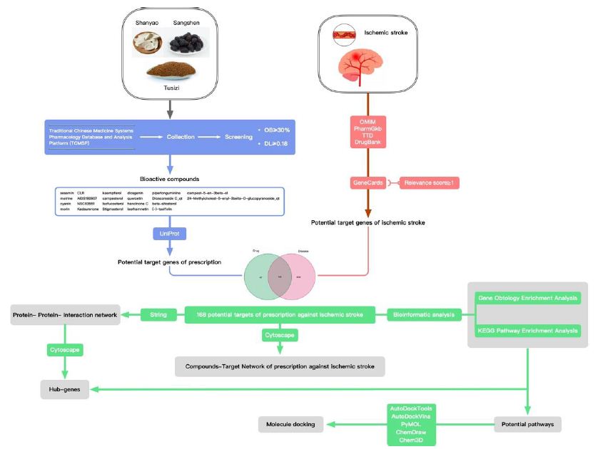
Figure 1: Network pharmacology and molecular docking workflow of bioactive compounds in Guishen Decoction for the treatment of ischemic stroke.

Materials and Methods

Identification and Screening of Bioactive Compounds in Guishen Decoction

Search separately using the Traditional Chinese Medicine Systems Pharmacology Database (TCMSP; https: //tcmsp-e.com/tcmsp.php, accessed on 20 February 2024) [45]. The chemical composition of the three-flavored Guishen Decoction of yam, mulberry and dodder. Guishen Decoction is an effective prescription for the treatment of ischemic stroke. The active ingredients in the drug should be able to normally complete the absorption, distribution, metabolism and excretion processes in the human body. Accordingly, we searched and evaluated the absorption, distribution, metabolism and excretion (ADME) [46] properties of the three Guishen Decoction drugs in the model of human body through the TCMSP database, and screened out compounds without ADME information. According to the standards established by the TCMSP database, compounds should meet the oral bioavailability (OB) ≥ 30% and drug-likeness (DL) ≥ 0.18 before they can be considered as potential bioactive ingredients of Guishen Decoction. Accordingly, OB and DL were used as key parameters to screen the compounds again.

Prediction of Target Genes in Guishen Decoction Bioactive Compounds

The TCMSP database was used to identify target genes of bioactive compounds found in Guishen Decoction, using the Universal Protein Knowledgebase in 2023 (UniProt; https: //www.uniprot.org/, accessed on 20 February 2024) [47] database normalizes the target gene names, deletes non-Homo sapiens category entries and duplicate entries, and organizes the final target gene list.

Prediction of Target Genes in Ischemic Stroke

Use “Ischemic stroke” as the keyword to search in the GeneCards ( https: //www.genecards.org/, accessed on 21 February 2024) [48] database, and filter the results based on Relevance score ≥ 1; use the same keyword to search in the Online Mendelian Inheritance in Man (OMIM; https: //omim.org/, accessed on 21 February 2024) [49], The Pharmacogenomics Knowledgebase (PharmGKB; https: //www.pharmgkb.org/, accessed on 21 February 2024) [50], Therapeutic Target Database (TTD; http: //db.idrblab.net/ttd/ , accessed on 21 February 2024) [51] and DrugBank (https: //go.drugbank.com/, accessed on 21 February 2024) [52] databases. The target gene lists of each database are summarized and redundant content is removed to ensure that the result list is comprehensive and not duplicated. The searches from each database were presented as Venn diagrams using R software.

Construction of Venn Diagram

Use the venn package of R 4.3.2 software to examine the targets of the ingredients in Guishen Decoction and the targets associated with ischemic stroke, and clarify the drug ingredient targets of Guishen Decoction that are related to ischemic stroke, the findings of the analysis are illustrated using a Venn diagram.

Construction of “Compound-Target-Disease” Network

Use Cytoscape 3.10.1 (https: //cytoscape.org/, accessed on 23 February 2024) software to illustrate the interaction between the drug ingredients of Guishen Decoction and ischemic stroke. The specific method is: classify and adjust the activated ingredient information of the compositions in Guishen Decoction and integrate the data into Cytoscape software enables the active ingredients of drugs to be visually displayed in Cytoscape; taking the drug ingredients of Guishen Decoction and the common target genes of ischemic stroke as network nodes, the relationship between the active ingredients of the drug and the common target genes is Displayed in the form of connecting lines, a “compound-target-disease” network is formed.

Protein-Protein-Interaction (PPI) Network Analysis

The STRING 12.0 (https: //cn.string-db.org/, accessed on 23 February 2024) database was used to analyze the intersection of bioactive compounds of Guishen Decoction and ischemic stroke target genes, and the analysis content was limited to human species, the confidence level is 0.7 and above, perform network visualization on the obtained protein, and enter the network into Cytoscape software. After integrating the data from the STRING database on the Cytoscape platform, use the CytoHubba plug-in [53] to calculate the Maximum Clique Centrality (MCC), Maximum Neighborhood Component (MNC) and degree values of each target gene, respectively determine the highest-ranking 10 genes with three algorithm scores, thereby determining potential central target genes in the network. The results of these three algorithms were copied into a set of target genes that were used as potential key targets in the network.

Gene Ontology and Pathway Enrichment Analysis

For the purpose of exploring the potential mechanism of action between the medicinal ingredients of Guishen Decoction and ischemic stroke, the Database for Annotation, Visualization, and Integrated Discovery database (DAVID), version 2021 (https: //david.ncifcrf. gov/home.jsp, accessed on 25 February 2024) [54] was deployed to analyze biological functions, cellular structures, molecular activities and signaling cascades [55-57] to obtain GO, and the first 15 results of KEGG analysis, using the Bioinformatic (https: //www.bioinformatics. com.cn, accessed on 25 February 2024) platform to visualize the results. After calculating the P value, statistically significant results have a P value of less than 0.05.

Molecular Docking

To analyse the interaction between the Guishen Decoction and the main genes specified via the UniProt database molecular connection is used to find the UniProt ID of the main gene., and the crystal structure of the core gene was searched in the RCSB protein database (https: //www.rcsb.org/, accessed on 25 February 2024) [58] based on the protein ID, resulting in IL6 (UniProt ID: P05231; PDB ID: 1ALU), TNF (UniPort ID: P01375; PDB ID: 1TNF), IL1B (UniProt ID: P01584; PDB ID: 1HIB). Import the three-dimensional structure of the core gene into PyMOL 2.5 (https: //pymol.org/, accessed on 25 February 2024) software, remove water molecules and existing small molecule ligands, and obtain the protein ligand file; use the PubChem database (https: //pubchem.ncbi.nlm.nih.gov/, accessed on 25 February 2024) [59] to search Guishen Decoction active ingredients matrine, quercetin, kaempferol For 2D structure, ChemOffice 22 (https: // www.chemdraw.com.cn/, accessed on 25 February 2024) software was used to convert the 2D structure of the active ingredient into a three-dimensional structure, and the three-dimensional structure was optimized using minimum energy. Using the active ingredient as the ligand and the core gene as the receptor, AutoDockTools 4.2 (http: // autodock.scripps.edu/, accessed on 25 February 2024) [60] software was used to predict the binding conformation between the ligand and the receptor, and AutoDockVina 1.2.0 (https: //vina.scripps.edu/, accessed on 25 February 2024) [61,62] software was used to visualize the molecular interaction between the protein and the ligand.

Results

Identification and Screening of Active Compounds in Guishen Decoction

Search the active compounds of the Guishen Decoction drug according to the TCMSP website, and a total of 191 bioactive compounds were obtained. All results were screened and sorted using OB≥30% and DL≥0.18 as the filtering conditions. The UniProt website was adopted to identify the protein target names. After annotation, a total of 33 active ingredients were found in the whole prescription. After excluding the ingredients whose corresponding targets could not be found, the remaining 22 kinds were found. There were a total of 210 corresponding target proteins, which can be used for further analysis (Table 1).

Table 1: The pharmacokinetic properties of the bioactive ingredients of Guishen Decoction based on Traditional Chinese Medicine Systems Pharmacology (TCMSP) database and Uniprot database.

Drug

Mol ID Ingredient Oral Bioavailability OB (≥30%) Drug-Likeness DL (≥0.18)

Target number

mulberry

MOL010300

dIDP

41.08

0.57

none

mulberry MOL002372 (6Z,10E,14E,18E)-2,6,10,15,19,23-hexamethyltetracosa- 2,6,10,14,18,22-hexaene

33.55

0.42

none

mulberry MOL006209 cyanin

47.42

0.76

2

mulberry MOL000737 morin

46.23

0.27

14

mulberry MOL002773 beta-carotene

37.18

0.58

none

mulberry MOL000098 quercetin

46.43

0.28

136

yam MOL000310 DenudatinB

61.47

0.38

none

yam MOL000322 Kadsurenone

54.72

0.38

22

yam MOL000449 Stigmasterol

43.83

0.76

27

yam MOL000546 diosgenin

80.88

0.81

15

yam MOL000953 CLR

37.87

0.68

3

yam MOL001559 piperlonguminine

30.71

0.18

8

yam MOL001736 (-)-taxifolin

60.51

0.27

3

yam MOL005429 hancinol

64.01

0.37

none

yam MOL005430 hancinoneC

59.05

0.39

17

yam MOL005435 24-Methylcholest-5-enyl-3belta-O-glucopyranoside_qt

37.58

0.72

1

yam MOL005438 campesterol

37.58

0.71

1

yam MOL005440 Isofucosterol

43.78

0.76

9

yam MOL005458 DioscoresideC_qt

36.38

0.87

2

yam MOL005461 Doradexanthin

38.16

0.54

none

yam MOL005463 Methylcimicifugoside_qt

31.69

0.24

none

yam MOL005465 AIDS180907

45.33

0.77

10

dodder seed MOL001558 sesamin

56.55

0.83

20

dodder seed MOL000184 NSC63551

39.25

0.76

1

dodder seed MOL000354 isorhamnetin

49.6

0.31

29

dodder seed MOL000358 beta-sitosterol

36.91

0.75

27

dodder seed MOL000422 kaempferol

41.88

0.24

54

dodder seed MOL005043 campest-5-en-3beta-ol

37.58

0.71

1

dodder seed MOL005440 Isofucosterol

43.78

0.76

9

dodder seed MOL005944 matrine

63.77

0.25

10

dodder seed MOL006649 sophranol

55.42

0.28

none

dodder seed MOL000953 CLR

37.87

0.68

3

dodder seed MOL000098 quercetin

46.43

0.28

136

Target Gene Prediction of Ischemic Stroke

Through a comprehensive search of five databases: GeneCards, Online Mendelian Inheritance in Man (OMIM), The Pharmacogenomics Knowledgebase (PharmGKB), Therapeutic Target Database (TTD), and DrugBank, 4212 target genes related to ischemic stroke were collected (Figure 2).

Figure 2: The target genes of ischemic stroke based on Genecards, Online Mendelian Inheritance in Man (OMIM), The Pharmacogenomics Knowledgebase (PharmGkb), Therapeutic Target Database (TTD) and DrugBank databases.

Common Target of Guishen Decoction and Ischemic Stroke

Venn diagram was used to show the potential targets of Guishen Decoction for ischemic stroke (Figure 3). This figure shows 168 intersection target genes of Guishen Decoction and ischemic stroke.

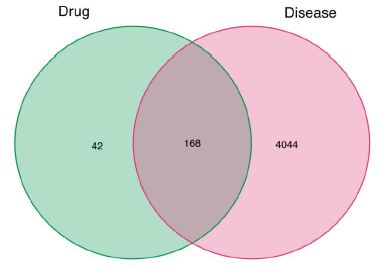
Figure 3: The potential targets of between bioactive compounds of Guishen Decoction against ischemic stroke via Venn diagram. There are 168 target genes in Guishen Decoction bioactive compounds and ischemic intersection (the middle part).

Compound-Target Network Constructions

Cytoscape software was used to describe the interaction between the bioactive components of Guishen Decoction and its potential targets for the treatment of ischemic stroke (Figure 4).

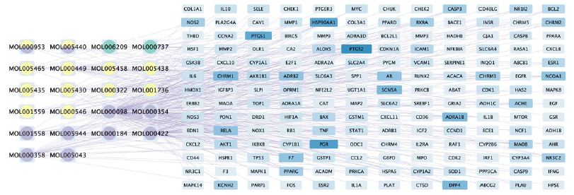
Figure 4: The compound-target network of Guishen Decoction bioactive compounds against ischemic stroke by using Cytoscape. The blue nodes represent the interacting target genes between the compound (yellow, green and purple nodes) and the ischemic stroke. The yellow nodes represent the bioactive compounds belong to yam, the green ones belong to mulberry, the purple ones belong to dodder seed; the nodes containing multicolor indicate that the compound is shared by two or three drugs. The color and width of the square in gene targets represent the number of genes, with darker colors and wider widths indicating a greater number of genes.

Protein-Protein Interaction (PPI) Network

By employing the STRING database and Cytoscape software, we depicted the interaction network between the active compounds of Guishen Decoction and their potential targets for treating ischemic stroke through Protein-Protein Interaction (PPI) analysis (Figure 5). The resulting network has 168 nodes, with 1271 edges after hiding unconnected nodes representing proteins and protein-protein associations, compared to the expected 335 edges, meaning that network interactions for this 168 targets are significantly enriched, and indicates that the proteins are at least partially biologically connected, as a group. Use the CytoHubba plug-in in the Cytoscape software to assign scores to the Betweenness, Closeness, Degree, Eigenvector, Information and LAC values of the PPI network nodes, and filter out each score to be greater than the median. Targets were finally identified as 21 core targets of Guishen Decoction on ischemic stroke (Figure 6).

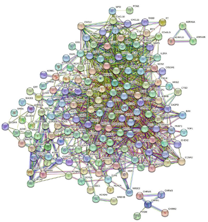
Figure 5: Protein-protein interaction networks are constructed using the STRING database. The network nodes represent proteins, each node represents all the proteins produced by a single, protein-coding gene locus. The colored nodes represent query proteins and first shell of interactors, and the white nodes representing the second shell of interactors. The edges represent protein- protein associations, the confidence levels of interaction are visually represented in the network diagram using color edges, including from curated databases, experimentally determined, gene neighborhood, gene fusions, gene co-occurrence, text-mining, co-expression and protein homology.

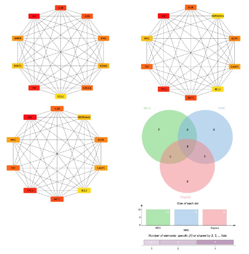
Figure 6: By combining 3 algorithms, MCC, MNC, degree, the key gene network of Guishen Decoction bioactive compounds with scores above the median for ischemic stroke was screened out. The genes with the highest values are considered the most important key genes and are indicated in yellow. Conversely, genes with lower values are considered less important and are indicated in blue, indicating each gene’s ranking position in the network.

Gene Ontology and Pathway Enrichment Analysis

In order to further understand the biological processes, cellular components, molecular functions and pathways involved in the therapeutic effect of Guishen Decoction bioactive compounds on ischemic stroke, a Gene Ontology (GO) study and Kyoto Encyclopedia of Genes and Genomes (KEGG) pathway enrichment analysis was conducted using DAVID bioinformatics resources and the Bioinformatics platform. The results of GO analysis (Figure 7) show that the targets of the bioactive compounds in Guishen Decoction are the most significant biological processes related to xenobiotic stimulus and lipopolysaccharide. Analysis of cellular components showed that these targets mainly involved membrane raft, membrane microdomain and caveola. In terms of molecular function, the top 10 significantly enriched terms include DNA-binding transcription factor binding, G protein-coupled amine receptor activity and transcription coregulator binding. The results of KEGG analysis (Figure 8) show that many targets of the bioactive compounds of Guishen Decoction on ischemic stroke are enriched in signaling pathways, such as lipid and atherosclerosis, AGE-RAGE signaling pathway in diabetic complications, fluid shear stress and atherosclerosis and prostate cancer. Through the analysis of metabolic pathways, signaling pathways such as pathways of neurodegeneration are directly related to ischemic stroke. Find the potential targets and mechanisms of Guishen Decoction to treat ischemic stroke by regulating Pathways of neurodegeneration (Figure 9).

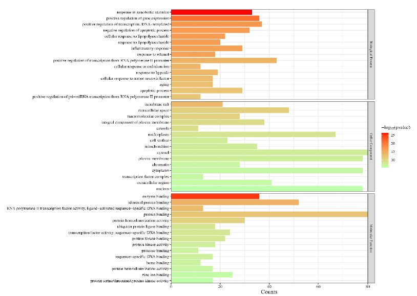
Figure 7: Gene ontology (GO) enrichment analysis. The bar chart represents the most significantly enriched GO terms in biological processes, cellular components, and molecular function, comprising the top 10 terms related to the target genes.

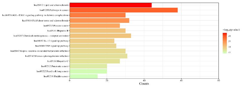
Figure 8: Kyoto Encyclopedia of Genes and Genomes (KEGG) pathway enrichment analysis. The bar chart visualizes the top 30 enriched KEGG pathways of Guishen Decoction against ischemic stroke.

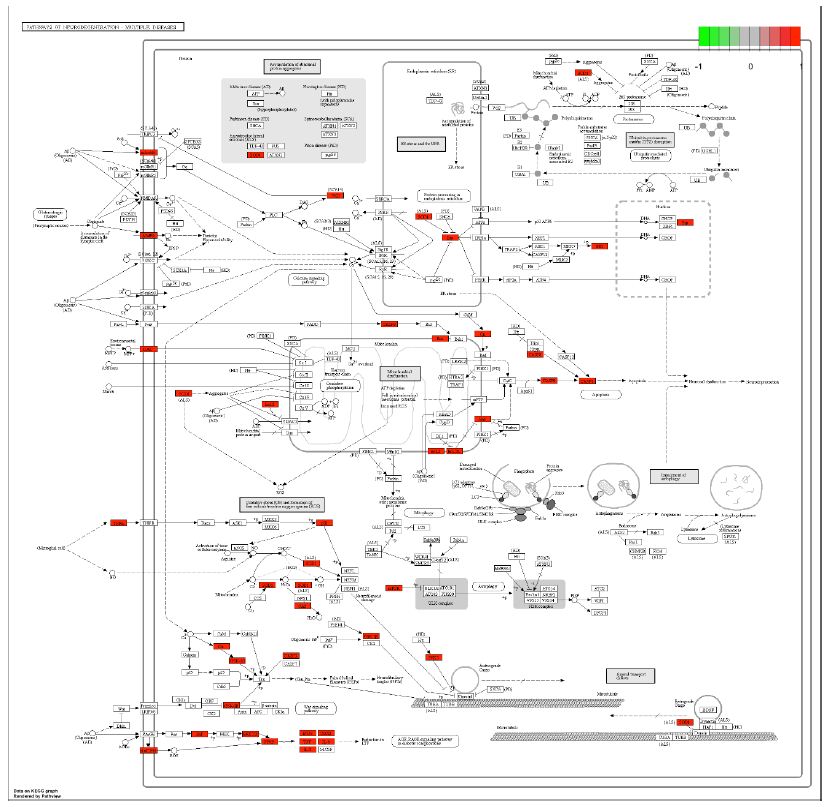
Figure 9: Potential targets and mechanism of bioactive compounds in Guishen Decoction against ischemic stroke. The figure represents the neurotrophin signaling pathway. The red ndoes represent the genes targeted within the pathways of neurodegeneration.

Molecular Docking

Through molecular docking research, the interaction relationship between the three active compounds selected in Guishen Decoction (matrine, kaempferol, quercetin) and three potential target genes (IL6, IL1B, TNF) was analyzed. AutoDockTools software was used to conduct molecular docking studies, and a total of 9 docking results were obtained, 7 of which had binding energies that resisted -5.0 kcal/ mol (Figure 10). The docking scores are shown in Table 2. The lower the binding energy, the tighter the binding between the compound and the protein. The binding energy of kaempferol and quercetin to each target gene is lower than -5.0 kcal/mol. Therefore, kaempferol and quercetin are important in the treatment of ischemic stroke. Play a particularly important role. The results show that most active ingredients have strong binding ability to the target gene TNF. The docking scores of kaempferol, matrine and quercetin combined with TNF are -6.6, -4.9 and -6.7 respectively. This result suggests that the bioactive compounds target TNF of Guishen Decoction may play a key role in the treatment of ischemic stroke. Use PyMOL to construct visualizations of the lowest binding energies between targets and bioactive compounds.

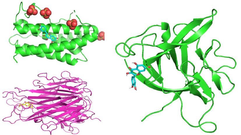
Figure 10: Molecular docking results of the lowest binding energy in each target with the bioactive compound of Guishen Decoction (a) IL6-quercetin, (b) IL1B-quercetin, (c) TNF-quercetin.

Table 2: Binding energy between active compounds and three core targets of Guishen Decoction.

Compound

Binding Energy (kcal/mol)

IL 6 IL 1 B

TNF

kaempferol

-5. 5

-5. 4

-6. 6

matrine

-4. 7

-4. 3

-4. 9

quercetin

-5. 6

-5. 6

-6. 7

Discussion

The complexity of ischemic stroke’s mechanism is profound, and the process of damage to neurons and brain tissue is related to various programmed cell deaths such as apoptosis, pyroptosis, and ferroptosis. The basic process of this disease is that internal and external factors damage the brain, resulting in the interruption or reduction of local blood supply to the brain, and brain cells die due to the inability of the brain tissue to obtain oxygen and other nutrients [63]. Although r-tPA remains the most efficacious medication for treating this condition, only 11% of patients are eligible for its use due to the constraints imposed by the time window for intervention [64]. Therefore, finding alternative treatments for ischemic stroke from TCM is one of the important directions for the development of TCM in the new era.

Clinical work and related literature research have validated that Guishen Decoction has a definite therapeutic effect on ischemic stroke. In previous studies, researchers have confirmed the correlation between ischemic stroke and kidneys [65], TCM prescriptions can not only be used in the treatment of kidney disease [66], but also through the method of nourishing kidney and yin indirectly affects the brain [67], achieving the effect of protecting brain neurons. As a kidney- tonifying and essence- replenishing prescription, the medicinal composition of Guishen Decoction has the effect of inhibiting the activity of oxygen free radicals, suppressing the activation of microglia and astrocytes, and reducing inflammatory reactions. In order to verify the therapeutic effect of Guishen Decoction on ischemic stroke through further research, and to understand as fully as possible the target and mechanism of Guishen Decoction in the treatment of ischemic stroke, this study Network pharmacology and molecular docking technology were employed to study the positive effect of Guishen Decoction on the treatment of ischemic stroke.

After network pharmacology analysis, this study identified 191 potential bioactive compound targets of Guishen Decoction; in the network diagram, this article focused on three central targets: IL6, IL1B, and TNF. To elucidate the pharmacokinetic characteristics of Guishen Decoction bioactive decoder compounds in ischemic brain injury, this study used oral bioavailability (OB) ≥ 30% and drug-likeness (DL) ≥ 0.18 as the screening criteria, and Discard the remaining compounds whose corresponding targets cannot be obtained, and only summarize the remaining compounds as potential bioactive compounds. Three compounds, matrine, quercetin, and kaempferol, were selected from Guishen Decoction for further research because they meet the criteria for drug screening and may play an important role in the treatment of ischemic stroke.

Interpreting the KEGG pathway results in the process of GO and KEGG enrichment analysis shows that Apoptosis, Neurotrophin signaling pathway, Pathways of neurodegeneration, Glioma, lipids, IL-17 signaling pathway, HIF-1 signaling pathway, etc. have all been confirmed and lacking [68-70]. This article conducts visual research on related signaling pathways, and conducts molecular docking verification of 3 key targets and 3 active ingredients. The results show that quercetin and kaempferol play an important role in the treatment of ischemic stroke, as these compounds can be closely bound in the active pouch of key targets with low binding energy. At the same time, TNF looks relatively well with most of guishen’s active analgesics.

This article uses network pharmacology and molecular docking to study the potential active ingredients, possible relevant targets and important biological pathways of Guishen Decoction in the treatment of ischemic stroke, providing a preliminary theory for further experimental research. It must be noted that the pharmacological mechanisms proposed in this article are all derived from network pharmacology technology and require further verification by pharmacological and clinical studies.

Conclusions

In summary, this article uses network pharmacology and molecular docking methods to study the potential mechanism of the bioactive compounds of Guishen Decoction in the treatment of ischemic stroke. The results show that matrine, quercetin, Compounds such as kaempferol may have a therapeutic effect on ischemic stroke through key biological targets such as IL6, IL1B, and TNF. Molecular docking studies have shown that the active ingredients of Guishen Decoction can interact with these targets relatively effectively. These conclusions provide a valuable basis for further clinical and pharmacological research.

References

  1. Wang L, Zhang X, Xiong X, Zhu H, Chen R, et (2022) Nrf2 Regulates Oxidative Stress and Its Role in Cerebral Ischemic Stroke. Antioxidants (Basel, Switzerland) 11: 2377. [crossref]
  2. Merel S Ekker, Jamie I Verhoeven, Mijntje MI Schellekens, Esther M Boot, Mayte E van Alebeek, et al. (2023) Risk Factors and Causes of Ischemic Stroke in 1322 Young Stroke 54: 439-447. [crossref]
  3. Wen-Jun Tu, Zhenping Zhao, Peng Yin, Lei Cao, Jingsheng Zeng, et (2023) Estimated Burden of Stroke in China in 2020. JAMA Network Open 6: e231455. [crossref]
  4. Chuan Qin, Sheng Yang, Yun-Hui Chu, Hang Zhang, Xiao-Wei Pang, Lian Chen, et (2022) Signaling pathways involved in ischemic stroke: Molecular mechanisms and therapeutic interventions. Signal Transduction and Targeted Therapy 7: 215. [crossref]
  5. Gao L, Liu F, Hou P-P, Manaenko A, Xiao Z-P, Wang F, et (2022) Neurons Release Injured Mitochondria as ‘Help-Me’ Signaling After Ischemic Stroke. Frontiers in Aging Neuroscience 14: 785761. [crossref]
  6. Han Y, Li X, Yang L, Zhang D, Li L, Dong X, et (2022) Ginsenoside Rg1 attenuates cerebral ischemia-reperfusion injury due to inhibition of NOX2-mediated calcium homeostasis dysregulation in mice. Journal of Ginseng Research 46: 515-525. [crossref]
  7. Jurcau A, Simion A (2021) Neuroinflammation in Cerebral Ischemia and Ischemia/ Reperfusion Injuries: From Pathophysiology to Therapeutic Strategies. International Journal of Molecular Sciences 23:14. [crossref]
  8. Zhang B, Zhang H-X, Shi S-T, Bai Y-L, Zhe X, et al. (2019) Interleukin-11 treatment protected against cerebral ischemia/reperfusion Biomedicine & Pharmacotherapy Biomedecine & Pharmacotherapie 115: 108816. [crossref]
  9. Shi Y, Han L, Zhang X, Xie L, Pan P, et (2022) Selenium Alleviates Cerebral Ischemia/Reperfusion Injury by Regulating Oxidative Stress, Mitochondrial Fusion and Ferroptosis. Neurochemical Research 47: 2992-3002. [crossref]
  10. Li X, Ma N, Xu J, Zhang Y, Yang P, et (2021) Targeting Ferroptosis: Pathological Mechanism and Treatment of Ischemia-Reperfusion Injury. Oxidative Medicine and Cellular Longevity 2021: 1587922. [crossref]
  11. Zhao Y, Zhang X, Chen X, Wei Y (2022) Neuronal injuries in cerebral infarction and ischemic stroke: From mechanisms to treatment (Review). International Journal of Molecular Medicine 49: 15. [crossref]
  12. Powers WJ, Rabinstein AA, Ackerson T, Adeoye OM, Bambakidis NC, et al. (2019) Guidelines for the Early Management of Patients With Acute Ischemic Stroke: 2019 Update to the 2018 Guidelines for the Early Management of Acute Ischemic Stroke: A Guideline for Healthcare Professionals From the American Heart Association/ American Stroke Stroke 50. [crossref]
  13. Benjamin EJ, Blaha MJ, Chiuve SE, Cushman M, Das SR, et al. (2017) Heart Disease and Stroke Statistics-2017 Circulation 135: e146-e603. [crossref]
  14. Aleksandra Steliga, Przemysław Kowiański, Ewelina Czuba, Monika Waśkow, Janusz Moryś, et (2020) Neurovascular Unit as a Source of Ischemic Stroke Biomarkers— Limitations of Experimental Studies and Perspectives for Clinical Application. Translational Stroke Research 11: 553-579. [crossref]
  15. Derek Barthels, Hiranmoy Das (2020) Current Advances in Ischemic Stroke Research and Therapies. Biochimica et Biophysica Acta. Molecular Basis of Disease 1866: 165260. [crossref]
  16. Lena Bourhy, Aurélien Mazeraud, Fernando A Bozza, Guillaume Turc, Pierre-Marie Lledo, et (2022) Neuro-Inflammatory Response and Brain-Peripheral Crosstalk in Sepsis and Stroke. Frontiers in Immunology 13: 834649. [crossref]
  17. Ji Ma, Shaofeng Shui, Xinwei Han, Dong Guo, Tengfei Li, Lei Yan (2017) microRNA- 200a silencing protects neural stem cells against cerebral ischemia/reperfusion PloS One 12: e0172178. [crossref]
  18. Karantali E, Kazis D, Papavasileiou V, Prevezianou A, Chatzikonstantinou S, et (2021) Serum BDNF Levels in Acute Stroke: A Systematic Review and Meta-Analysis. Medicina (Kaunas, Lithuania) 57: 297. [crossref]
  19. Maria Calvo-Rodriguez, Elena Hernando-Pérez, Sara López-Vázquez, Javier Núñez, Carlos Villalobos, et al. (2020) Remodeling of Intracellular Ca2+ Homeostasis in Rat Hippocampal Neurons Aged In Vitro. International Journal of Molecular Sciences 21:1549. [crossref]
  20. SK Kritas, A Saggini, G Cerulli, A Caraffa, P Antinolfi, et (2014) Corticotropin- Releasing Hormone, Microglia and Mental Disorders. International Journal of Immunopathology and Pharmacology 27: 163-167. [crossref]
  21. Sebastian Zimmer, Philip Roger Goody, Matthias Oelze, Alexander Ghanem, Cornelius F Mueller, et (2021) Inhibition of Rac1 GTPase Decreases Vascular Oxidative Stress, Improves Endothelial Function, and Attenuates Atherosclerosis Development in Mice. Frontiers in Cardiovascular Medicine 8: 680775. [crossref]
  22. An-rong Wang, Lian-fang Mi, Zhi-lei Zhang, Ming-zhe Hu, Ze-yu Zhao, et (2021) Saikosaponin A improved depression-like behavior and inhibited hippocampal neuronal apoptosis after cerebral ischemia through p-CREB/BDNF pathway. Behavioural Brain Research 403: 113138. [crossref]
  23. Ling S-S, Pan R-H, Zhan L-C, Li M, Yang Z-J, et al. (2022) Ruyi Zhenbao Pills for Patients with Motor and Sensory Dysfunction after Stroke: A Double-Blinded, Randomized Placebo-Controlled Clinical Trial. Chinese Journal of Integrative Medicine 28: 872-878. [crossref]
  24. Li L, Liu Y, Zheng Y, Zhu J, Wu D, et al. (2023) Exploring the mechanisms under Zuogui Pill’s treatment of ischemic stroke through network pharmacology and in vitro experimental Frontiers in Pharmacology 14: 1153478. [crossref]
  25. Tang N-Y, Liu C-H, Liu H-J, Li T-C, Liu J-C, et al. (2012) Chinese medicine patterns in patients with post-stroke dementia. Journal of Traditional and Complementary Medicine 2: 123-128. [crossref]
  26. Saji SS, Abhishek DRR, Venkatesh KR (2016) Anti-Oxidant activity of various leaf extracts of mulberry species in rotenone induced oxidative stress model of rat. J Chem Pharm Sci 9: 2732-2736.
  27. Nade VS, Yadav AV (2010) Anti-stress effect of ethyl acetate soluble fraction of Morus alba in chronic restraint Pharmaceutical Biology 48: 1038-1046. [crossref]
  28. Nade VS, Kawale LA, Yadav AV (2010) Protective effect of Morus alba leaves on haloperidol-induced orofacial dyskinesia and oxidative stress. Pharmaceutical Biology 48: 17-22. [crossref]
  29. Turgut NH, Mert DG, Kara H, Egilmez HR, Arslanbas E, et al. (2016) Effect of black mulberry (Morus nigra) extract treatment on cognitive impairment and oxidative stress status of D-galactose-induced aging mice. Pharmaceutical Biology 54: 1052-1064. [crossref]
  30. Bauomy AA (2014) The potential role of Morus alba leaves extract on the brain of mice infected with Schistosoma mansoni. CNS & Neurological Disorders Drug Targets 13: 1513-1519. [crossref]
  31. Shih P-H, Chan Y-C, Liao J-W, Wang M-F, Yen G-C (2010) Antioxidant and cognitive promotion effects of anthocyanin-rich mulberry (Morus atropurpurea ) on senescence-accelerated mice and prevention of Alzheimer’s disease. The Journal of Nutritional Biochemistry 21: 598-605. [crossref]
  32. Rebai O, Belkhir M, Boujelben A, Fattouch S, Amri M (2017) Morus alba leaf extract mediates neuroprotection against glyphosate-induced toxicity and biochemical alterations in the brain. Environmental Science and Pollution Research International 24: 9605-9613. [crossref]
  33. El-Baz KF, Aly H, Khalil W, Booles H (2016) Neuroameliorative effects of berry extracts in Alzheimer induced Int J Pharma Bio Sci 7.
  34. Wattanathorn J, Muchimapura S, Thukhammee W, Tong-un T, Wannanon P, et (2012) Mulberry Fruits Protects Against Age-Related Cognitive Decline. American Journal of Applied Sciences 9: 1503-1511.
  35. Samuel SS, Desai A, Raghubir R, Kumar VR (2016) Neuroprotective profile of mulberry leaf extract in focal cerebral ischemia model in Indian Journal of Research in Pharmacy and Biotechnology 4: 116.
  36. Kang TH, Oh HR, Jung SM, Ryu JH, Park MW, et (2006) Enhancement of neuroprotection of mulberry leaves (Morus alba L.) prepared by the anaerobic treatment against ischemic damage. Biological & Pharmaceutical Bulletin 29: 270-274. [crossref]
  37. Kaewkaen P, Tong-Un T, Wattanathorn J, Muchimapura S, Kaewrueng W, et al. (2012) Effects of Mulberry Fruit Powder in Animal Model of American Journal of Agricultural and Biological Sciences 7: 322-329.
  38. Tam DNH, Nam NH, Elhady MT, Tran L, Hassan OG, et al. (2021) Effects of Mulberry on the Central Nervous System: A Literature Current Neuropharmacology 19: 193-219. [crossref]
  39. Hou L, Yang L, Zhu C, Miao J, Zhou W, Tang Y, et (2023) Cuscutae semen alleviates CUS-induced depression-like behaviors in mice via the gut microbiota- neuroinflammation axis. Frontiers in Pharmacology 14: 1107781. [crossref]
  40. Ju IG, Kim N, Choi JG, Lee JK, Oh MS (2019) Cuscutae Japonicae Semen Ameliorates Memory Dysfunction by Rescuing Synaptic Damage in Alzheimer’s Disease Nutrients 11: 2591. [crossref]
  41. Kim S, Shin S, Hyun B, Kong H, Han S, et (2012) Immunomodulatory Effects of Dioscoreae Rhizome Against Inflammation through Suppressed Production of Cytokines Via Inhibition of the NF-κB Pathway. Immune Network 12: 181-188. [crossref]
  42. Lee S-C, Tsai C-C, Chen J-C, Lin C-C, Hu M-L, et (2002) The evaluation of reno- and hepatoprotective effects of huai-shan-yao (Rhizome Dioscoreae). The American Journal of Chinese Medicine 30: 609-616. [crossref]
  43. Li S, Zhang B (2014) Traditional Chinese medicine network pharmacology: Theory, methodology and application. Chinese Journal of Natural Medicines 11: 110-120. [crossref]
  44. Liu C, Liu L, Li J, Zhang Y, Meng D-L (2021) Virtual screening of active compounds from jasminum lanceolarium and potential targets against primary dysmenorrhea based on network pharmacology. Natural Product Research 35: 5853-5856. [crossref]
  45. Ru J, Li P, Wang J, Zhou W, Li B, et (2014) TCMSP: A database of systems pharmacology for drug discovery from herbal medicines. Journal of Cheminformatics 6: 13. [crossref]
  46. Williams J, Siramshetty V, Nguyễn Ð-T, Padilha EC, Kabir M, et (2022) Using in vitro ADME Data for Lead Compound Selection: An Emphasis on PAMPA pH 5 Permeability and Oral Bioavailability. Bioorganic & Medicinal Chemistry 56: 116588. [crossref]
  47. Consortium U (2023) UniProt: The Universal Protein Knowledgebase in Nucleic Acids Research 51: D523-D531. [crossref]
  48. Safran M, Dalah I, Alexander J, Rosen N, Stein TI, et (2010) GeneCards Version 3: The human gene integrator. Database: The Journal of Biological Databases and Curation 2010: baq020. [crossref]
  49. Amberger JS, Bocchini CA, Schiettecatte F, Scott AF, Hamosh A (2015) org: Online Mendelian Inheritance in Man (OMIM®), an online catalog of human genes and genetic disorders. Nucleic Acids Research 43: D789-D798. [crossref]
  50. Barbarino JM, Whirl-Carrillo M, Altman, RB, Klein TE (2018) PharmGKB: A worldwide resource for pharmacogenomic information. Wiley Interdisciplinary Systems Biology and Medicine 10: e1417. [crossref]
  51. Zhou Y, Zhang Y, Zhao D, Yu X, Shen X, et (2024) TTD: Therapeutic Target Database describing target druggability information. Nucleic Acids Research 52: D1465-D1477. [crossref]
  52. Knox C, Wilson M, Klinger CM, Franklin M, Oler E, et (2024) DrugBank 6.0: The DrugBank Knowledgebase for 2024. Nucleic Acids Research 52: D1265-D1275. [crossref]
  53. Chin C-H, Chen S-H, Wu H-H, Ho C-W, Ko M-T, et al. (2014) CytoHubba: Identifying hub objects and sub-networks from complex interactome. BMC Systems Biology 8: S11. [crossref]
  54. Sherman BT, Hao M, Qiu J, Jiao X, Baseler MW, et (2022) DAVID: A web server for functional enrichment analysis and functional annotation of gene lists (2021 update). Nucleic Acids Research 50: W216-W221. [crossref]
  55. Dennis G, Sherman BT, Hosack DA, Yang J, Gao W, Lane HC, et (2003) DAVID: Database for Annotation, Visualization, and Integrated Discovery. Genome Biology 4: R60. [crossref]
  56. Huang DW, Sherman BT, Lempicki RA (2009) Systematic and integrative analysis of large gene lists using DAVID bioinformatics Nature Protocols 4: 44-57. [crossref]
  57. Kanehisa M, Furumichi M, Sato Y, Kawashima M, Ishiguro-Watanabe M (2023) KEGG for taxonomy-based analysis of pathways and Nucleic Acids Research 51: D587-D592. [crossref]
  58. Berman HM, Westbrook J, Feng Z, Gilliland G, Bhat TN, et al. (2000) The Protein Data Nucleic Acids Research 28: 235-242.
  59. Kim S, Chen J, Cheng T, Gindulyte A, He J, et (2021) PubChem in 2021: New data content and improved web interfaces. Nucleic Acids Research 49: D1388-D1395. [crossref]
  60. Morris GM, Huey R, Lindstrom W, Sanner MF, Belew RK, et (2009) AutoDock4 and AutoDockTools4: Automated docking with selective receptor flexibility. Journal of Computational Chemistry 30: 2785-2791. [crossref]
  61. Eberhardt J, Santos-Martins D, Tillack AF, Forli S (2021) AutoDock Vina 1.2.0: New Docking Methods, Expanded Force Field, and Python Bindings. Journal of Chemical Information and Modeling 61: 3891-3898. [crossref]
  62. Trott O, Olson AJ (2010) AutoDock Vina: Improving the speed and accuracy of docking with a new scoring function, efficient optimization, and multithreading. Journal of Computational Chemistry 31: 455-461. [crossref]
  63. Rui Mao, Ningning Zong, Yujie Hu, Ying Chen, Yun Xu (2022) Neuronal Death Mechanisms and Therapeutic Strategy in Ischemic Neuroscience Bulletin 38: 1229-1247. [crossref]
  64. Zhao F, Peng C, Li H, Chen H, Yang Y, et (2023) Paeoniae Radix Rubra extract attenuates cerebral ischemia injury by inhibiting ferroptosis and activating autophagy through the PI3K/Akt signaling pathway. Journal of Ethnopharmacology 315: 116567. [crossref]
  65. T Olsson, N Marklund, Y Gustafson, B Näsman (1992) Abnormalities at different levels of the hypothalamic-pituitary-adrenocortical axis early after stroke. Stroke 23: 1573-1576. [crossref]
  66. Yang L, Wang B, Ma L, Fu P (2022) Traditional Chinese herbs and natural products in hyperuricemia-induced chronic kidney Frontiers in Pharmacology 13: 971032. [crossref]
  67. Liu Y, Wu D, Yan X, Xu X, Zhu J, et al. (2024) Zuogui Pill Promotes Neurite Outgrowth by Regulating OPN/ IGF-1R/PTEN and Downstream mTOR Signaling Combinatorial Chemistry & High Throughput Screening. [crossref]
  68. Cui Y, Zhang Y, Zhao X, Shao L, Liu G, Sun C, et al. (2021) ACSL4 exacerbates ischemic stroke by promoting ferroptosis-induced brain injury and Brain, Behavior, and Immunity 93: 312-321. [crossref]
  69. Ghosh MK, Chakraborty D, Sarkar S, Bhowmik A, Basu M (2019) The interrelationship between cerebral ischemic stroke and glioma: A comprehensive study of recent Signal Transduction and Targeted Therapy 4: 42. [crossref]
  70. Yang C, Hawkins Ke, Doré S, Candelario-Jalil E (2019) Neuroinflammatory mechanisms of blood-brain barrier damage in ischemic stroke. American Journal of Physiology. Cell physiology 316. [crossref]

Exploration of Phenomenology and Dimensionality of Happiness: A Qualitative Study

DOI: 10.31038/PSYJ.2025714

Abstract

Happiness is characterized by joy and satisfaction, and the two are essential for both society and personal wellbeing. The aim of this study was to investigate the phenomenology and dimensionality of happiness in university students using thematic analysis. Our goal is to explore subjective experiences and multifaceted components of happiness in university students, to identify their primary source of happiness, to investigate the strategies and approaches employed by individuals to achieve happiness, to assess the perspective of participants on the measurability of happiness and to explore the influence of interpersonal relationships and environment on happiness. Our target population was university students both male and female, age ranging from 18 to 26. We used convenience samples to collect data from open ended questionnaire (43) and took semi-structured interviews (6) by thematic analysis to investigate the phenomenology and dimensonality of happiness. Our results highlighted the individualized and variable aspect of happiness emphasizing the subjective nature of happiness. Happiness incorporates a lot of thrilling emotions such as pleasure, satisfaction, contentment, fulfillment and relaxation. The study emphasizes the huge influence of interpersonal relationships and environment on happiness. The concept of happiness varies from person to person, stressing that happiness is highly complicated and unique concept. Our research identified various strategies to achieve happiness such as taking part in fun activities, fostering meaningful relationships and achieving goals. The study adds to our understanding of variables affecting individual happiness, the influence of relationships on our happiness and emphasizes the importance of living in better environment to promote wellbeing.

Keywords

Happiness, Thematic analysis, Relationships, Environment, Wellbeing, University students

Introduction

Concept of happiness is a unique yet highly individual concept which has been fascinating many philosophers, psychologists and researchers. Happiness is essential to both the harmony of the society and the wellbeing of a person. It is characterized by joy, satisfaction, fulfillment and contentment. This study explores the phenomenology and dimensionality of happiness among university students by thematic analysis to understand its complex nature.

Happiness has several different facets. It is not a single, cohesive feeling. Our objective is to explore and investigate what pleasure means to university students by comprehending these elements. Happiness is a multidimensional concept so it is not studied merely as a single emotion but as a composite of different pleasant emotions and experiences. The aim of our study is to improve our knowledge of how happiness affect our everyday life, especially the life of university students because there experiences and developmental phases provide us new insights in happiness.

Number of variables that affect happiness have been identified in previous studies. These variables includes fulfilling relationships, achieving objectives and taking part in worthwhile experiences. Watson et al. [1] developed the Positive And Negative Affect Schedule (PANAS). According to Watson, PANAS serves as a fundamental instrument to measure the wide range of components that affect happiness.

Furthermore, other important and comprehensive theories of happiness were developed by Csikszentmihalyi [2] and Seligman [3] which gave the idea of flow and positive psychology have enhanced our knowledge of well being and positive emotions by providing the difference between Hedonia (pleasure) and eudaimonia (meaning and self-actualization). These concepts were explored by Ryan and Deci [4] and further evoloved by Huda and Waterman (2014).

This study examines adolescent happiness through thematic analysis of data from surveys and semi-structured interviews. This approach makes it possible to conduct a comprehensive, in-depth analysis of the participants’ experiences, emphasizing the value of early experiences and connections as well as common sources of pleasure like reaching objectives and having fun. The results demonstrate the relevance of social relationships and personal accomplishments in influencing happiness, underscoring the role that supportive environments play in fostering well-being.

Literature Review

To better comprehend the multifaceted nature of happiness, a wide range of domains and perspectives have been covered in past studies on happiness. Seligman and Csikszentmihalyi [5] established positive psychology by defining the fundamental components of happiness, which include accomplishments, positive feelings, engagement, connections, and purpose.

Their ground-breaking study demonstrated the criticality of cultivating a feeling of purpose and fulfillment for enduring happiness. An important contribution to the field was made by Ryan and Deci (2001) when they distinguished between hedonic and eudaimonic well-being. They emphasized that although pleasure and enjoyment are essential to happiness (hedonia), a deeper sense of meaning and self-actualization are also vital for long-term wellbeing (eudaimonia).

Their conclusions highlighted how crucial it is to have sincere connections and pursue noble goals in order to enjoy life to the fullest. Diener et al. [6] conducted a ground-breaking study on subjective well-being in which they looked at people’s affective and cognitive assessments of their own lives. According to their research, personality traits, financial situation, and social ties all play a big role in happiness. Diener’s work laid the groundwork for comprehending subjective well-being as an essential component of overall contentment.

The field was enhanced by Kahneman at al. [7] by generating the concept of affective forecasting, which studies how people anticipate and assess their future emotional experiences. Their research highlighted systematic flaws in human judgment and highlighted how hard it is to foresee what will ultimately make us happy. The findings of this study have important ramifications for treatments meant to enhance wellbeing and judgment Additionally, research on how to improve subjective well-being delves into the science of happiness [8]. Through experimental and long-term studies, they discovered that feeling thankful, showing kindness to others, and having wonderful experiences are all effective strategies to boost happiness.

Their findings have informed interventions aimed at improving well-being and resilience. Stress causing factors in university students are related to well-being [9]. Those can be replaced with happiness and positive emotions. In addition to these basic studies, ongoing research endeavours’ continue to explore various facets of pleasure, including its cultural diversity, biological underpinnings, and social implications. Scholars endeavor to enhance their comprehension of the intricate characteristics of pleasure and its role in human wellbeing by integrating viewpoints from other disciplines, including psychology, neurology, sociology and others.

Objective

  • To explore the subjective aspect and multifaceted components of happiness.
  • To explore the source of happiness.
  • To investigate the strategies and approaches employed by individuals to achieve happiness.
  • To assess the perspective on measurability of happiness.
  • To explore the impact of relationships and environment on happiness.

Method

Qualitative research design was used in current research. This study involves in depth interviews to understand the opinions and viewpoint of individuals on phenomenology and dimensionality of happiness. Furthermore, open ended questionnaire was used in qualitative analysis to measure the frequency of happiness promoting variables.

Procedure

54 university students participated in the study. Participants ranged from 18 to 26 years old. 44 participants completed open ended questionnaires and 7 participants were engaged in semi-structured interviews. Participants were not organized based on ethnicity and socioeconomic status. Participants were given the right to engage in interviews and questionnaire voluntarily. After verbal consents were taken, we recorded the interview. Confidently of the participant data was strictly maintained throughout the study. Thematic analysis was used in our research. Our research was formulated in themes and sub themes. Participants who took part in open ended questionnaire were coded as O.E.Q and those who gave interviews were coded as S.I.

Findings

Several significant findings have come from our research on the phenomenology and dimensionality of happiness in university students. Through the analysis of data from (6) semi-structured interviews and (43) class open ended questionnaire, we were able to find common patterns that highlight the complex nature of happiness. The participants in the study were people with a variety of age, gender, and socioeconomic backgrounds. The complexity of human experiences was reflected in the wide range of viewpoints on happiness that descriptive statistics revealed. Our study showed that the source of happiness of female participants were their friends and the source of happiness of male were their mothers. Following are the themes and sub themes of our study.

Subjective Aspect and Multifaceted Components of Happiness

The participants highlighted the individualized and variable aspect of happiness, emphasizing its subjective nature. Wide variations in responses suggest that everyone experiences and perceives happiness in various ways. Pleasure, satisfaction, fulfillment, and good emotions were all mentioned as parts of the complex emotion that is happiness. The various facets of happiness were expressed by the participants, highlighting its complexity and richness.

  • “Feeling of pleasure and contentment.” (O.E.Q, 3)
  • “Happiness to me is a profound sense of contentment and fulfillment derived from living authentically, fostering meaning full connections, pursuing passion and finding joy in the present moment.” (O.E.Q, 37)

Following are the sub themes:

Personal Perspectives on Happiness

According to participants, Happiness is a complex yet highly individualized concept. It has variable aspects emphasizing its subjective nature. According to our participants, happiness is characterized by joy, satisfaction, fulfillment and contentment.

  • “A state of satisfaction and relaxation, fulfillment.” (O.E.Q, 12)

Diverse Factors Contributing to Well-being

Wide variations in responses suggest that everyone experiences and perceives happiness in various ways. According to our participants, wide range of factors contribute to their happiness such as goal attainment, achievements and pursuing their interests.

  • Completing my task makes me happy”. (S.I, 6)

Goal Attainment and Achievement

Taking part in worthwhile activities and reaching goals were mentioned as significant factors in happiness. Participants who completed tasks and pursued their interests showed feelings of contentment and happiness.

  • Achieving goals that I have set for myself makes me happy.” (S.I, 3)

Source of Happiness

According to our findings, The primary source of happiness for our participants are their mothers. They emphasized that spending time with their mothers and seeing them happy is their greatest happiness. Their social circle and interests and their achievements all comes later. Following are the sub themes:

Primary Source of Happiness

For a considerable number of participants, their mother was the source of their greatest happiness. It was said that they felt love, support, and emotional fulfillment from their mother. Participants emphasized how their happiness and general well-being were shaped by their mothers’ love and nurturing.

  • “For me, making my mother happy ultimately makes me happy.” (O.E.Q, 13)
  • “Although there are a lot factors that makes me happy but seeing me mother happy bring me the most happiness.” (S.I, 5)

Secondary Source of Happiness

According to the data we collected from our interviews, the social circle of the participants, their goals and interests, their achievements played the role of secondary source of happiness.

Psychological Impact of Happiness

While the participants’ approaches to happiness varied, similar elements included goal-setting and achievement, cultivating deep connections, and engaging in mindfulness practices. Finding joy in the present, following passions, and leading an authentic life were found to be important routes to happiness. A happy existence, according to the participants, also requires having inner peace, being grateful, and spending quality time with loved ones. In order to experience lasting enjoyment, participants stressed the need of emotional and psychological well-being.

  • I can achieve happiness by doing things I like.” (S.I, 3)

Well-being

Our participants stressed upon the need of emotional and psychological well being. According to them, being happy requires positive emotions, having inner peace, taking part in activities and doing things that improve our personal well being.

Mindfulness

Although participants response to happiness varied, similar factors were included in order to attain happiness such as goal achievement, improving interpersonal relationships and engaging in mindfulness practices like meditation, positive thinking and self compassion.

  • “Inner peace and our well being plays a crucial role in being happy so I would focus on achieving these to make myself happy.” (S.I, 9)

Participant’s Perspective on Happiness Measurability

Most participants said that they didn’t think it was possible to quantify happiness. Happiness is difficult to measure due to its complexity and subjective character, even though it may be subjectively felt and observed. Participants underlined that conventional assessment techniques fall short of truly capturing happiness since it is a highly subjective and unique feeling. According to participants, happiness is an emotion that can’t be measured though there were some who said that they can quantify their emotions.

  • “I don’t think happiness can be measured.” (S.I, 5)

Happiness can be Measured

According to the data we collected from our research, happiness has a subjective nature. According to most of our participants, their happiness can’t be measured. Happiness is a complex feeling, it can’t be quantified.

  • “Emotions are not something that can be measured.” (S.I, 7)

Happiness Can’t be Measured

Though most of the participants said that emotions can’t be quantified. There were some who said that they can quantify their emotions. They said their emotions can be rated on a scale.

I think emotions can be measured. I can rate my happiness.” (S.I, 1)

Social Impact on Happiness

Participants highlighted the satisfaction and contentment that come from spending time with loved ones, and relationships were shown to be a major source of happiness. Having deep relationships with friends and family was thought to be essential to happiness in general. According to the participants, their environment had a huge influence on their happiness. The emotions of people around them had a significant effect on their own emotion and determined their happiness.

  • “Spending time with my family makes me happy.” (O.E.Q, 4)
  • “When I am not feeling good internally, my emotions are messed up. If I spend time with people like my friends or family, it eventually brings me happiness.” (S.I, 1)

Role of Interpersonal Relationship

Our participants stressed upon the fact how their happiness is affected by their interpersonal relationships. According to them interpersonal relationships play huge role in attaining happiness. Spending time with their loved loves especially with family gives them feelings like satisfaction and contentment.

  • “Being with my friends and family makes me the most happy.” (S.I, 5)

Role of Environment

According to the data we collected, environment of people have huge influence on their feelings and emotions. The emotions of people around them had a significant impact on their happiness, Especially their family and friends. Seeing them happy made happy and seeing them sad made them sad.

  • “People around me have a huge influence on my emotions. There mood determine my mood.” (S.I, 4)

According to our research, adolescent happiness is a complex and highly individualized experience. Several factors, like as one’s accomplishments, relationships, and mindfulness exercises, have an impact on it. The main sources of happiness usually involve one’s family, especially mothers, and the satisfaction that comes from achieving one’s own objectives. Even though it’s widely acknowledged that happiness is essential to wellbeing, its complexity makes it difficult to measure. In the end, the study emphasizes how important relationships and psychological wellness are to adolescents’ happiness judgments.

Discussion

The phenomenology and dimensionality of happiness in university students have been studied, and the results provide fascinating new perspectives on how university students perceive and comprehend happiness. University students of different ages, genders, and socioeconomic backgrounds provide a wide spectrum of opinions for the study, which is conducted using semi-structured interviews and class open ended questionaire. We collected our data through thematic analysis.

The first objective of our study was to explore the subjective nature and multifaceted components of happiness. According to our findings, Happiness is a complex yet highly individualized concept. Wide variations in responses suggest that everyone experiences and perceives happiness in various ways. According to our participants, wide range of factors contribute to their happiness such as goal attainment, achievements and pursuing their interests. Taking part in worthwhile activities and reaching goals were mentioned as significant factors in happiness. Many researchers have given their studies on subjective nature of happiness. The study “The crosscultural corelates of life satisfaction and self-esteem” by Diener and Diener [10] highlights the subjective nature of happiness across diverse culture context.

The second objective of our study was to investigate the source of happiness. According to our participants, The primary source of happiness for our participants are their mothers. They emphasized that spending time with their mothers and seeing them happy is their greatest happiness, the social circle of the participants, their goals and interests, their achievements played the role of secondary source of happiness. There are numerous previous studies on this topic, Argyle [11] stresses upon the significance of interpersonal relationships, particularly with family members in attaining happiness.

The third objective of our study was to investigate the strategies and approaches employed by individuals to achieve happiness. While the participants’ approaches to happiness varied, similar elements included goal-setting and achievement, cultivating deep connections, and engaging in mindfulness practices. Our participants stressed upon the need of emotional and psychological well being. There are previous literatures that align with our findings. Emmons [12] highlighted the personal goals centre’s upon individual’s identity and life purpose, contributing to attaining happiness. Locke and Lathem [13] proposed that setting specific and challenging goals leads to higher performance and satisfaction leading to achieve happiness. Brown and Ryan [14] found that individuals who practice mindfulness tend to have higher level of well-being and reduce stress.

The fourth objective of our study was to assess the participant’s perspective on measurability of happiness. Most participants said that they didn’t think it was possible to quantify happiness. Happiness is difficult to measure due to its complexity and subjective character, even though it may be subjectively felt and observed. Participants underlined that conventional assessment techniques fall short of truly capturing happiness since it is a highly subjective and unique feeling. According to participants, happiness is an emotion that can’t be measured though there were some who said that they can quantify their emotions. Diener (1984) emphasized that happiness varies signficantly for individuals based on their personal experiences and perspectives that makes the qunatification of happiness challenging. Lyobomirsky and Lipper [15] developed the Subjective Happiness Scale to measure subjective happiness globaly but acknowledged its limitations.

The fifth objective of our study was to explore the impact of relationships and environment on happiness. Participants highlighted the satisfaction and contentment that come from spending time with loved ones, and relationships were shown to be a major source of happiness. Having deep relationships with friends and family was thought to be essential to happiness in general. According to the participants, their environment had a huge influence on their happiness. The emotions of people around them had a significant effect on their own emotion and determined their happiness. Diener and Seligman [16] highlighted that strong social bonds play huge role in attaining happiness and subjective well-being. Similarly, Barmiest and Leary [17] argued that forming meaningful bonds and the need to belong is a crucial human motivation which is essential for emotional well-being. Fowler and Christakis [18] discovered that happiness can spread through social networks, stressing that emotional state of others in one’s environment can influence their happiness and emotional well-being. Happiness has cultural and religious bases [19].

Our study explores the Phenomenology and Dimensionality of happiness through thematic analysis among university students. We collected data from semi-structured interviews and open ended questionnaires. According to our findings, happiness is a complex concept, it has subjective nature which makes the quantification of happiness challenging. Relationship especially with family and the surroundings have huge impact on happiness.

Conclusion, Limitations and Recommendation

In conclusion, a thorough thematic analysis of this study has highlighted the intricate and varied aspects of university students’ happiness. According to our research, happiness is a mixture of several emotions like joy, fullness, and contentment rather than a single, universal experience. The accomplishment of personal objectives and taking part in fun activities were found to be the main sources of happiness, with a strong focus on the importance of family bonds, especially those with mothers. The significance of relationships and supportive circumstances in augmenting individuals’ sense of well-being was emphasized by the participants. This study’s dependence on self-reported data, which could be biased since participants could modify their answers to reflect what they think is socially acceptable, is one of its limitations. Furthermore, even though the sample size was diverse, it was small and limited to a particular demographic, which might have limited how broadly the results could be applied. Subjectivity is introduced by the qualitative character of the interviews and the subjective interpretations that are part of the overall concept analysis, which may have an impact on the outcomes.

To improve the generalizability of the results, larger and more varied sample sizes should be the goal of future study. Long term studies might also be helpful in seeing how happiness varies over time and how multiple factors affect happiness over time. Furthermore, combining quantitative and qualitative approaches could offer a more thorough understanding of happiness and solve the difficulties associated with quantifying such a subjective emotion. Future research can help further understand the complexity of adolescent happiness and develop more potent interventions and techniques for fostering young people’s well-being by pursuing these paths of inquiry.

References

  1. Watson D, Clark LA, Tellegen A (1988) Development and validation of brief measures of positive and negative affect: The PANAS scales. Journal of Personality and Social Psychology 54: 1063-1070. [crossref]
  2. Csikszentmihalyi M (1990) Flow: The psychology of optimal experience. Harper & Row.
  3. Seligman MEP (2002) Authentic happiness: Using the new positive psychology to realize your potential for lasting fulfillment. Free Press.
  4. Ryan RM, Deci EL (2001) On happiness and human potentials: A review of research on hedonic and eudaimonic well-being. Annual Review of Psychology 52: 141-166.
  5. Seligman MEP, Csikszentmihalyi M (2000) Positive psychology: An introduction. American Psychologist 55: 5-14.
  6. Diener E (1984) Subjective well-being. Psychological Bulletin 95: 542-575.
  7. Kahneman D, Krueger AB, Schkade D, Schwarz N, Stone AA (2004) A survey method for characterizing daily life experience: The day reconstruction method. Science 306: 1776-1780. [crossref]
  8. Lyubomirsky S, Sheldon KM, Schkade D (2005) Pursuing happiness: The architecture of sustainable change. Review of General Psychology 9: 111-131.
  9. Javaid ZK, Faizi DW.un-N, Lodhi AK, Kamran M (2024a) Conceptualization of Happiness Across Cultures and Role of Religion: A Thematic Analysis on Young Adults. Journal of Arts & Social Sciences 11: 25-33.
  10. Diener E, Diener M (1995) Cross-cultural correlates of life satisfaction and self-esteem. Journal of Personality and Social Psychology 68: 653-663. [crossref]
  11. Argyle M (1999) Causes and correlates of happiness. In D. Kahneman, E. Diener, & N. Schwarz (Eds.), Well-being: The foundations of hedonic psychology (pp. 353373) Russell Sage Foundation.
  12. Emmons RA (1986) Personal strivings: An approach to personality and subjective well-being. Journal of Personality and Social Psychology 51: 1058-1068.
  13. Locke EA, Latham GP (2002) Building a practically useful theory of goal setting and task motivation: A 35-year odyssey. American Psychologist 57: 705717.
  14. Brown KW, Ryan RM (2003) The benefits of being present: Mindfulness and its role in psychological well-being. Journal of Personality and Social Psychology 84: 822-848. [crossref]
  15. Lyubomirsky S, Lepper H S (1999) A measure of subjective happiness: Preliminary reliability and construct validation. Social Indicators Research 46: 137-155.
  16. Diener E, Seligman MEP (2002) Very happy people. Psychological Science 13: 81-84. [crossref]
  17. Baumeister RF, Leary M R (1995) The need to belong: Desire for interpersonal attachments as a fundamental human motivation. Psychological Bulletin 117: 497-529.
  18. Fowler JH, Christakis NA (2008) Dynamic spread of happiness in a large social network: Longitudinal analysis over 20 years in the Framingham Heart Study. BMJ 337: a2338. [crossref]
  19. Javaid ZK, Chen Z, Ramzan M (2024b) Assessing stress causing factors and language related challenges among first year students in higher institutions in Pakistan. Acta Psychologica 248: 104356. 

Efficacy of a BioMin F Toothpaste Compared to Conventional Toothpastes in Remineralisation and Dentine Hypersensitivity: An Overview

DOI: 10.31038/JDMR.2025811

Abstract

Dentine hypersensitivity (DH) and enamel demineralisation are prevalent oral health concerns. BioMin toothpaste, containing fluoro-calcium phosphosilicate (FCPS), has emerged as a potential alternative to conventional toothpastes for remineralisation and DH management. This literature overview analyses the efficacy of a BioMin F toothpaste compared to selected conventional toothpastes in addressing DH and remineralisation. The review examines in vitro and clinical studies (in vivo) to assess the relative benefits and limitations of both approaches. Analysis of controlled clinical trials utilising VAS scores, Schiff Cold Air sensitivity scores (SCASS), and SEM imaging revealed BioMin F exhibits a distinctive temporal efficacy gradient: modest immediate tubular occlusion (18.1% sensitivity reduction at one-minute) but superior long-term efficacy (61.1% reduction at 6 weeks). Scanning electron microscopy confirmed progressive mineralisation within the dentinal tubules (50% occlusion at three weeks increasing to 75% at 6 weeks) through formation of acid-resistant fluorapatite crystals. This overview concludes that BioMin F may be appropriate for sustained rather than immediate relief, although it is recognised that more standardised long-term studies are required to validate its efficacy.

Introduction

DH is a prevalent clinical condition defined short, sharp pain that arises from exposed dentine in response to thermal, tactile, evaporative, osmotic, or chemical stimuli, in the absence of any other dental disease [1]. The pain is usually localised and self-limiting but can significantly impact an individual’s quality of life by affecting routine oral functions such as eating, drinking, and toothbrushing [2]. The average global prevalence from all studies reported by Zeola et al. [3] is around 33.5% (ranging from 4.8% to 62.3% with an estimated prevalence of 11.5%) in adults. West et al. [4] also reported that 41.9% of participants experienced DH in a multi-centre European study of young adults. DH occurs when the dentinal tubules become exposed due to loss of enamel via erosion, abrasion, abfraction or gingival recession. This allows external stimuli to affect the pulpal nerve endings resulting in a pain response. Brannstrom and Astrom’s hydrodynamic theory is the widely accepted mechanism, which proposes that movement of tubular fluid within the open dentinal tubules activates nociceptors near the pulp, resulting in pain. Management strategies for DH therefore aim to occlude the dentinal tubules, prevent fluid movement, or by nerve desensitisation which ultimately reduces their responsiveness to stimuli. Over the counter (OTC) desensitising toothpastes are the most accessible and commonly used form of treatment, and their efficacy depends on the active ingredients and mechanism of actionof the individual toothpaste formulations. BioMin F (Biomin Technologies Limited UK) is a novel bioactive glass toothpaste containing 5% fluoro-calcium phosphosilicate (FCPS), which slowly releases calcium, phosphate, and fluoride ions to promote acid-resistant fluorapatite (FAP) crystals, leading to sustained occlusion of the dentinal tubules and potential long-term relief from DH [5]. Although other desensitising toothpastes are available in the UK market, long term comparative clinical efficacy comparing the formulations are limited. Several studies have reported short term (<6 months) improvements in DH but variations in methoodology is somewhat problematic when drawing direct comparisions between the toothpastes used for DH management. DH therefore presents a persistent clinical challenge due to its high prevalence and effects on patients’ quality of life and currently there does not appear to be one ideal desensitising product for everyone [6]. Various methods have been adopted to manage DH symptoms; however, the multifactorial nature and complexity of its aetiology, coupled with the rising global prevalence, underscore the need for research and development of more effective and long-term solutions

Conventional Treatment Approaches

The primary aims of conventional DH management in the industry aims to mitigate pain symptoms by reducing fluid movement within the exposed dentinal tubules. This is achieved through physical and chemical tubule occlusion, nerve desensitisation and emerging techniques such as photobiomodulation [7]. Despite these available treatments, a gold standard treatment is not established due to the complexity of DH’s multifactorial aetiology.

Grossman [8] statedthe ideal characteristics ofa dentine desensitising agent to be one that has a rapid onset of action, sustained long term efficacy and minimal pulpal irritation. Additionally, factors such as ease of use, painless application and free from causing discolouration to the tooth are also of benefit to patients. These characteristics were updated by Gillam [9] from the clinical perspective and more recently from a chemical viewpoint by Hill and Gillam [10] (Table 1).

Table 1: An Ideal Desensitiser based on Chemical Characteristics.

Rapid apatite formation (<6 Hrs) in the mouth
Formation of FAp rather than HCA
Particle size distribution that includes small <3 micron particles for entering the dentine tubules that give rise to tubule occlusion and larger particles for more sustained release
Strontium for its caries inhibition and re-mineralizing potential as well as for its possibility of binding to the odontoblast surface.
Fluoride releasing
Potassium releasing to reduce nerve sensation.
pH rise <8.0.
No harder than enamel at 3.5 GPa.
Inexpensive to produce (Note that strontium compounds are expensive)

Focusing on over the counter (OTC) toothpastes (dentifrices), which include potassium salts, sodium fluoride, strontium chloride, dibasic sodium citrate, formaldehyde, sodium monofluorphosphate and stannous fluoride. Their mechanisms of action are designed to address dentine hypersensitivity by targeting specific pathways [11] (Table 2).

Table 2: Mechanisms of action of active ingredients in over-the-counter toothpastes for dentin hypersensitivity[12]

Active Ingredient

Mechanism of Action

Potassium salts Increase extracellular potassium ion concentration, reducing nerve excitability by depolarizing nerve endings, thereby minimizing pain transmission.
Sodium fluoride Enhances enamel remineralization by promoting the formation of fluorapatite, which reduces tubule exposure and strengthens tooth surfaces.
Strontium chloride Replaces calcium ions in hydroxyapatite with strontium, accelerating mineralization and forming less soluble strontium apatite to occlude dentinal tubules.
Tricalcium phosphate Mimics natural remineralization by depositing calcium and phosphate within dentinal tubules, forming apatite minerals and effectively sealing tubules at a deeper level.
Stannous fluoride Combines tubule occlusion through precipitation with antimicrobial activity, reducing plaque and associated sensitivity triggers.
Dibasic sodium citrate Precipitates proteins within the dentinal tubules to reduce fluid movement and alleviate sensitivity.
Formaldehyde Forms protein coagulates within dentinal tubules, creating a barrier that reduces fluid flow and sensitivity.

There are several commercially available over the counter (OTC) toothpastes directed towards the reduction of DH which utilise various formulations for efficacy, including toothpastes incorporating strontium acetate and Pro-Argin™ technology. These have a varied mechanism which ultimately aim to occlude the dentinal tubules and have fast-acting desensitising properties. The effect of strontium salts has been attributed to their ability to absorb onto the connective tissue of dentine and to form strontium apatite, which may occlude the dentine tubules although the action of dentine tubule occlusion by strontium salts has not been proven [13]. Pro-Argin technology combines both calcium carbonate and arginine to create a positively charged complex that binds to the negatively charged dentine surface, effectively resulting in the occlusion of the dentinal tubules [14]. These agents provide varying levels of DH relief but may have limitations such as requiring multiple applications or exhibiting short-term effects. The emergence of a new toothpaste BioMin F toothpaste employs an advanced mechanism for managing DH by leveraging its fluoride releasing bioactive glass technology, with the active ingredient of 5% fluoro-calcium phosphosilicate (FCPS) [15]. FCPS gradually dissolves to release calcium, phosphate, and fluoride ions, which combine to form acid-resistant fluorapatite (FAP) crystals. These crystals lead to occlusion within the exposed dentinal tubules, reducing fluid movement and alleviating sensitivity. Additionally, this formulation has three times higher phosphate content and a much lower silica content with smaller particles, which aims to infiltrate into the tubules to alleviate DH symptoms [16]. Hence the use of a BioMin F toothpaste could prove to be a highly effective option in the long-term management for DH as well as in the treatment of the early carious lesion (e.g. white spots) (remineralisation).

Considering the rising prevalence of DH, this present study will attempt to evaluate and compare the efficacy of BioMin F with other commercially available toothpastes by analysing the temporal efficacy patterns at various time intervals to establish the optimal application in DH management.

Methods

Aims of the Study

This literature overview attempts to evaluates the long-term efficacy of BioMin F toothpaste in managing dentine hypersensitivity (DH) compared to conventional desensitising agents. It explores mechanisms of action, clinical effectiveness and identifies gaps in research to inform future studies.

Search Strategy

A structured literature search was conducted using PubMed, ScienceDirect, and Google Scholar to identify clinical trials, systematic reviews, and in vitro studies published from 2013 to 2025. Search terms included “BioMin F”, “bioactive glass toothpaste”, “dentine hypersensitivity”, and “desensitising agents”. No language restrictions were applied, and reference lists of key articles were screened for additional relevant studies. Grey literature was excluded to ensure the inclusion of only peer-reviewed sources.

Inclusion Criteria

Studies were selected based on the following criteria:

  • Inclusion Criteria: Peer-reviewed clinical trials, systematic reviews, and in vitro studies (2013–2025). Research comparing BioMin F with conventional desensitising
  • Studies evaluating tubule occlusion, DH relief, and remineralisation

Exclusion Criteria:

  • Non-peer-reviewed sources, case reports, and anecdotal
  • Studies on non-dental applications of bioactive glass. Research on in-office treatments unless directly comparing BioMin F with conventional

PICO Criteria

A structured framework was applied to guide the selection of the published literature (Table 3).

Table 3: PICO Criteria.

PICO Component

Description

Patient and Population (P) Adults (≥18 years) diagnosed with dentine hypersensitivity (DH) due to mechanical or thermal stimuli.
Intervention (I) Use of a BioMin F toothpaste containing fluoro-calcium phosphosilicate (FCPS) for DH management.
Comparator or Control (C) Conventional desensitising toothpastes containing potassium nitrate, fluoride (stannous/sodium), or strontium chloride, or a placebo,
Outcomes (O) Reduction in dentine hypersensitivity, assessed using Schiff Cold Air sensitivity scores (SCAS/ SCASS), visual analogue scales (VAS), and scanning electron microscopy (SEM) analysis of tubule occlusion.

Results

In Vitro Studies

To supplement the clinical evidence of the efficacy of Biomin F toothpastes in the management of various clinical conditions such as dentine hypersensitivity, remineralisation and the early carious lesion as well in bleaching sensitivity, tables were constructed to compare the effectiveness of Biomin F with other recognised toothpastes (Table 4).

Table 4: Comparison of selected in vitro studies evaluating the effectiveness of desensitising products.

Study No

Author Study design

Conclusion


1


Alhussain et al.2018

The aim of this study was to assess the efficacy of a toothpaste based on novel fluoride incorporated bioactive glass in remineralizing artificial carious lesions in human enamel and compare it with a standard fluoride-containing toothpaste. Twenty-four human extracted teeth were sectioned at the cementoenamel junction to obtain enamel blocks. These blocks (n=24) were randomly divided into 3 groups, with each group containing 8 specimens: group 1 (negative control group; distilled water), group 2 (positive control group; fluoride toothpaste) and group 3 (test group; BioMin™ F toothpaste).  


The BioMin F group outperformed the other two groups in terms of remineralizing the demineralized enamel structure.


2


da Cruz et al. 2018


The aim of the present study, therefore, was to compare several bioactive glass formulations to investigate their effectiveness in an established in vitro model. A 45S5 glass was synthesized in the laboratory together with several other glass formulations: (1) a mixed glass (fluoride and chloride), (2) BioMinF, (3) a chloride glass, and (4) an amorphous chloride glass.

The dentine samples were analyzed using scanning electron microscopy (SEM), and observation of the SEM images indicated that there was good surface coverage following artificial saliva immersion. Furthermore, although the acid challenge removed the hydroxyapatite layer on the dentine surface for most of the samples, except for the amorphous chloride glass, there was evidence of tubular occlusion in the dentine tubules. The conclusions from the study would suggest that the inclusion of bioactive glass into a toothpaste formulation may be an effective approach to treat DH.


3


Pereira et al, 2018

In-vitro, 45 dentine specimens

The purpose of this study was to evaluate the in vitro effectiveness of two different bioglass- containing commercial desensitizing toothpastes together with a fluoride containing toothpaste as a control on dentinal tubule occlusion before and after a citric acid challenge and immersion in artificial saliva.

Forty-five dentine specimens with patent tubules were randomly divided into 3 groups (n=15), Group A: brushing with Biomin (Elsenz®); Group B: brushing with Novamin (Sensodyne Repair®); and control Group C: brushing with fluoride (Colgate Total®). In each group, treated specimens were further subdivided into Subgroup A: directly underwent SEM, Subgroup B and C soaked in 0.3% citric acid and artificial saliva (Wet mouth®) for 5 minutes respectively. The percentage of tubule occlusion (%OCT) of representative images from each group was analyzed using an environmental scanning electron microscopy and were scored by blind review.


The %OCT with BioMin® containing toothpaste was significantly higher than NovaMin® and a control i.e., fluoride containing toothpaste. Biomin ® and Novamin® containing toothpastes showed significant citric acid resistant compared to the fluoride containing toothpaste although the BioMin® containing toothpaste significantly showed better resistant to a citric acid challenge than the NovaMin® containing toothpaste. Immersion in artificial saliva resulted in an increase in tubular occlusion for all groups which was insignificant.


4


Farooq et al. 2021


This study aimed to analyze the enamel remineralization efficacy of a novel fluoridated bioactive glass (F-BG) toothpaste compared to a standard fluoride toothpaste. Seventy-two enamel blocks (N=72) were divided into groups of twenty-four blocks according to the toothpaste exposure— group 1: brushed with distilled water, group 2: brushed with fluoride toothpaste (Colgate™), and group 3: brushed with F-BG toothpaste (BioMinF™).

The F-BG toothpaste, in comparison to fluoride toothpaste, showed greater surface micro-hardness (VHN), a smoother enamel surface (low surface roughness), and better enamel volume restoration (remineralization) within the limitations of the experiment. Future in vitro studies and in vivo trials validating the formation of FAP and clinical remineralization potential of F-BG toothpaste are recommended.


5


Khare et al. 2022

The aim of the study was to compare and evaluate the efficacy of BioMin F and Propolis containing toothpastes on dentinal tubule occlusion with and without the use of an adjunct 810 nm Diode Laser. Forty-five freshly extracted teeth were extracted of which thirty were sectioned into halves and divided into four test groups BioMin F, Propolis, BioMin F + Laser, and Propolis + Laser and control group. All the specimens were treated twice a day for 7 days and then evaluated under scanning electron microscope for partial and complete dentinal tubule occlusion A significantly higher number of completely occluded tubules were seen in BioMin F + laser group followed by Propolis + laser, Biomin F, and Propolis. A combination approach of desensitizing agent and laser provided a better result than the desensitizing agent alone and when compared individually Biomin F was a more effective desensitizer as compared to Propolis.


6


Chen et al. 2023

The crowns of 23 extracted sound teeth were removed leaving their roots only. Subsequently, each root was divided into four parts. A total of 15 sound root dentine (SRD) was left untreated as baseline. The ARCLs were developed for the remaining roots using a demineralisation solution (pH-4.8). 15-ARCLs samples were then left untreated. The rest of samples were divided into four groups (n=15 each) and treated with Group-1(BG with 540 ppm-F); Group-2(5000 ppm-F); Group-3(1450 ppm-F) and Group-4(deionised water) All toothpastes were promising in fluorapatite formation. BG with a 540 ppm-F toothpaste released more ions (Ca2+and P) and reharden the artificial root carious lesions when compared to other groups. However, 1450 ppm-F toothpaste showed more fluoride- substituted apatite formation whereas 5000 ppm-F toothpaste had more fluorapatite formation.


7


Ergucu et al 2023

This study investigated the application of toothpaste either containing calcium sodium phospho- silicate bioglass (NovaMin) or calcium fluorosilicate bioglass (BioMinF) on the surface mineral composition and morphology of enamel after bleaching procedure. Thirty extracted noncarious human teeth were allocated into five groups (n=6). Group 1: Bleaching using 40% hydrogen peroxide (HP) and fluoridated toothpaste containing bioactive glass (1450 ppm fluoride). Group 2: Bleaching using 40%HP and toothpaste containing calcium fluorosilicate bioglass (540 ppm fluoride). Group 3: Bleaching using 40%HP and fluoridated toothpaste (1450 ppm fluoride). Group 4: Bleaching alone using 40%HP. Group 5: Negative control with distilled water alone. Within the limitations of this laboratory-based study, there was no significant decrease in the Ca%, P% values and surface properties of enamel after the bleaching procedure following the use of different formulations of toothpastes for a period of 45 days. However, the Ca% and P% values were significantly high for the toothpaste containing calcium fluorosilicate bioglass (BioMinF) on the bleached enamel.


8


Chen et al. 2024

The aim of this study was to investigate the potential mineral exchange and fluorapatite formation within artificial root carious lesions (ARCLs) using different toothpastes containing 5,000 ppm F, 1,450 ppm F or bioactive glass (BG) with 540 ppm F. The crowns of three extracted sound tooth were removed. The remaining roots were divided into four parts (n = 12). Each sample was randomly allocated into one of four groups: Group 1 (Deionised water); Group 2 (BG with 540 ppm F); Group 3 (1,450 ppm F) and Group 4 (5,000 ppm F). Within the limitation of this laboratory-based study, all toothpastes were potentially effective to increase the mineral density of artificial root caries on the surface, however there was evidence of mineral loss within the subsurface for Groups 1(Deionised water), 3 (1,450 ppm F) and 4 (5,000 ppm F).
 

9


Doura Alomari et al 2024


This in vitro study was accomplished to demonstrate the antibacterial efficacy of BioMin F and NovaMin toothpastes against the recently isolated Streptococcus Mutans in comparison with a commonly used fluoride toothpaste.

BioMin F toothpaste showed superior antibacterial effect against Streptococcus mutans to Signal and NovaMin toothpastes.

Novamin showed the lowest antibacterial effect. This in vitro study suggests that BioMin F toothpaste shows encouraging potential to be recommended as a preventive measure to reduce the caries risk.


10


Eldeeb et al. 2024

This in vitro study was conducted to assess the combined effect of Biomin F toothpaste and Diode laser on remineralization of white spot lesions. 30 premolars were divided into three groups: Group A (Biomin F Toothpaste), Group B (Biomin F with laser application for 30 sec), Group C (Negative control) Within the limitation of the present study, we concluded that Biomin F toothpaste is promising in the repairing of white spot lesions on the surface of the demineralized enamel. Th diode laser did not affect the remineralizing ability of Biomin F toothpaste.


11


Thoutam et al 2024

To assess and compare the remineralization potential of Elsenz™ and Shy-NM™ toothpaste on artificially induced carious lesions on permanent teeth, using the Vickers microhardness measuring method and scanning electron microscope (SEM) connected to energy dispersive X-ray analysis after laboratory stimulation of the oral environment employing the pH cycling model. A total of 30 sound human premolar teeth were divided into six groups for both parameters. Group I-Elsenz™ toothpaste, group II-Shy-NM™ toothpaste, and group III-control. Within the scope of this study, the incorporation of fluoride in bioactive glass (BAG) in Elsenz™ had the potential to remineralize enamel better than Shy-NM™ toothpaste. It can, therefore, be concluded that Elsenz™, when compared with Shy-NM™, would be effective in inhibiting demineralization.


12


Poopirom et al. 2025

This study aimed to compare the remineralization effect of a fluoride bioactive glass (FBG) toothpaste with different concentrations of sodium fluoride toothpaste based on the surface microhardness (SMH) in artificial enamel carious lesions of primary teeth. Fifty sound primary incisors were allocated into five groups (n=10): Group DI (deionized water); Group FBG (Biomin® F); Group 500 ppmF (Jordan®); Group 1000 ppmF (Kodomo®); and Group 1500 ppmF (Systema®) The remineralization effect of the FBG toothpaste was comparable to that of 1500 ppmF toothpaste and had a greater efficacy than those of 500 and 1000 ppmF based on SMH testing on enamel carious lesions in primary teeth. It offers an effective alternative option for toothpaste with a lower risk of systemic fluoride toxicity, offering a safer, effective option for caries prevention in children.

Comparative Clinical Efficacy

Immediate Effects (1 Minute 24 Hours)

To evaluate the immediate effects of BioMin F toothpastes on DH reduction Arshad et al. demonstrated that at immediate application of one minute, when BioMin was applied. The first post-treatment measures were accessed using the Schiff Cold Air Sensitivity Score (SCASS) and VAS scores, where the group BioMin F displayed a reduction of 18.1% in the mean SCASS score compared to the baseline [17]. Whereas there was a greater reduction of 45.7% with Pro-Argin™ and strontium acetate showed the lowest reduction at 8%. These results indicated that Pro-Argin™ provided the most immediate relief within one minute of application indicating it was the most effective for rapid symptom relief compared to BioMin F, although BioMin F demonstrated a lower immediate reduction in DH compared to the other toothpastes within the first minute, its longer exposure to saliva significantly enhances its effectiveness. This observation was supported by the results from Da Cruz et al., which showed that after one hour of immersion in artificial saliva, BioMin F exhibited almost complete dentinal tubule occlusion (Figure 1).

Figure 1: SEM images of dentine before and after BioMin F treatment. At 5000× (top) and 10,000× (bottom) magnifications, control images (left) show open dentinal tubules, while the images on the right are treated with BioMin F followed by 1-hour immersion Adapted from Da Cruz et al. (2018).

Short-Term Effects and Long-Term Effects

Hussain et al. [18] conducted a study comparing the efficacy of a 5% fluoro-calcium phosphosilicate (FCPS) toothpaste compared with a calcium sodium phosphosilicate (NovaMin) and a fluoride- based toothpaste in managing DH. The study assessed VAS scores at 15 days, 30 days, and 60 days, with results demonstrating BioMin F’s superior short-term effects. By 15 days, there was a greater reduction in mean VAS score for the FCPS group when compared to the calcium sodium phosphosilicate and fluoride group, with patients reporting immediate improvement in sensitivity to thermal stimuli. By way of comparison, Hamouda et al. evaluated the efficacy of BioMin F toothpaste in the management of DH in patients with non-carious cervical lesions (NCCL). The study compared BioMin F with other specialist desensitising agents over a 12-week period. DH reduction was assessed using the Schiff Cold Air Sensitivity Scale (SCASS) Scores, VAS and Scanning Electron Microscopy (SEM) to evaluate tubule occlusion. Interestingly at the 3-week interval the group using BioMin F showed only a slight reduction in hypersensitivity, with VAS scores and SCASS score remaining comparable to the baseline. The mean SCASS score remained the same at 2.14, while the mean VAS score showed a marginal decrease from 4.71 to 4.14. The SEM analysis revealed at three weeks there was partial occlusion of tubules with 50% occlusion. However, at six weeks there was a notable difference to the baseline scores for BioMin F with SCASS scores decreasing significantly to 0.71 and VAS scores decreasing to 2.71. With SEM analysis also confirming occlusion of 75% of tubules (Table 5).

Table 5: Summary of selected clinical studies evaluating the effectiveness of Desensitising toothpastes.

Study No

Author

Study design

Conclusion


1


Gautam, & Halwai, 2017

To compare the clinical efficacy of four different commercially available toothpastes in the management of dentinal hypersensitivity. 160 patients clinically diagnosed with dentinal hypersensitivity (93 males and 67 females) participated in this study. The participants were randomly divided into four groups: Group 1 – toothpaste containing 5% potassium nitrate; Group 2 – toothpaste containing 5% fluoro calcium phosphosilicate; Group 3 – toothpaste containing 10% strontium chloride; and Group 4 – a herbal formulation. ie patients’ DH scores for tactile, thermal, and evaporative stimuli were recorded on a visual analogue scale at baseline, 2 weeks, 1 month, and 2 months.  

This study demonstrated that the fluoro calcium phosphosilicate group showed significantly better results compared to either potassium nitrate, strontium chloride, or a herbal toothpaste in reducing dental hypersensitivity symptoms.


2


Ashwini et al, 2018

Two months’ randomized clinical trial compared the desensitizing efficacy of toothpaste containing 5% fluoro calcium phosphosilicate versus 5% calcium sodium phosphosilicate in participants with sensitive teeth. A total of 60 participants above 18 years of age with a history of DH who displayed a visual analogue scale (VAS) score of ≥ 4 to both subjective and thermal sensitivity in at least two teeth at the qualifying as well as baseline visit were considered eligible. Participants were randomly allocated to one of the following toothpastes: 5% fluoro calcium phosphosilicate; 5% calcium sodium phosphosilicate; and a standard toothpaste containing fluoride. Sensitivity scores (VAS) were measured at baseline, immediately after scaling and root planning, at 15, 30, and at 60 days. The fluoro calcium phosphosilicate group showed a higher degree of effectiveness in reducing DH, followed by calcium sodium phosphosilicate then standard fluoride toothpastes. Under the conditions of a clinical trial, the fluoro calcium phosphosilicate group showed a comparable reduction in the symptoms of DH.


3.


Aggarwal et al, 2019

This study aimed to evaluate and compare the clinical effectiveness of toothpastes containing fluoro calciumphosphosilicate, calcium sodium phosphosilicate, and strontium chloride hexahydrate for the treatment of dentin hypersensitivity (DH) when applied twice daily.: Participants with a history of DH and with visual analogue scale (VAS) scoreof ≥5 to a painful test stimuli response (dental explorer) in at least one tooth at the qualifying baseline visit were enrolled in this four-week randomized study. Participants (n=93) were randomly allocated to one of the following groups: Group 1––fluoro calciumphosphosilicate (BioMin™), Group 2––calcium sodium phosphosilicate (NovaMin®), and Group 3––strontium chloride hexahydrate. Clinical effectiveness (VAS), perceived sensation score (verbal rating scale [VRS]), participants’ subjective assessment (four-item questionnaire) and oral health-related quality of life (Oral Health Impact Profile-14 [OHIP-14]) questionnaire) were assessed  

Fluoro calcium phosphosilicate bioactive glass containing desensitizing toothpaste treatment may provide better treatment response for the treatment of DH because of its early onset of action in relieving hypersensitivity symptoms as compared with other toothpastes


4.


Hussain et al, 2019

This study was designed as randomized clinical trial to compare the efficacy of Biomin and Novamin in reducing the subjective and provoked (thermal) experience of dentinal hypersensitivity. Sixty subjects were randomly prescribed three toothpastes after oral prophylaxis Group A (20 patients): toothpaste containing 5% fluoro calcium phosphosilicate (Biomin); group B (20 patients): toothpaste e containing 5% calcium sodium phosphosilicate (Novamin); Group C (20 patients): standard toothpaste containing fluoride.

Subjective and thermal sensitivity was assessed using a 10-point VAS score at baseline, at 15 days, 30 days and 60 days of treatment.

Within the limitations of this study, it can be concluded that flouro calcium phosphosilicate is a promising agent for the management of dentinal hypersensitivity, as evidenced by the earlier reduction in patient perceived as well as objective experience of sensitivity as compared to the conventionally used calcium sodium phosphosilicate.


5


Patel et al, 2019


A randomised clinical trial compared and evaluated the efficacy of 5% fluorocalcium phosphosilicate with an 8% arginine and calcium carbonate and placebo toothpaste. 75 patients clinically diagnosed with DH were randomly divided into three groups: Group A, 5% fluorocalcium phosphosilicate; Group B, 8% arginine and calcium carbonate; and Group C, placebo. The DH was evaluated by tactile and evaporative stimuli, and a visual analogue scale (VAS) was used for evaporative stimuli at pre-baseline, baseline (15 days) and post-baseline (1 month).

5% fluorocalcium phosphosilicate showed a better reduction of DH than arginine and calcium carbonate & placebo. The results showed symptoms of DH were reduced in all three groups. However, Group A showed a better reduction of DH than the other two groups. The toothpaste containing 5% fluorocalcium phosphosilicate was reported to be more efficacious than the other two toothpastes in managing DH.


6


Reddy et al, 2019


To compare the efficacy of four commercially available toothpastes in the treatment of dentinal hypersensitivity (DH). In a single-centered clinical trial, a total of 160 subjects were divided equally into four groups: group 1 – a toothpaste containing 5% fluoro calcium sodium phosphosilicate with fused silica (Biomin); group 2 – a toothpaste containing 5% CSPS (NovaMin); group 3 – herbal formulation; and group 4 – a toothpaste containing 5% potassium nitrate. Patient’s DH scores for tactile, evaporative stimuli were recorded on a visual analogue scale at baseline, 2 weeks, and at the end of 4 weeks.

All the four desensitizing toothpastes containing different active agents were effective in relieving DH. However, the Biomin group showed a better clinical response at the end of 4 weeks when compared with others. The Biomin group showed significantly better results compared with either NovaMin, herbal, and potassium nitrate toothpastes in the treatment of dental hypersensitivity symptoms.
 

7


Arshad et al, 2021

A randomised, controlled, triple-blinded clinical trial was conducted with 140 participants clinically diagnosed with DH and equally randomized into four groups with parallel treatment assignment of FCPS, Pro-Argin™, 8% strontium acetate, and sodium fluoride-based OTC toothpastes, and tested for DH with air blast, mechanical, and water jet stimuli on SCHIFF cold air sensitivity scale (SCASS) and visual analogue scale (VAS) at interim efficacy intervals of one minute, three days, two, four, and six weeks, subsequently. OTC toothpastes with Pro-argin™ and strontium acetate are effective for immediate pain relief from DH, and FCPS could be the best possible treatment option for long term management of DH.

Aggarwal et al. [19] conducted a randomised, single-blind clinical study to evaluate the effectiveness of BioMin F in reducing DH compared to NovaMin and strontium chloride over a four-week period. At Week 2, BioMin F exhibited a 58.19% reduction in VAS scores, showing greater short-term relief than NovaMin (49.18%) and strontium chloride (52.69%). The results suggested that BioMin F provided faster relief than NovaMin and comparable long-term efficacy to strontium chloride, making it a promising treatment for DH. Saha et al. [20] also compared BioMin F with strontium chloride in dentinal tubule occlusion. The study measured tubule occlusion at Day 7 and Day 14 using SEM. The results at Day 7 for BioMin F alone revealed moderate occlusion, with 33.85% of tubules completely sealed, indicating limited short-term efficacy. However, by Day 14, tubule occlusion had significantly increased to 51.14%, suggesting a progressive remineralisation effect. BioMin F however exhibited lower occlusion compared to strontium chloride (SC), which reached 63.07% occlusion by Day 14) (see also Table 5).

The Arshad et al. (2021) study reported after three days that the BioMin F toothpaste demonstrated a 22.9% reduction in SCASS scores, which is slightly lower than Pro-Argin™ (25.5%) and strontium acetate (24.4%), indicating limited short-term efficacy. However, by week 2, the group using BioMin F observed a significant increase in DH reduction with a 48.9% change from baseline and now exceeding Pro Pro-Argin™ (46.9%) and strontium acetate (37.2%). And similarly for weeks 4 and 6, BioMin F continued to outperform the other dentifrices in DH reduction, reaching 61.1% at the end of the six-week period. Additionally, Patel et al. [21] conducted a randomised clinical trial to evaluate the efficacy of 5% FCPS (BioMin F) in comparison to 8% Pro-Argin and a placebo toothpaste for the management of (DH). After one month of use, BioMin F demonstrated the greatest reduction in DH, with VAS scores decreasing from 5.58 to 0.51, a reduction of 90.86% (Patel et al., 2019). Whereas, Pro-Argin showed moderate improvement, reducing VAS scores from 5.42 to 2.36 (56.46%), while the placebo had minimal effect (5.25 to 4.02), reinforcing the need for active desensitising agents in DH management. Finally, a randomised Controlled Study Trial (RCT) by Reddy et al. [22] reported on BioMin F, NovaMin, herbal-based, and potassium nitrate-based dentifrices for DH reduction over four weeks. At the Week 4 interval, BioMin F demonstrated the greatest reduction in DH, with VAS scores decreasing from 7.58 to 1.74 (76.6%), outperforming NovaMin (61.2%), herbal (37.5%), and potassium nitrate-based toothpaste (27.0%). Similar trends were observed for air blast sensitivity reduction, with BioMin F reducing scores from 8.24 to 2.16 (73.8%) (See also Table 5).

Discussion

The results of this review demonstrate that BioMin F displays a distinct pattern of efficacy in managing DH, characterised by gradually increasing effectiveness over time rather than immediate relief. This temporal pattern supports BioMin F’s proposed mechanism of action and has significant implications for clinical practice particularly in terms of prevention of 1) erosion in DH) and 2) the early carious lesion (white spots) in children and young adults.

Mechanism of Action and Temporal Efficacy Pattern

The comparative analysis of clinical studies reveals a consistent pattern wherein BioMin F demonstrates moderate immediate effectiveness but superior long-term efficacy. This pattern can be explained by examining BioMin F’s unique bioactive glass composition and its interaction with the oral environment.

BioMin F contains calcium, phosphate, and fluoride ions within a phosphosilicate glass matrix, which slowly dissolves in saliva to release these ions at a controlled rate [23]. This is unlike other toothpastes such as Pro-Argin™, which seals exposed dentinal tubules via Arginine- CaCO3 group interactions [24]. Whereas BioMin F requires sustained exposure to the oral environment to achieve maximum efficacy. This may explain the findings of Arshad et al., where BioMin F showed superior efficacy after 6 weeks compared to Pro-Argin in reducing the VAS scores. This outcome aligns with Patel et al., which also reported superior clinical efficacy with BioMin F compared to Pro-Argin™ in reducing air-blast stimulated DH on the VAS scale after one month of application. This suggests that for patients requiring immediate relief, Pro-Argin might be more suitable initial treatment, however, for long term management the effectiveness of BioMin F is significantly enhanced with the remineralisation and long-term occlusion, reinforcing its efficacy over time in DH management. The slow-release mechanism of BioMin F facilitates fluorapatite (FAP) crystals rather than hydroxyapatite (HA) crystals due to the presence of fluoride in its composition. FAP has superior acid resistance compared to carbonated HA, providing longer-lasting protection against DH [25]. This property explains the progressive improvement observed in studies such as Hamouda et al., where SEM analysis showed an increase from 50% tubule occlusion at three weeks to 75% at six weeks.

Gautam and Halwai’s [26] in vivo study further support this claim, who proposed a 4-step mechanism to explain the superiority of 5 % FCPS toothapste: initial chemical bonding of particles to dental structures, controlled dissolution releasing calcium, phosphate and fluoride ions into the oral environment, subsequent precipitation and crystallisation forming FAP crystals, and sustained remineralisation through prolonged fluoride release. This mechanism provides deeper and more complete tubule occlusion through the formation of acid- resistant FAP crystals within the dentinal tubules, creating a more durable and stable barrier against hydrodynamic stimuli compared to other desensitizing agents. A further notable characteristic of BioMin F is the higher phosphate content, with greater content of P₂O₅ compared to conventional bioactive glass material. Mneimne et al. also confirmed that increase in phosphate content results in rapid degradation of the glass lattice and increases the pH in the environment, favouring the formation of FAP crystals rather than fluorite. The results demonstrated that increasing phosphate contents from 1 mol% P₂O₅ to 6 mol%, accelerated the FAP formation from 3 days to 6 hours. Ultimately, this increased deposition of FAP results in improved tubule occlusion thus a faster relief from DH for patients.

Clinical Relevance and Therapeutic Implications

The temporal efficacy pattern of BioMin F has important clinical implications for DH management strategies. The observed delayed onset but superior long-term efficacy suggests that BioMin F may be most appropriate for patients seeking sustained relief rather than immediate symptom management. Hence for patients with acute, severe DH requiring immediate relief, the results from Arshad et al. would indicate that a Pro-Argin™-based toothpaste may be more suitable initial treatment. However, the progressive tubule occlusion demonstrated in SEM analyses across multiple studies suggests that BioMin F not only provides symptomatic relief but also addresses the underlying structural aetiology of DH by physically blocking exposed dentinal tubules. This dual action a) symptomatic relief and b) structural modification represents a comprehensive approach to DH management (Table 5). The studies included in the review would suggest that a Biomin F toothpaste may be beneficial in the management of several clinical conditions such as dentine hypersensitivity, white spot lesions as observed with orthodontic patients (remineralisation). More recently an in vitro study (enamel carious lesions in primary teeth) by Poopirom et [27] compared the remineralization effect of a 530 ppm FBG (Biomin F) 500 ppmF toothpaste to 1000 ppmF and 1500 ppmF (Sodium Fluoride) toothpastes The results indicated that the remineralization effect of the FBG toothpaste was comparable to that of the 1500 ppmF toothpaste and had a greater efficacy than those of the 500 ppmF and 1000 ppmF respectively (based on the surface microhardness (SMH) testing). According to these authors in view of the risk of fluoride toxicity or dental fluorosis in children [28] using a FBG toothpaste may be an alternative option for enhancing remineralization in children (Table 5).

Critical Analysis of Study Limitations

Despite consistent findings demonstrating BioMin F’s efficacy, methodological heterogeneity across studies limits this review. Firstly, the variability in assessment methods, for example Hamouda et al. used multiple stimulation methods including thermal stimuli to assess hypersensitivity, whereas Patel et al. employed VAS for evaluating DH using an air blast stimulus. These variations in stimulus type may significantly influence reported efficacy rates, as different stimuli activate distinct hydrodynamic mechanisms within the dentinal tubules. Another limitation observed was the relatively short time (generally 2 to 6 weeks) intervals used for the follow up period for subjects for most studies and given BioMin F’s gradual formation of FAP crystals over time, longer term studies (>6 months) are required to assess sustained efficacy and long-term benefits. Furthermore, the advantage of the bio- active glass in dissolving faster in an acidic environment enabling the pH to rise towards a neutral pH may be relevant in the prevention of the early carious lesion (white spots) and erosion from dietary acids etc. The studies in this review had relatively small sample sizes and those participants lost to follow up in some studies may also further reduce the sample size, potentially limiting the statistical power and subsequently the outcomes from the studies Finally, evaluating the cost-effectiveness of BioMin F compared to other desensitising agents would aid clinical decision- making especially in resource-limited settings. Future research would therefore, benefit from larger, standardised assessment trials comparing BioMin F directly to other desensitising products over a longer period (>6-8 months) to validate long-term outcomes and the durability of the fluorapatite crystals formed.

Conclusion

The observation from this review indicates that BioMin F exhibits a distinct pattern of efficacy in managing DH, characterised by gradually increasing effectiveness over time rather than immediate relief. This pattern aligns with its proposed mechanism of action involving the slow release of calcium, phosphate, and fluoride ions and the formation of acid-resistant fluorapatite crystals. The clinical implications of these findings suggest that BioMin F may be more appropriate for long-term DH management rather than acute symptom relief. Its superior efficacy in reducing DH over extended periods, as evidenced by multiple clinical studies, positions it as a promising agent for comprehensive DH management. However, methodological limitations in the current evidence base necessitates further research, particularly studies with longer follow-up periods, larger sample sizes, and standardised assessment protocols. Such research would, therefore, enhance our understanding of BioMin F’s long-term efficacy and optimise its clinical application in DH management and remineralisation. In conclusion, while BioMin F demonstrates promising efficacy in managing DH, particularly over extended periods, a more nuanced understanding of its temporal efficacy pattern is essential for optimising its clinical application and maximising patient benefit.

Disclosure

One of the Authors (DG) has one or more patents on bioactive glass formulations as well as cofounder, Non-Executive Director and Clinical Consultant for BioMin Technologies Ltd, UK.

References

  1. Davari A, Ataei E, Assarzadeh H (2013) Dentin hypersensitivity: etiology, diagnosis and treatment; a literature J Dent (Shiraz) 14: 136-45. [crossref]
  2. Gillam DG (2021) The Impact of Dentine Hypersensitivity on the Quality of Life: A Clinical Oral Science and Dentistry 4: 1-6.[crossref]
  3. Zeola LF, Soares PV,Cunha-Cruz J (2019) Prevalence of dentin hypersensitivity: Systematic review and meta-analysis. J Dent, 81: 1-6. [crossref]
  4. West NX, Sanz M, Lussi A, Bartlett D, Bouchard P, et (2013) Prevalence of dentine hypersensitivity and study of associated factors: A European population-based cross- sectional study. J Dent, 41: 841-851. [crossref]
  5. Hamouda Y, Elsayed H, Etman W (2021) Biomimetic Mineralization Approach of Dentin Hypersensitivity in Patients with Early Non-Carious Cervical Lesions. Egyptian Dental Journal /Egyptian Dental Journal, 67: 3693-3710.
  6. Gillam D, Chesters R, Attrill D, Brunton P, Slater M, et al. (2013) Dentine hypersensitivity–guidelines for the management of a common oral health problem. Dent Update 40: 514-6,518-20,523-4. [crossref]
  7. Schmidlin PR, Sahrmann P (2012) Current management of dentin Clinical Oral Investigations, 17: 55-59.
  8. Grossman LI (1935) A systematic method for the treatment of hypersensitive J Am Dent Assoc. 22: 592-602.
  9. Gillam DG (1997) Clinical Trial Designs for Testing of Products for Dentine Hypersensitivity – A Review. Periodontal Abstracts 45: 37-46.[crossref]
  10. Hill R , Gillam D (2024) Future Strategies for the Development of Desensitising In Dentine Hypersensitivity Advances in Diagnosis, Management, and Treatment.147-172. Springer Nature Switzerland.
  11. Miglani S, Aggarwal V, Ahuja B (2010) Dentin hypersensitivity: Recent trends in Journal of Conservative Dentistry, 13: 218. [crossref]
  12. Jang JH, Oh S, Kim HJ, Kim DS (2023) A randomized clinical trial for comparing the efficacy of desensitizing toothpastes on the relief of dentin Scientific Reports, 13.
  13. Arnold WH, Prange M, Naumova EA (2015) Effectiveness of various toothpastes on dentine tubule occlusion, J Dent, 43: 440-449.[crossref]
  14. Lavender SA, Petrou I, Heu R, Stranick MA, Cummins D, et (2010) Mode of action studies on a new desensitizing dentifrice containing 8.0% arginine, a high cleaning calcium carbonate system and 1450 ppm fluoride. Am J Dent. Spec No A: 14A-19A. [crossref]
  15. Da Cruz LPD, Hill RG, Chen X, Gillam DG (2018) Dentine tubule occlusion by novel bioactive Glass-Based toothpastes. International Journal of Dentistry, 1-10. [crossref]
  16. Samueli A, Hill RG, Gillam DG (2017) Bioactive glasses in the Management of dentine Hypersensitivity: a review. Dental Health Current Research.
  17. Arshad S, Zaidi SJA, Farooqui WA (2021) Comparative efficacy of BioMin-F, Colgate Sensitive Pro-relief and Sensodyne Rapid Action in relieving dentin hypersensitivity: a randomized controlled BMC Oral Health. [crossref]
  18. Hussain H, Jan SM, Behal R (2019) CLINICAL COMPARISON OF 5% FLUORO CALCIUM PHOSPHOSILICATE VERSUS 5% CALCIUM SODIUM PHOSPHOSILICATE IN THE TREATMENT OF DENTINAL International Journal of Medical and Biomedical Studies.
  19. Aggarwal S, Borkar A, Borse N, Acharya A (2019) Comparative evaluation of fluoro calcium phosphosilicate, calcium sodium phosphosilicate, and strontium chloride hexahydrate containing dentifrice for the treatment of dentin hypersensitivity: A randomized single-blind Journal of International Oral Health, 11: 404.
  20. Saha S, Malagi SK, Abraham DV, Johnson L, Mahilkar S et (2024) ‘Comparative evaluation of the efficacy of strontium chloride and Biomin F with and without diode laser in the treatment of dentinal hypersensitivity: A scanning electron microscopy study’, Journal of Microscopy and Ultrastructure.
  21. Patel VR, Shettar L, Thakur S, Gillam D, Kamala DN (2019) A randomised clinical trial on the efficacy of 5% fluorocalcium phosphosilicate‐containing novel bioactive glass toothpaste. Journal of Oral Rehabilitation, 46: 1121-1126. [crossref]
  22. Reddy GV, Surakanti JR, Vemisetty H, Doranala S, Hanumanpally JR, et (2019) Comparative assessment of effectiveness of Biomin, NovaMin, herbal, and potassium nitrate desensitizing agents in the treatment of hypersensitive teeth: A clinical study. Journal of Dr NTR University of Health Sciences, 8: 24.
  23. Mneimne M, Hill RG, Bushby AJ, Brauer DS (2010) High phosphate content significantly increases apatite formation of fluoride-containing bioactive glasses. Acta Biomaterialia, 7: 1827-1834. [crossref]
  24. Uraz A, Erol-Simsek O, Pehlivan S, Suludere Z, Bal B (2012) The Efficacy of 8% Arginine-CaCO3 applications on dentine hypersensitivity following periodontal therapy: A clinical and scanning electron microscopic study. Medicina Oral Patología Oral Y Cirugía Bucal,e298-e305. [crossref]
  25. Brauer DS, Karpukhina N, O’Donnell MD, Law RV, Hill RG (2010) Fluoride- containing bioactive glasses: Effect of glass design and structure on degradation, pH and apatite formation in simulated body Acta Biomaterialia, 6: 3275-3282. [crossref]
  26. Gautam V, Halwai H (2017) Comparison of clinical efficacy of four dentifrices in the management of dentinal hypersensitivity. INTERNATIONAL JOURNAL OF SCIENTIFIC RESEARCH, 6.
  27. Poopirom C, Yimcharoen V, Rirattanapong P (2025) Comparative Analysis of Application of Fluoride Bioactive Glass and Sodium Fluoride Toothpastes for Remineralization of Primary Tooth Enamel J Int Soc Prev Community Dent 15: 34-41.
  28. Martinez-Mier EA (2012) Fluoride: Its metabolism, toxicity, and role in dental J Evid Based Complement Alternat Med 17: 28-32.

Progress Research on Wnt/β-Catenin Signaling Pathway

DOI: 10.31038/IMROJ.20251011

Abstract

The Wnt/β-catenin signaling pathway is a key signal pathway. Its occurrence and development are closely related to biological mechanisms such as inflammation and angiogenesis. This article systematically elaborates on the current research progress of the Wnt/β-catenin signaling pathway from several aspects, including the activation process of the Wnt/β-catenin signaling pathway, the controllable disease spectrum, the research status in the field of cerebrovascular disease, typical receptors, agonists, inhibitors of the signaling pathway, research progress on inflammatory effects, and the crosstalk of the NF-κB signaling pathway. In order to provide a basis for subsequent studies on the correlation between diseases or drugs in this pathway.

Keywords

Wnt/β-catenin signaling pathway, Activation, Disease spectrum, Cerebral vascular disease, Inflammation, NF-κB

Wnt/β-Catenin Signaling Pathway Activation

The Wnt/β-catenin signaling pathway is a classic pathway in current disease research. It plays a very important role in regulating the normal development of embryos and participating in cell proliferation and differentiation. The typical Wnt/β-catenin signaling pathway is used in the signal transmission of the cdkey cell regulator, the β-catenin protein, which is mainly found in the cytoplasm, and its level determines the activation of the pathway. The mechanism of the β-catenin dependence is regulated by the cytoplasmitic complex, including the Gsk3β \ Axin \CK1/2 \PP2A\Apc factor etc. In the absence of the Wnt ligands signal, the β-catenin in the cytoplasm is phosphorylated by Gsk3β, which in turn is modified, which is eventually degraded, so that the β-catenin in the cytoplasm remains low level. In the case of the Wnt ligands signal, they combine with the transmembrane receptors, destroy destruction complex including GSK3β, inhibit Gsk3β activity, and lost its phosphorylation to β-catenin, thus result in the accumulation of unphosphorylated β-catenin in the cytoplasm and subsequent translocation to the nucleus, where it binds to various transcription factors. This promotes the transcription of the Wnt target gene in the downstream Wnt target of the Wnt signal transduction and vascular growth.

The Disease Spectrum of Wnt/β-Catenin Signaling Pathway and the Research Status of Cerebrovascular Disease

Wnt/β-Catenin Signaling Pathway Can Regulate the Spectrum of Diseases

The Wnt/β-catenin signaling pathway is now found to be associated with a variety of diseases. ① Neurological disease: [1]. In the study of the animal model of the mice Alzheimer’s disease, the mechanism of the memory and particle cells of mice may be improved by downregulating DKK1 to activate the Wnt/β-catenin pathway, which can improve the shortening and lack of cognition of neuronal dilatations. ② Hepatic disease: [2] In the study of non-alcoholic fatty liver disease, the multi-pathway analysis platform showed that the Wnt-signaling was a common biological pathway associated with non-alcoholic fatty liver disease and non-alcoholic fatty hepatitis. For the first time, the activation of the classic Wnt signal may be one of the main ways of the two diseases associated with gender type 2. ③ Renal disease: [3] Dong Xiangnan et al. found that long-chain non coding RNA-H19 mediates the fibrosis process from acute kidney injury to chronic kidney disease by regulating the miR-196a/Wnt/β-Catenin signaling pathway. ④ Tumor disease: The overactivation of β-catenin caused by mutations in APC, Axin, or β-catenin is a well-known cancer-related high-risk factor, such as colon cancer [4]. ⑤ Metabolic disease: [5-8] The imbalance of lrp6 has a strong correlation with coronary artery disease (CAD) and atherosclerosis. Through whole genome analysis of CAD patients, it was found that multiple residue mutations such as r473q in LRP6 are associated with the pathogenesis of CAD, which is determined by the levels of hyperglycemia, hyperlipidemia, and low- density lipoprotein in blood vessels [9]. The impaired activity of lrp6 is highly correlated with coronary heart disease, mainly through pdgf signaling transduction. Studies have found that miRNA-17-92 clusters targeting LRP6 can downregulate wnt/β-catenin signal transduction, and the lack of miRNA17-92 in endothelial cells can improve blood flow and atherosclerosis. ⑥ Inflammatory disease: [10]cytokines can regulate the Wnt/lrp6 signal. For example, the cell kinetic interferon or tumor necrosis factor, which is exposed in the state of inflammation for a long period of time, induces the expression of dkk1, inhibiting the transmission of Wnt/β-catenin signaling and increasing the incidence of intestinal inflammation [11]. Dendritic cells (DCs) – Specific knockout of LRP5/6 can promote the differentiation of effector T cells, inhibit the differentiation of regulatory T cells, thereby enhancing anti-tumor immunity and inhibiting tumor growth, both of which indicate that the fine regulation of LRP6 is crucial for appropriate immune responses. ⑦ skeletal muscle disease: [12] LRP5 mutations typically lead to decreased bone mass and osteoporosis, which is caused by downregulation of the Wnt/β-catenin signaling pathway. ⑧ Blood disease: [13] Hematopoietic stem cells are the best mammal stem cells. Many studies have shown that the Wnt signaling pathway is an important regulatory factor for hematopoietic stem cells and progenocytes. The hematopoietic stem cells themselves and the bone marrow microenvironment can produce Wnt protein. The above lists some of the diseases related to the Wnt/β-catenin signaling pathway that have been discovered. In addition, there are still many related diseases that need to be explored.

Research Status of Wnt/β-Catenin Signaling Pathway in the Field of Cerebrovascular Diseases

Animal experiments have found that miR-124 can affect neuronal apoptosis during cerebral infarction through the Wnt/β-catenin signaling pathway [14]. [15] Zhizhun et al. observed abnormal activation of Wnt signaling in ischemic stroke, accompanied by blood- brain barrier disruption, neuronal apoptosis, and neuroinflammatory symptoms in the central nervous system. Through cell experiments, it has been proposed that the Wnt/β-catenin signaling pathway can serve as a therapeutic target for ischemic stroke. [16] Satchakorn et al. found through animal experiments and motor function tests that after reperfusion injury, quercetin can significantly reduce the infarct size, blood-brain barrier leakage, and apoptotic cells after injury. The main mechanism involved is angiogenesis, and the Wnt/β-catenin signaling pathway may run through it. [17] Wenyong et al. pointed out that celastrol mediates the Wnt/β-catenin signaling pathway to alleviate cerebral ischemia-reperfusion injury in rats. [18] Donya et al. believe that the FoxO1 and Wnt/β-catenin signaling pathways are molecular targets for protecting against cerebral ischemia/reperfusion injury. [19] Other studies have shown that NPD1 inhibits excessive autophagy in cerebral ischemia-reperfusion injury by targeting the RNF146 and Wnt/β-catenin pathways; [20] Dexmedetomidine hydrochloride has a protective effect on the Wnt/β-catenin signaling pathway in cerebral ischemia-reperfusion injury; [21] The involvement of Wnt/β-catenin signaling pathway in cerebral vascular reperfusion injury may be related to the transforming growth factor β 1/Smad3 signaling pathway. The Wnt/β-catenin signaling pathway is closely related to the occurrence, development, and treatment of various cerebrovascular diseases, especially ischemic cerebral perfusion injury, which can serve as a new therapeutic target.

Wnt/β-Catenin Signaling Pathway Receptors, Agonists, and Inhibitors

Wnt signaling receptors [22] can bind to frizzled (fz) proteins, which are seven transmembrane receptors characterized by an extracellular cysteine rich N-terminal domain (crd). The current research results show that the surface expression of LRP5/6 receptors is a necessary condition for initiating Wnt signaling. The transmembrane tyrosine kinase receptor Derailed is also a Wnt signaling receptor.

The main agonists of the Wnt signaling pathway have been found to be Norrin, r-spondins, and others. Norrin [23] binds with high affinity to frizzled4 and activates typical signaling pathways in an LRP5/6-dependent manner. Other factors that activate the typical Wnt signaling pathway include r-spondins, which are proteins containing thrombin reactive protein. In previous studies [24], it was confirmed that r-spondin-2 is a Wnt agonist that can synergistically activate β-catenin with Wnt. Moreover, cell experiments have shown that r-spondins can physically interact with the extracellular regions of LRP6 and frizzled8, thereby activating Wnt signaling [25].

The main inhibitors of the Wnt signaling pathway have been found to be DKK, WISE, SFRPS, and WIFS. Secretory DKK protein inhibits Wnt signaling by directly binding to LRP5/6 [26]. The secretory Wnt inhibitor WISE also acts by binding to lrp [27], such as its member SOST [28,29]. The soluble frizzled related protein (SFRPS) is similar to the ligand binding crd domain of frizzled family Wnt receptors [30]. WIF protein is a secreted molecule that is similar to the extracellular portion of Derailed/ryk transmembrane Wnt receptors [31]. SFRPS and WIFS are considered to have the function of extracellular Wnt inhibitors [32,33].

There are many receptors, agonists, and inhibitors of Wnt that have been discovered, and here are only some typical proteins. With the continuous deepening of research, more and more protein targets will be discovered in the future.

Research Progress on the Inflammatory Effects of Wnt/ β-Catenin Signaling Pathway

The Wnt/β-catenin pathway has dual anti-inflammatory and pro- inflammatory effects. Its anti-inflammatory and pro-inflammatory effects vary with different conditions, and the regulatory mechanisms are also different. The anti-inflammatory effect of Wnt/β-catenin signaling pathway: On the one hand, studies have found that Wnt/β-catenin signaling can downregulate the production of pro- inflammatory cytokines such as IL-1 β and IL-6 when stimulated by lipopolysaccharides, cytokines, viruses, and bacteria [34-40]. The anti-inflammatory effect of β-catenin may be due to the induction of PI3K/Akt signaling transduction and the reduction of TLR4 driven inflammatory response in DCs. Liu Yongsheng et al. [41] found that in the process of atherosclerosis, PAR2 plays an anti-inflammatory role in ox-LDL treated macrophages through Dkk 1/Wnt/β-catenin signaling pathway.

There are also studies indicating that the deficiency of β-catenin leads to increased inflammatory response and disease onset [42]. The Wnt/β-catenin pathway not only has anti-inflammatory effects but also pro-inflammatory effects [43]. The β-catenin signal can induce inflammatory responses in liver cells, participate in direct transcriptional regulation, and activate the NF-κB pathway. This β-catenin signaling may indirectly promote tumor associated inflammatory responses by altering cellular components in the microenvironment. Another study also reported the positive effect of β-catenin on lipopolysaccharide induced production of pro- inflammatory cytokines in human bronchial epithelial cells [44].

Interference  Between  Wnt/β-Catenin  Signaling Pathway and NF-κB Signaling Pathway

The Wnt/β-catenin and NF-κB signaling pathways are two very important inflammatory signaling pathways. Numerous literature studies have confirmed the inhibition of inflammatory response by interfering with the Wnt/β-catenin or NF-κB pathways. Zhang Tao et al. [45] found through experiments that metformin reduces inflammation and cell apoptosis by activating the Wnt/β-catenin signaling pathway. Similarly, studies have found that curcumin can alleviate asthma symptoms and inflammatory responses by activating the Wnt/β-catenin signaling pathway[46]. Puerarin inhibits atherosclerotic inflammatory response in rabbits by inhibiting NF-κB signaling pathway[47]. In addition, many effective traditional Chinese medicine monomers have been experimentally proven to rely on the NF-κB signaling pathway to exert anti-inflammatory effects, such as gastrodin, quercetin, baicalin, and so on.

The crosstalk between Wnt/β-catenin and NF-κB signals is bidirectional, indicating that these two pathways regulate each other. In the hair follicle development model [48], members of the tumor necrosis factor-α family bind to their receptor EDAR to induce NF- κB nuclear translocation and activation in developing hair follicles. EDAR is a direct target of Wnt/β-catenin and can activate the Wnt/β- catenin signaling pathway. The literature suggests that the localization expression of Wnt10b/Wnt10a requires NF-κB signaling transduction, and Wnt10b is a direct transcriptional target gene of NF-κB. In addition, Wnt/β-catenin signaling antagonist DKK4 is a target gene of the EDAR/NF-κB pathway and can act as a negative feedback to limit β-catenin signaling transduction [49]. Other studies have found that high expression of β-catenin can block NF-κB-mediated cell apoptosis, while endotoxin induced NF-κB can promote β-catenin expression and β-catenin regulated cell proliferation [50]. Xi Yang et al. [51] revealed that esomeprazole can inhibit the activation of MAPK and Wnt/β-catenin induced by IL-1 β, as well as inhibit the process of p65 entering the nucleus from the cytoplasm induced by IL-1 β. The progression of rheumatoid arthritis model in rats can be delayed in vivo, providing new treatment ideas for clinical treatment of rheumatoid arthritis. Some studies have also found that curcumin can inhibit the inflammatory response of acute lung injury by suppressing the Wnt/β- catenin and NF-κB signaling pathways. Tang Bi et al. [52] found that Circ 0001434 RNA inhibits the inflammatory response of acute lung injury models by regulating miR-625-5p, NF-κB, and Wnt/β-catenin signaling pathways. Suo Tao et al. [53] found that MicroRNA-1246 inhibits acute lung injury induced lung inflammation and apoptosis by suppressing NF-κB and Wnt/β-catenin pathway activation.

In summary, Wnt/β-catenin and NF-κB signaling are mutually regulated in various cells and tissues, and play an important role in maintaining environmental balance within cells/tissues.

The cross regulation of Wnt/β-catenin and NF-κB also links inflammation and tumorigenesis, not only within cells but also between cells. Carcinogenic inflammation has been recognized as one of the biomarkers of cancer [54]. The positive regulation of Wnt/β- catenin by the NF-κB pathway in tumor models may contribute to tumor development. For example, in colon cancer models, activated NF-κB and β-catenin/Tcf4 act as transcriptional co activators, inducing a series of stem cell genes and subsequently promoting tumor cell growth [55]. In a gastric tumor model, macrophages activated by Helicobacter pylori infection induce NF-κB-mediated TNF-α production, thereby enhancing the oncogenic Wnt/β-catenin signaling pathway [56,57]. In addition to creating favorable tumor microenvironments composed of various pro-inflammatory cells, NF- κB mediated inflammation can enhance the tumorigenic potential of cancer cells by upregulating Wnt/β-catenin signaling. Therefore, NF- κB may be a therapeutic target for inflammation related cancers.

Author Contributions

HYZ: designed this work of review; TLW: performed the literature search of the databases; YL: wrote the manuscript of this paper; YW, QNY, and CCY: revised the manuscript; All authors approved the paper for publication.

Funding

This paper was supported by Major national projects(No. 2019ZX09201004-002-092) and Shanghai University of Traditional Chinese Medicine research project (No. Y2021085)

Acknowledgments

The authors gratefully acknowledge funding support.

References

  1. Tong Xin-Kang, Royea Jessika, Hamel Edith, Simvastatin rescues memory and granule cell maturation through the Wnt/β-catenin signaling pathway in a mouse model of Alzheimer’s Cell Death Dis, 2022. [crossref]
  2. Yeh Matthew M, Shi Xiuhui, Yang Jingxuan et al. Perturbation of Wnt/β-catenin signaling and sexual dimorphism in non-alcoholic fatty liver disease. Hepatol Res, 2022. [crossref]
  3. Dong Xiangnan, Cao Rui, Li Qiang et al. The Long Noncoding RNA-H19 Mediates the Progression of Fibrosis from Acute Kidney Injury to Chronic Kidney Disease by Regulating the miR-196a/Wnt/β-Catenin Signaling. Nephron, 2022. [crossref]
  4. Bugter JM, Fenderico N, Maurice Mutations and mechanisms of WNT pathway tumour suppressors in cancer. Nat Rev Cancer, 2021. [crossref]
  5. Mani A, Radhakrishnan J, Wang H, Mani A, Mani MA, Nelson-Williams C, et LRP6 mutation in a family with early coronary disease and metabolic risk factors. Science, 2007. [crossref]
  6. Liu W, Mani S, Davis NR, Sarrafzadegan N, Kavathas PB, Mani Mutation in EGFP domain of LDL receptor-related protein 6 impairs cellular LDL clearance. Circ Res, 2008. [crossref]
  7. Ye ZJ, Go GW, Singh R, Liu W, Keramati AR, Mani LRP6 protein regulates low- density lipoprotein (LDL) receptor-mediated LDL uptake J Biol Chem, 2012. [crossref]
  8. Jeong Wonyoung, Jho Eek-Hoon. Regulation of the Low-Density Lipoprotein Receptor-Related Protein LRP6 and Its Association With Disease: Wnt/β-Catenin Signaling and Front Cell Dev Biol 2021. [crossref]
  9. Landskroner-Eiger S, Qiu C, Perrotta P, Siragusa M, Lee MY, Ulrich V, et Endothelial miR-17~92 cluster negatively regulates arteriogenesis via miRNA-19 repression of WNT signaling. Proc Natl Acad Sci USA, 2015. [crossref]
  10. Nava P, Koch S, Laukoetter MG, Lee WY, Kolegraff K, Capaldo CT, et Interferon- gamma regulates intestinal epithelial homeostasis through converging beta-catenin signaling pathways. Immunity, 2010. [crossref]
  11. Hong Y, Manoharan I, Suryawanshi A, Shanmugam A, Swafford D, Ahmad S, et Deletion of LRP5 and LRP6 in dendritic cells enhances antitumor immunity. Oncoimmunology, 2016. [crossref]
  12. Gong Y, Slee RB, Fukai N, Rawadi G, Roman-Roman S, Reginato AM, et al. LDL receptor-related protein 5 (LRP5) affects bone accrual and eye Cell, 2001. [crossref]
  13. Clevers Wnt/β-catenin signaling in development and disease. Cell, 2006. [crossref]
  14. Q-Q Che, T Huang, Y-D Zhang et al. Effect of miR-124 on neuronal apoptosis in rats with cerebral infarction through Wnt/β-catenin signaling Eur Rev Med Pharmacol Sci, 2019. [crossref]
  15. Zhizhun Mo, Zhongyi Zeng, Yuxiang Liu et Activation of Wnt/Beta-Catenin Signaling Pathway as a Promising Therapeutic Candidate for Cerebral Ischemia/ Reperfusion Injury. Front Pharmacol 2022. [crossref]
  16. Satchakorn Khamchai, Wijitra Chumboatong, Janejira Hata et al. Morin Attenuated Cerebral Ischemia/Reperfusion Injury Through Promoting Angiogenesis Mediated by Angiopoietin-1-Tie-2 Axis and Wnt/β-Catenin Neurotox Res, 2022. [crossref]
  17. Wenyong Pan, Zhiming Xu. Triptolide mediates Wnt/β-catenin signalling pathway to reduce cerebral ischemia-reperfusion injury in Folia Neuropathol, 2021. [crossref]
  18. Donya Nazarinia, Masoomeh Sharifi, Mojtaba Dolatshahi et FoxO1 and Wnt/β- catenin signaling pathway: Molecular targets of human amniotic mesenchymal stem cells-derived conditioned medium (hAMSC-CM) in protection against cerebral ischemia/reperfusion injury J Chem Neuroanat, 2021. [crossref]
  19. Mu Qiong, Zhou Hailong, Xu Yingning et NPD1 inhibits excessive autophagy by targeting RNF146 and Wnt/β-catenin pathway in cerebral ischemia-reperfusion injury. J Recept Signal Transduct Res, 2020. [crossref]
  20. Li Ping, Zhang Yongfang, Liu The role of Wnt/β-catenin pathway in the protection process by dexmedetomidine against cerebral ischemia/reperfusion injury in rats. Life Sci 2019. [crossref]
  21. Zhang G, Ge M, Han Z, Wang S, Yin J, Peng L, Xu F, Zhang Q, Dai Z, Xie L, Li Y, Si J, Ma Wnt/β-catenin signaling pathway contributes to isoflurane postconditioning against cerebral ischemia-reperfusion injury and is possibly related to the transforming growth factorβ1/Smad3 signaling pathway. Biomed Pharmacother, 2019. [crossref]
  22. Bhanot P, Brink M, Samos CH, Hsieh JC, Wang Y, Macke JP, Andrew D, Nathans J, Nusse R. A new member of the frizzled family from Drosophila functions as a Wingless receptor. Nature, 1996. [crossref]
  23. Xu Q, Wang Y, Dabdoub A, Smallwood PM, Williams J, Woods C, Kelley MW, Jiang L, Tasman W, Zhang K, Nathans Vascular development in the retina and inner ear: control by Norrin and Frizzled-4, a high-affinity ligand-receptor pair. Cell, 2004. [crossref]
  24. Kazanskaya O, Glinka A, del Barco Barrantes I, Stannek P, Niehrs C, Wu R-Spondin2 is a secreted activator of Wnt/β-catenin signaling and is required for Xenopus myogenesis. Dev Cell, 2004. [crossref]
  25. Nam JS, Turcotte TJ, Smith PF, Choi S, Yoon Mouse Cristin/R-spondin family proteins are novel ligands for the frizzled 8 and LRP6 receptors and activate β-catenin-dependent gene expression. J Biol Chem, 2006. [crossref]
  26. Glinka A, Wu W, Delius H, Monaghan AP, Blumenstock C, Niehrs Dickkopf-1 is a member of a new family of secreted proteins and functions in head induction. Nature 1998. [crossref]
  27. Itasaki N, Jones CM, Mercurio S, Rowe A, Domingos PM, Smith JC, Krumlauf Wise, a context-dependent activator and inhibitor of Wnt signaling. Development, 2003. [crossref]
  28. Li X, Zhang Y, Kang H, Liu W, Liu P, Zhang J, Harris SE, Wu Sclerostin binds to LRP5/6 and antagonizes canonical Wnt signaling. J Biol Chem, 2005. [crossref]
  29. Semenov M, Tamai K, He SOST is a ligand for LRP5/LRP6 and a Wnt signaling inhibitor. J Biol Chem, 2005. [crossref]
  30. Hoang B, Moos M, , Vukicevic S, Luyten FP. Primary structure and tissue distribution of FRZB, a novel protein related to Drosophila frizzled, suggest a role in skeletal morphogenesis. J Biol Chem, 1996. [crossref]
  31. Hsieh JC, Kodjabachian L, Rebbert ML, Rattner A, Smallwood PM, Samos CH, Nusse R, Dawid IB, Nathans A new secreted protein that binds to Wnt proteins and inhibits their activities. Nature, 1999. [crossref]
  32. Logan CY, Nusse The Wnt signaling pathway in development and disease. Annu. Rev. Cell Dev Biol, 2004.
  33. Nusse R, Varmus Many tumors induced by the mouse mammary tumor virus contain a provirus integrated in the same region of the host genome. Cell 1982. [crossref]
  34. Ma B, van Blitterswijk CA, Karperien A Wnt/β-catenin negative feedback loop inhibits interleukin-1-induced matrix metalloproteinase expression in human articular chondrocytes. Arthritis Rheum, 2012. [crossref]
  35. Ma B, Fey M, Hottiger MO. WNT/β-catenin signaling inhibits CBP-mediated RelA acetylation and expression of proinflammatory NF-kappaB target J Cell Sci, 2015. [crossref]
  36. Sun J, Hobert ME, Duan Y, Rao AS, He TC, Chang EB, et al. Crosstalk between NF- kappaB and β-catenin pathways in bacterial-colonized intestinal epithelial Am J Physiol Gastrointest Liver Physiol, 2005. [crossref]
  37. Duan Y, Liao AP, Kuppireddi S, Ye Z, Ciancio MJ, Sun β-catenin activity negatively regulates bacteria-induced inflammation. Lab Invest, 2007. [crossref]
  38. Die L, Yan P, Jun Jiang Z, Min Hua T, Cai W, Xing Glycogen synthase kinase-3β inhibitor suppresses Porphyromonas gingivalis lipopolysaccharide-induced CD40 expression by inhibiting nuclear factor-kappa B activation in mouse osteoblasts. Mol Immunol, 2012. [crossref]
  39. Kim SJ, Lim JY, Lee JN, Choe SK, Kim YI, Song SR, et Activation of β-catenin by inhibitors of glycogen synthase kinase-3 ameliorates cisplatin induced cytotoxicity and pro-inflammatory cytokine expression in HEI-OC1 cells. Toxicology, 2014. [crossref]
  40. Hao HP, Wen LB, Li JR, Wang Y, Ni B, Wang R, et LiCl inhibits PRRSV infection by enhancing Wnt/β-catenin pathway and suppressing inflammatory responses. Antiviral Res, 2015. [crossref]
  41. Liu Yongsheng, Wei Mei, Liu Gang et Silencing Protease-Activated Receptor-2 alleviates ox-LDL-induced lipid accumulation, inflammation and apoptosis via activation of Wnt/β-catenin signaling. Gen Physiol Biophys, 2020. [crossref]
  42. Martin M, Rehani K, Jope RS, Michalek Toll-like receptor-mediated cytokine production is differentially regulated by glycogen synthase kinase 3. Nat Immunol, 2005. [crossref]
  43. Manicassamy S, Reizis B, Ravindran R, Nakaya H, Salazar-Gonzalez RM, Wang YC, et Activation of β-catenin in dendritic cells regulates immunity versus tolerance in the intestine. Science 2010. [crossref]
  44. Jang J, Ha JH, Chung SI, Yoon β-catenin regulates NF-kappaB activity and inflammatory cytokine expression in bronchial epithelial cells treated with lipopolysaccharide. Int J Mol Med 2014. [crossref]
  45. Zhang Tao, Wang Fang, Li Kang et al. Therapeutic effect of metformin on inflammation and apoptosis after spinal cord injury in rats through the Wnt/β- catenin signaling pathway. Neurosci Lett, 2020. [crossref]
  46. Yang Xia, Lv Jian-Ning, Li Hui et al. Curcumin reduces lung inflammation via Wnt/β- catenin signaling in mouse model of asthma. J Asthma, 2017. [crossref]
  47. Ji Ling, Du Qiang, Li YunTao et Puerarin inhibits the inflammatory response in atherosclerosis via modulation of the NF-κB pathway in a rabbit model. Pharmacol Rep, 2016. [crossref]
  48. Zhang Y, Tomann P, Andl T, Gallant NM, Huelsken J, Jerchow B, et Reciprocal requirements for EDA/EDAR/NF-kappaB and Wnt/β-catenin signaling pathways in hair follicle induction. Dev Cell, 2009. [crossref]
  49. Fliniaux I, Mikkola ML, Lefebvre S, Thesleff Identification of dkk4 as a target of Eda-A1/Edar pathway reveals an unexpected role of ectodysplasin as inhibitor of Wnt signalling in ectodermal placodes. Dev Biol, 2008. [crossref]
  50. Wen F, Liu Y, Wang W, Li M, Guo F, Sang Y, et Adenomatous polyposis coli genotype- dependent toll-like receptor 4 activity in colon cancer. Oncotarget 2016. [crossref]
  51. Xi Yang, Huang Xiaojian, Tan Genmei et Protective effects of Erdosteine on interleukin-1β-stimulated inflammation via inhibiting the activation of MAPK, NF-κB, Wnt/β-catenin signaling pathways in rat osteoarthritis. Eur J Pharmacol, 2020. [crossref]
  52. Tang Bi, Xu Qingmei, Xuan Ling et Circ 0001434 RNA reduces inflammation in acute lung injury model through Wnt/β-catenin and NF-κB by miR-625-5p. Int J Clin Exp Pathol, 2019. [crossref]
  53. Suo Tao, Chen Guo-Zhong, Huang Yi et al. miRNA-1246 suppresses acute lung injury-induced inflammation and apoptosis via the NF-κB and Wnt/β-catenin signal Biomed Pharmacother 2018. [crossref]
  54. Hanahan D, Weinberg Hallmarks of cancer: the next generation. Cell, 2011. [crossref]
  55. Schwitalla S, Fingerle AA, Cammareri P, Nebelsiek T, Goktuna SI, Ziegler PK, et al. Intestinal tumorigenesis initiated by dedifferentiation and acquisition of stem-cell- like Cell 2013. [crossref]
  56. Oguma K, Oshima H, Aoki M, Uchio R, Naka K, Nakamura S, et Activated macrophages promote Wnt signalling through tumour necrosis factor-alpha in gastric tumour cells. EMBO J, 2008. [crossref]
  57. Ben-Neriah Y, Karin M. Inflammation meets cancer, with NF-kappaB as the matchmaker. Nat Immunol 2011. [crossref]

Strong Isotope Fractionation Between 13C and 12C in the Supercritical Fluids Related to the Variscan Mineralizations in Erzgebirge, Slavkovský Les(Kaiserwald), and Lusatian Mountains, Germany, and the Czech Republic and Some Remarks on the Low-Pressure Formation of Diamond

DOI: 10.31038/GEMS.2025722

Abstract

The document shows strong isotope fractionation between 13C and 12C of diamonds transported and formed by supercritical fluids related to Variscan mineralizations in Germany and the Czech Republic at a crustal level. Furthermore, we give evidence that the formation of diamonds at low pressure and temperature at a crustal level is also possible. Beyond that, diamonds can form at the interface of metals and graphite crucibles by catalytic activation of methane at low pressure. On an exotic example, the growth of diamond during the Stockbarger growth of fluorite, we show that the range of diamond formation is vast in pressure and temperature.

Keywords

13C-rich diamond, Supercritical fluid, Strong isotope fractionation, Variscan mineralization, Extensive range of diamond formation

Introduction

According to Cartigny (2005) [1], the abundance for 12C is about 98.9%, and for 13C, 1.1% in natural diamonds formed in Earth’s mantle. Therefore, the ratios of both isotopes are expressed in parts per thousand relative to an internationally accepted standard. However, the finding of very high 13C values in natural diamonds is a novum and needs an acceptable interpretation (e.g., Thomas, 2025). The low 13C concentration of diamonds is more or less the same for the principal diamond types: peridotitic or P-type and eclogitic or E-type. That is also true for the strange diamonds carbonado and framesite [2]. Traditionally, the formation of diamonds is associated with extremely high pressures and temperatures deep in the Earth’s crust or upper mantle. However, in the last years, this canonical view has been challenged by recent data and interpretations that suggest metastable growth of diamonds in low-pressure environments [3]. Diamonds were found in various regions of the world embedded in geological structures that do not exhibit the typical conditions for diamond formation. For example, in recent years, diamonds have been discovered in the Brazilian state of Bahia in rock layers that have experienced only low pressure and moderate temperatures. These diamonds demonstrate that the rock environment can play a crucial role in stabilizing the diamond structure, even in the absence of the usual extreme conditions regarding pressure and temperature. Thomas et al. (2023a) [4] have shown that diamonds can transported fast via supercritical fluids from the mantle region into the crust. Such diamonds, mostly spherical with a very smooth surface, are entirely out of the formation place. However, further studies show unusual diamond and lonsdaleite crystals for which a high-pressure and high-temperature formation is at least unlikely [5] because some such crystals are on growth zones of fluorite from Zinnwald. Also, the occurrence of moissanite whiskers (together with nano-diamonds) in beryl crystals [6] speaks against the high-pressure and high- temperature formation. Melt inclusion studies [7] yield data for the pressure and temperature of about ≤ 3 kbar and ≤ 750°C. This data is very different from the classic results.

Recently, Gong et al. (2024) [8] have produced diamonds under normal pressure and high temperatures of about 1175°C in a metal bath in a graphite crucible under a low-pressure methane atmosphere. Together with our results and the results from Gong et al. (2024) [8] and Pujol-Solà et al. (2020) [3], there are obviously more possibilities for the formation of diamonds in nature and technique: (i) classic way at high pressure and high temperature, (ii) via high-pressure and high-temperature transport of diamonds via supercritical fluids into the moderate pressure and temperature range in the Earth’s crust (iii) direct formation at crustal conditions and (iv) formation at high temperature and very low pressure at technical processes (e.g., Gong et al. (2024) [8] and this work).

Sample Material and Methodology

Sample Material

A brief description of the sample material used and basic results is provided in a row of old and recent publications [4-7,9-13] and the references cited in them.

Here, we want to restrict ourselves to two different samples, which have not been described up to now; however, they give new views on the formation of diamonds under very different conditions.

The beryl-quartz sample (Figure 1) from Schlaggenwald (Slavkovský les) is from a small symmetric shaped vein (Mining Academy Freiberg, old archive material from an underground mine from the 1930s years) – see René (2018) [14] and Sejkora et al. (2006) [15].

Figure 1: Beryl-quartz sample from Schlagenwald (Slavkovský les). Brl: beryl, Qtz: quartz, Mol: molybdenite. Scale is in cm.

Synthetic fluorite (Figure 2) grown in graphite crucible after the Stockbarger method (in Jena, Germany) – see Leeder 1979 [16].

Figure 2: Synthetic water-clear and very pure fluorite grown by the Stockbarger method at atmospheric pressure.

Beryl Crystals from SLAVKOVSKÝ les (Kaiserwald)

The beryl-quartz sample contains the primary minerals beryl, quartz, and molybdenite, as well as many tiny graphite and moissanite crystals in beryl. The thickness of the small vein is about 7 cm. Genetically, the beryl crystals are earlier than the quartz. Beryl grows from the wall to the center of the vein (The sample looks like the beryl sample from the Sauberg mine near Ehrenfriedersdorf [6,10]. The graphite is characterized by nano-, micro-diamonds, and moissanite, which are obviously formed during the graphite crystallization.

Synthetic Fluorite Crystal, Grown After the Stockbarger Method

Sample 2, a water-clear fluorite aggregate, is a cropped piece from a larger one grown using the Stockbarger method (Figure 2). This sample contains a very small number of tiny spherical melt inclusions (~20µm in diameter) and has never dealt with diamonds for preparation.

Microscopy and Raman Spectroscopy

We performed all microscopic and Raman spectroscopic studies with a petrographic polarization Microscope (BX 43) with a rotating stage coupled with an EnSpectr RamMics M532 Raman spectrometer. Raman spectra were recorded in the spectral range of 0–4000 cm-1 using a 50 mW single mode 532 nm laser, an entrance aperture of 20 µm, a holographic grating of 1800 g/mm, and a spectral resolution of 4–6 cm-1. Depending on the grain size, we used microscope objectives with magnifications between 3.2× and 100×. For most measurements on diamonds, lonsdaleite, and moissanite, we used a long-distance LMPLFLN 100× objective from Olympus. The laser energy on the sample was continuously adjusted down to 0.02 mW. The position of the Raman bands was controlled before and after each series of measurements of the Si band, using a single crystal chip of semiconductor-grade silicon. The run-to-run repeatability of the line position (from 20 measurements each) was ± 0.3 cm-1 for Si (520.4 ± 0.3 cm-1) and 0.5 cm-1 for diamond (1332.3 ± 0.5 cm-1 over the range 0–2000 cm-1), respectively. For diamond reference, we used a water-clear natural diamond crystal from Brazil (Mining Academy Freiberg, No 2453/37). For azimuth-dependent measurements, we used a scaled rotating microscope stage. For the identification of the different mineral phases using Raman micro-spectroscopy, we used the data from Hurai et al. 2015 [17] and the RRUFF database (Lafuente et al. (2016) [18]. We also routinely determined the zero point of the Raman spectrometer to test the first-order band position of the diamonds. The position of the first-order Raman bands of the used diamond sprays 0.25 and 1 µm (Struers A/S, Pederstrupvej 84, DK-2750 Ballerup/Denmark) for polishing, measured with Raman spectrometer M532 (n = 10 diamond grains each):

DP-Spray 0.25 µm, 1332.1 ± 1.9 cm-1, FWHM = 65.7 ± 12.8 cm-1,

DP-Spray 1.0, µm, 1332.6 ± 0.7 cm-1, FWHM = 75.4 ± 4.4 cm-1.

FWHM is the Full-Width at Half Maximum.

Results

Slavkovský les (Kaiserwald)

The beryl sample from Slavkovský les is similar to the beryl samples from Ehrenfriedersdorf [10]. However, diamond and moissanite are significantly rarer. In some of the graphite needles, there are also calcite crystals that hint at the starting of a hydrothermal reworking.

Raman measurements on diamond and graphite of such graphite needles give the following values (see also Figures 3 and 4):

Diamond: 1326.8 ± 2.6 cm-1 and Graphite: 1570.0 ± 7.2 cm-1 (n = 18 each).

Figure 3: Graphite (Gr) crystals (arrows) at the rim of the graphite/carbon needle in beryl (Brl) and diamond cluster (D).

In contrast to the spherical diamond crystals, which came with the supercritical fluid from the mante region into the crust, the graphite needles crystallized together with beryl on the spot.

Figure 4: Raman spectrum of nano-diamond (1327.4 cm-1) in the graphite needle (Figure 3) shows the strong G-band of graphitic carbon (1565.3 cm-1).

Generalizing the Finding of Diamonds in a Crustal Level

During the Raman spectrometric study of different minerals (mostly beryl, cassiterite, fluorite, quartz, and topaz) in the Variscan tin-tungsten mineralizations of the Slavkovsky les and Erzgebirge as well as in quartz of quartz veins and granites in the Lusation Mountains we often found nano- and micro-diamonds. To avoid contamination by the preparation [19], the samples were carefully cleaned in an ultrasonic bath, and only diamond, lonsdaleite, and SiC grains that lay under the polished surface were generally used. Cropped pieces that have never seen diamonds for preparation are also suitable. Polishing with Al2O3 or a suspension of silica gel in an alkaline solution is an alternative, which we used in the case of cassiterites. In Figure 5, the results are plotted for diamonds and belonging graphite. Conspicuously, the measuring points often accumulate around 1330 cm-1 for diamonds and 1570 cm-1 for graphite. The most spherical diamonds, transported via supercritical fluids (as inclusions), belong to this group. Noteworthy are the trends A and B. Both trends show a decrease in the first-order Raman position of the diamond with a decline in the carbon band position. The diamonds of the B-trend are more strongly related to the 13C-rich graphite/carbon than trend A. Note, however, that the Raman band of pure 13C-graphite is at 1519 cm-1. The points under these values correspond to the D-band of carbon or can be attributed to vibrations of trans-polyacetylene molecules (see Zaitsev 2001) [20]. The small group of diamonds (point C) represents, according to Zaitsev (2001) [20], a low-temperature formation.

Figure 5: Correlation of the first-order diamond line with the belonging graphite band (475 points). A, B: show the correlations of diamond vs. graphite; C: diamond cluster, grown according to Zaitsev (2001) [20] at low temperatures (below 500°C).

Figure 6 shows the frequency distribution of the 13C content in diamonds (475 sample points) – calculated according to Enkovich et al. 2016 [21]. Such extreme isotope fractionation is remarkable (see Thomas 2025b) [22] and was, up to now, never found. An explanation is a strong isotope fractionation of CH4 or CO2 (12C/13C) in the supercritical fluid and the crystallization of the 13C-rich diamond from it in the crust. Because most 13C-rich diamonds are related to beryl crystals, another explanation should be discussed: Does beryllium, maybe as an intermediate and metastable salt-like carbide [23] at supercritical or near supercritical conditions, have a catalytic meaning for the crystallization of moissanite (often as whiskers) and also favor the formation of 13C-rich diamonds? We know that the solubility of BeO (bromelite) under near-critical conditions is extreme (see Figure 5 in Thomas and Davidson 2010) [24]. It is also essential here that 13C-rich diamonds were never used for sample preparation because the effort to produce such material is too considerable.

Figure 6: Frequency distribution of 13C in diamond, calculated according to Enkovich et al. 2016 [21].

Two bands dominate most diamonds in crustal rock: the diamond band ≤ 1332.7 cm-1 and the G-band of graphitic carbon around 1584 cm-1 (see Figure 5). However, there are a small number of diamonds that show no or only a very small G-band (Figure 7). That means that the shielding effect of graphitic and amorphous carbon (see Al- Tamimi et al. 2019) [25] usually surrounding the nano-diamonds does not work. That means that the diamonds without the characteristic G-band are micro-diamonds greater than 1 µm.

Figure 7: Raman spectrum of 13C-rich diamond in cassiterite (Sn-23) from Zinnwald [12]. The Raman band at 1527 cm-1 is the G band from the 13C-rich graphite [26].

Figure 8 gives the frequency distribution of diamonds without the graphite/carbon band, corresponding to about 30% of all measurements. Table 1 shows the Gaussian fitting results of Figure 8.

Figure 8: Frequency distribution of the Raman first-order diamond band for diamonds without a graphite band (n = 145 measurements).

Table 1: Gaussian fitting results of the data plotted in Figure 9 (R2 = 0.99344).

Peak

Area

Center

Width

Height

1

386.68

1333.7

5.42

56.88

2

226.11

1315.5

11.26

23.10

The difference between peaks 1 and 2 is significant. A classification of these diamonds as lonsdaleite, according to Shumilova et al. (2011) [27], is not convincing.

Some Remarks on the Low-Pressure Formation of Diamonds

We have shown that at least two different diamonds occur in Variscan minerals in the Earth’s crust: (i) diamonds transported via supercritical fluids from the mantle region into the crust (mostly very smooth spherical crystals) and (ii) diamonds (often together with moissanite) formed directly in the crust level under low pressure (~3 kbar, ≤ 750°C). The possibility of the formation at low pressure and temperature of nano-diamonds has also been shown, for example, by Pujol-Solà et al. (2020) [3] at the serpentinization of ocean lithosphere under strong reducing conditions (350°C and 1 kbar). However, see Yang et al. 2020 [28], which does not accept this interpretation.

The Formation of Diamonds at Low Pressure and High Temperatures in Technological Processes

The formation of diamonds is traditionally associated with extremely high pressures and temperatures deep in the Earth’s crust or upper mantle. These conditions are also necessary for the technique of transforming carbon atoms into the dense, crystalline structure of diamonds. However, recent research and discoveries have shown that diamonds can also form under less extreme conditions. These findings shed new light on the diverse processes of diamond formation and expand our understanding of geological activities. Gong et al. (2024) [8] have shown through laboratory experiments that diamonds can form under less extreme conditions than previously thought. In controlled experiments, these authors were able to synthesize diamonds at lower pressures and temperatures by using specific chemical catalysts. These experiments confirm that diamond formation is not exclusively dependent on high pressures and temperatures but can also be facilitated by chemical processes. We show here that the unforeseen (and overlooked) formation of diamonds is possible at high temperatures (~1418°C) and very low (~10-4 Torr) pressures during the Stockbarger growing of optical fluorite (CaF2) in a graphite crucible [29]. Figure 2 shows such a water- clear cropped piece of optical fluorite fragment, which contains very rare melt inclusions. These inclusions contain calcium carbide (CaC2), graphite, and tiny diamond crystals (see Figure 9).

Figure 9: Melt inclusion in fluorite. CaC2: calcium carbide, D: diamond, Gr: graphite.

Figure 10 shows a Raman spectrum of diamond and carbon in such inclusion in optical fluorite. The strong band at 1875 cm-1 in some Raman spectra is characteristically for CaC2 [30]. From 9 measurements, the mean for the first-order Raman band of the diamond is 1320 ± 13.7 cm-1. The FWHM = 31.9 ± 13.4 cm-1.

Figure 10: Raman spectrum of diamond in synthetic CaF2 characterized by the 1303.6 cm-1 band and the strong band at 1446 cm-1 corresponding to Zaizev (2001) [20] to nondiamond carbon phases.

Conclusion

The traditional view that diamonds can only form under extreme conditions deep in the Earth’s crust or upper mantle is being challenged by new evidence and research findings. The discovery of diamonds in geological structures with less extreme conditions, such as the Variscan mineralizations represented by them, and experimental evidence expands our understanding of diamond formation processes. These insights open new perspectives for the search for diamonds and the exploration of geological activity on Earth, as well as the technique of production of synthetic diamonds.

Acknowledgment

This paper is dedicated to Paul Davidson (Hobart/Tasmania) for his 20 years of productive cooperation with the first author.

References

  1. Cartigny P (2005) Stable isotopes and origin of Elements 1: 77-84.
  2. Heaney PJ, Vicenzi ER, De S (2005) Strange diamonds: The mysterious origins of carbonado and Elements 1: 85-89.
  3. Pujol-Solà N, Garcia-Casco A, Proenza JA, González-Jiménez JM, delCampo A, et (2020) Diamond forms during low pressure serpentinisation of oceanic lithosphere. Geochemical Perspectives Letters 15: 19-24.
  4. Thomas R, Davidson P, Rericha A, Recknagel U (2023a) Ultrahigh-pressure mineral inclusions in a crustal granite: Evidence for a novel transcrustal transport mechanism. Geoscience 194: 1-13.
  5. Thomas R, Trinkler M (2024) Monocrystalline lonsdaleite in REE-rich fluorite from Sadisdorf and Zinnwald/E-Erzgebirge, Geol Earth Mar Sci 6: 1-5.
  6. Thomas R, Recknagel U, Rericha A (2023b) A moissanite-diamond-graphite paragenesis in a small beryl-quartz vein related to the Variscan tin-mineralization of the Ehrenfriedersdorf deposit, Aspects in Mining & Mineral Sci 11: 1310- 1319.
  7. Thomas R, Klemm W (1997) Micrometric study of silicate melt inclusions in Variscan granites from SE Germany: Volatile content and entrapment Journal of Petrology 38: 1753-1765.
  8. Gong Y, Luo D, Choe M, Seong WK, Bakharev P, et (2024) Grow of diamond in liquid metal at 1 atm pressure. Nature 629: 348-354.
  9. Thomas R (1982) Ergebnisse der thermobarogeochemischen Untersuchungen an Flüssigkeitseinschlüssen in Mineralen der postmagmatischen Zinn-wolfram- Mineralisation des Erzgebirges. Freiberger Forschungshefte C370, Pg: 85.
  10. Thomas R (2023) Growth of SiC whiskers in beryl by a natural supercritical VLS Aspects in Mining & Mineral Sciences 11: 1292-1297.
  11. Thomas R (2024) Rhomboeddric cassiterite as inclusions in tetragonal cassiterite drom Slavkovský les – North Bohemia (Czech Republic). Geol Earth Mar Sci 6: 1-6.
  12. Thomas R (2025a) Extremely 13C-rich diamond in orthorhombic cassiterites in the Variscan Erzgebirge, Saxony/Germany. Geol Earth Mar Sci 7: 1-5.
  13. Thomas R, Recknagel U (2024) Lonsdaleite, diamond, and graphite in a lamprophyre: Minette from East-Thuringia/Germany. Geol Earth Mar Sci 6: 1-4.
  14. René M (2018) Petrology, geochemistry and mineralogy of greisens associated with tin-tungsten mineralisation: Hub Stock deposit at Krásno–Horní Slavkov Ore District, Czech Chapter 1 in: Contributions to Mineralization, ed. by Ali Al-Juboury, IntechOpen Limited, London. 1-21.
  15. Sejkora J, Onduš P, Fikar M, Veselovský F, March Z, et (2006) New data on mineralogy of the Vysoký Kámen deposits near Krásno, Slavkovský les area, Czech Republic. Journal of the Czech Geological Society 51: 43-55.
  16. Leeder O (1979) VEB Deutscher Verlag für Grundstoffindustrie, Leipzig, Pg: 266.
  17. Hurai V, Huraiova M, Slobodnik M, Thomas R (2015) Geofluids—Developments in Microthermometr, Spectroscopy, Thermodynamics, and Stable Isotopes; Elsevier: Amsterdam, The Pg: 489.
  18. Lafuente B, Downs RT, Yang H, Stone N (2016) The power of databases: The RRUFF In: Highlights in Mineralogical Crystallography; Armbruster, T., Danisi, R.M., Eds.; De Gruyter: Berlin, Germany; München, Germany; Boston, MA, USA, pp: 1–30.
  19. Keller DS, Ague Jj (2022) Possibilities for misidentification of natural diamond and coesite in metamorphic rocks. N Jb Miner Abh 197: 253-261.
  20. Zaitsev AM (2001) Optical Properties of A Data Handbook. Springer, Pg: 502.
  21. Enkovich PV, Brahkin VV, Lyapin SG, Novikov AP, Kanda H, et (2016) Raman spectroscopy of isotopically mixed (12.5) diamond single crystals at ultrahigh pressures. Journal of Experimental and Theoretical Physics 123: 443-451.
  22. Thomas R (2025b) Orthorhobic cassiterite and 13C-rich diamond in a sample from Land’s End granite, SW England, and some remarks on the origin of the 13C-rich diamond and Geol Earth Mar Sci 7: 1-4.
  23. Everest DA (1964) The Chemistry of Elsevier. Pg: 151.
  24. Thomas R, Davidson P (2010) Hambergite-rich melt inclusions in morganite crystals from the Muiane pegmatite, Mozambique and some remarks on the paragenesis of Minr Petrol 100: 227-239.
  25. Al-Tamimi BH, Jabbar II, Al-Tamimi HM (2019) Synthesis and characterization of nanocrystalline diamond from graphite flakes via a cavitation-promoted process. Heliyon 5: 1-5.
  26. Gutierrez G, Le Normand F, Aweke F, Muller D, Speisser C, Antoni F (2014) Mechanism of thin layers graphite formation by 13C implantation and Appl Sci 4: 180-194.
  27. Shumilova TG, Mayer E, Isaenko SI (2011) Natural monocrystalline Doklady Earth Sciences 441: 1552-1554.
  28. Yang J, Lian D, Robinson PT, Qiu T, Xiong F, et (2020) Geological evidence does not support a shallow origin for diamonds in ophiolite. Acta Geologica Sinica 94: 70-72.
  29. Wilke K-Th, Bohm J (1988) Kristall Züchung. VEB Deutscher Verlag der Wissenschaften Pg: 1088.
  30. Konar S, Nylén J, Svensson G, Bernin D, Edén M, et al. (2016) The many phases of CaC2. Journal of Solid State Chemistry 239: 204-213.

Effectiveness of Cosmetic Rhinoplasty on Psychosocial Outcomes and Quality of Life: An Experimental Study of Iranian Women

DOI: 10.31038/AWHC.2025814

Abstract

Despite increase in the number of cosmetic rhinoplasty between women worldwide, its different aspects remained unclear. This study explores the impact of cosmetic rhinoplasty on psychosocial outcomes and quality of life among Iranian women attending cosmetic surgery clinics in Tehran. Participants, aged 18 to 56 years, completed questionnaires one month before surgery and three months post-surgery. The sample of 235 women was randomly selected from two private clinics in central and northern Tehran, with the sample size determined using Cochran’s formula. Out of 400 eligible participants, 301 patients completed the pre-surgery questionnaires, and 240 returned the post-surgery questionnaires. Results indicated significant improvements in quality of life (t=6.91, p < 0.001, d=0.41) and self-esteem (t=4.57, p < 0.001, d=0.63) following rhinoplasty, though no significant changes were observed in physical or mental health. The study highlights the need to address psychological factors that may limit the positive outcomes of cosmetic surgery. These findings are valuable for healthcare providers, emphasizing the importance of comprehensive pre- and post-operative counselling to optimize patient outcomes.

Keywords

Cosmetic rhinoplasty, Quality of life, Psychosocial outcomes, Physical health, Self-esteem, Women

Introduction

Cosmetic surgery, also known as aesthetic surgery, involves elective procedures designed to improve an individual’s appearance. These procedures include liposuction, breast augmentation, rhinoplasty (nose surgery), blepharoplasty (eyelid surgery), and rhytidectomy (facelift). Rhinoplasty, in particular, has seen significant popularity in recent years, both globally and in Iran. In 2022, liposuction was the most commonly performed cosmetic surgery worldwide, followed by breast augmentation, eyelid surgery, and abdominoplasty (tummy tuck). Among non-surgical procedures, Botox injections, hyaluronic acid (dermal fillers), laser hair removal, and chemical peels are among the most common [1-6].

In Iran, approximately 320,000 cosmetic surgeries were conducted in 2022, with rhinoplasty being one of the most popular. The desire for physical enhancement is natural across all human societies, but it holds particular significance for women, who are often more concerned with their appearance compared to men. Many women use cosmetic surgery to improve their social status and reinforce their personal identity [4]. This trend has been amplified in modern societies where women feel pressure to meet stringent and sometimes unattainable beauty standards. The dominant culture’s validation of these standards contributes to increased anxiety regarding appearance, leading many women to undergo costly and potentially harmful procedures [7-10].

This study specifically focuses on cosmetic rhinoplasty, one of the most commonly performed cosmetic surgeries in Iran. Despite its popularity, there is a limited body of research examining the psychosocial outcomes and quality of life impacts following this procedure. While previous studies have shown that many patients experience dissatisfaction with body image, social pressures, and cultural influences related to beauty standards, little research has specifically focused on the impact of rhinoplasty on quality of life, physical health, social well-being, and self-esteem in Iranian women [11].

Iran’s unique cultural and social factors, such as the influence of media, societal pressures, and the importance of beauty in shaping personal identity, have contributed to an increasing demand for cosmetic procedures, particularly rhinoplasty. These factors necessitate an in-depth exploration of the psychological and psychosocial effects of such surgeries on Iranian women. Thus, this study aims to evaluate the effectiveness of cosmetic rhinoplasty on the quality of life, physical health, social well-being, and self-esteem of Iranian women. By examining these factors before and after surgery, this research will contribute valuable insights into how rhinoplasty impacts the lives of women, addressing both the benefits and potential drawbacks of such a popular cosmetic procedure [12].

Given the limited data on this subject in Iran, the findings from this study could help inform medical practitioners, policymakers, and counsellors, enabling them to provide more effective psychological support and guidance for individuals considering or recovering from cosmetic rhinoplasty.

Methodology

Participants and Procedures

This study employed a semi-experimental design [11] to investigate the psychosocial outcomes and quality of life following cosmetic surgery. The participants were women aged 18 to 56 years who visited cosmetic surgery clinics in Tehran, Iran. They completed questionnaires at two time points: one month before and up to three months after their cosmetic procedures. Exclusion criteria included individuals with alcohol or drug addiction, mental illnesses, congenital nasal deformities (e.g., polyps and nasal deviation), prior nasal trauma, cleft lip and nose, previous rhinoplasty or septoplasty, obsessive–compulsive disorder, or major depression. Participants provided informed consent before being included in the study. Randomized sampling was employed, with participants selected based on clinic visitation days.

Sampling was conducted in two stages to ensure geographic diversity. First, two private clinics in central and northern Tehran were randomly selected. Second, patients who had undergone cosmetic surgeries within the prior one to three months were randomly chosen. The sample size was determined using Cochran’s formula, resulting in 235 distributed questionnaires, accounting for potential incomplete or missing data.

Measures

Several standardized instruments were employed to assess the impact of cosmetic surgery on quality of life, psychological well-being, and social functioning. These included the Glasgow Benefit Inventory (GBI), measuring changes in physical, mental, and social health dimensions; the Rhinoplasty Outcomes Evaluation (ROE), assessing satisfaction with rhinoplasty; and the Deriford Appearance Scale (DAS59), evaluating appearance-related concerns and their influence on self-esteem. Reliability coefficients for these tools were 0.85, 0.80, and 0.88, respectively. Additionally, the main questionnaire gathered demographic data and included 29 questions addressing appearance awareness, social support, and physical health, rated on a five-point Likert scale [13,14].

Data Analysis

Data were analysed using SPSS software. Descriptive statistics, including relative frequency and means, were calculated, and paired-sample t-tests were performed to assess differences between pre- and post-surgery measures. Participants provided voluntary informed consent and were thoroughly briefed about the study’s purpose, procedures, potential risks, and their right to withdraw at any time without penalty. Confidentiality was ensured by anonymizing data and securely storing it with access restricted to the research team. Measures were implemented to minimize psychological or emotional discomfort, with participants encouraged to seek counselling if needed.

Results

The demographic characteristics of the subjects are included gender, age, education level, occupation, the number of cosmetic surgeries, and marital status shown in Table 1. The comparison of the studied variables before and after the aesthetic rhinoplasty in the subjects studied is shown in Table 2. The results of the t-tests indicate significant changes in some psychosocial indicators following cosmetic surgery. Participants reported a notable improvement in quality of life, with mean scores increasing from 3.16 (SD=0.86) before surgery to 3.52 (SD=0.89) after surgery, (t (239)=6.91, p < .001, d=0.41), reflecting a moderate positive effect. Additionally, a significant increase in self-esteem was observed, with mean scores rising from 3.24 (SD=0.67) to 3.41 (SD=0.76), (t (239)=4.57, p < .001, d=0.63), indicating a large effect size. Social well-being also showed slight improvement, with mean scores increasing from 3.09 (SD=0.49) to 3.16 (SD=0.51), (t (239)=2.34, p=.02, d=0.15), though the effect size was small.

Table 1: Demographic characteristics of female participants before and after cosmetic surgery.

Characteristic

Before Surgery (n)

After Surgery (n)

Age Distribution    
15-24 years

72

75

25-34 years

32

70

35-44 years

53

47

45-56 years

21

31

Education Level    
Elementary/Associate

71

110

Bachelor’s Degree

68

81

Master’s Degree

18

36

Doctoral Degree

3

5

Marital Status    
Single

94

112

Married

36

52

Divorced

21

41

Widowed

10

16

Employment Status    
Employed

93

139

Unemployed

67

80

Number of Surgeries    
Two or fewer surgeries

113

113

More than two surgeries

122

122

Total Respondents

235

235

Table 2: Quality of life, physical health, social health, and self-esteem before and after surgery.

Variable

Mean (SD) Before Surgery Mean (SD) After Surgery Cohen’s d t

p

Quality of Life

3.16 (0.86)

3.52 (0.89) 0.41 6.91

<0.001

Physical Health

3.84 (0.61)

3.85 (0.58) 0.02 0.24

0.81

Social Health

3.09 (0.49)

3.16 (0.51) 0.15 2.34

0.02

Mental Health

1.57 (0.60)

1.52 (0.55) 0.08 1.24

0.22

Self-Esteem

3.24 (0.67)

3.41 (0.76)

0

4.5.637

<0.001

In contrast, no significant changes were observed in physical or mental health after surgery. Physical health scores remained nearly unchanged, with pre-surgery mean scores of 3.84 (SD=0.61) and post-surgery mean scores of 3.85 (SD=0.58), (t (239)=0.24, p=.81, d=0.02), suggesting no meaningful impact. Similarly, mental health scores showed no significant difference, with a slight decrease from 1.57 (SD=0.60) to 1.52 (SD=0.55) post-surgery, (t (239)=1.24, p=.22, d=0.08), indicating a minimal or negligible effect. These findings suggest that while cosmetic surgery significantly improved self-esteem, quality of life, and social well-being, it had no substantial impact on physical or mental health. This underscores the selective benefits of cosmetic surgery in addressing psychosocial dimensions without extending to physical or mental health outcomes.

Discussion

This study makes a significant contribution to the expanding literature on cosmetic surgery by specifically examining the psychosocial and quality-of-life outcomes of rhinoplasty in Iranian women, a population uniquely shaped by cultural and societal expectations. While rhinoplasty is globally recognized as one of the most common cosmetic procedures, its impacts on psychological and social well-being are underexplored, particularly in culturally distinct contexts like Iran. By evaluating outcomes such as self-esteem, quality of life, and social and mental health, this research provides a comprehensive understanding of the broader implications of rhinoplasty. Findings revealed marked improvements in self-esteem and quality of life, suggesting that rhinoplasty can positively influence psychosocial well-being. This aligns with previous studies that emphasize the role of cosmetic procedures in reducing social anxiety and fostering a positive self-concept. These insights provide a valuable framework for healthcare providers, enabling them to address the psychological dimensions of cosmetic surgery and support patients in achieving holistic benefits [15,16].

The demographic characteristics of the participants underscore important trends among rhinoplasty candidates, offering insights into their underlying motivations. This study identified a predominance of younger individuals, especially those aged 15–24, consistent with existing research that associates youth with heightened sensitivity to societal beauty standards and peer influences. Medical recommendations to delay cosmetic surgery until facial growth is complete may explain the lower representation of individuals under 20 years old. The decline in demand among older participants may reflect shifting priorities with age, as seen in other studies highlighting a focus on inner well-being over physical appearance in later life stages. These findings emphasize the role of societal norms and age-specific pressures in shaping cosmetic surgery trends, underscoring the need for tailored preoperative education that reflects patients’ developmental and social contexts [17-19].

Education and marital status trends observed in the study further reveal the evolving motivations for rhinoplasty. The significant representation of participants with higher education levels contrasts with earlier research that reported a dominance of candidates with lower educational attainment. This shift may reflect broader access to education and heightened awareness of cosmetic procedures among more informed populations. Additionally, the higher prevalence of single women undergoing rhinoplasty, consistent with findings (2022), suggests that societal pressures to enhance physical appearance may be more pronounced among individuals seeking romantic or social opportunities. However, the substantial representation of married women highlights the changing perceptions of cosmetic surgery as a form of self-care and empowerment across different life stages. Employment status also emerged as a key factor, with employed individuals comprising a large proportion of candidates, underscoring the increasing importance of physical appearance in professional and social domains [20-24].

The study further highlights the impact of rhinoplasty on quality of life, corroborating findings from previous research that emphasize the psychological benefits of aesthetic surgery. Significant improvements in self-image and reductions in emotional distress were reported, contributing to greater social confidence and life satisfaction [25,26]. Consistent with studies by [27,28], this research affirms that aesthetic rhinoplasty can enhance quality of life by addressing dissatisfaction with appearance. Moreover, studies by [29,30] suggest that satisfaction with cosmetic surgery outcomes increases over time, indicating that patients may benefit from long-term psychosocial improvements. However, some studies have challenged these findings, emphasizing the need for a nuanced understanding of the multi-dimensional nature of quality of life and its interplay with socio-economic variables [31]. Integrating psychological preparation into preoperative care could help patients set realistic expectations and maximize the benefits of rhinoplasty.

While this study observed no significant changes in physical health outcomes, this aligns with the nature of cosmetic surgeries, which are primarily aesthetic rather than functional. This finding underscores the importance of managing patient expectations regarding the procedure’s limitations, focusing instead on its psychological and social benefits. The modest improvements in social well-being, including enhanced interpersonal relationships and reduced social anxiety, suggest that rhinoplasty can serve as a catalyst for improved social functioning. However, these outcomes are influenced by cultural and individual factors, such as societal beauty norms and personal resilience. To maximize these benefits, a multidisciplinary approach that includes counselling and social skills training may be necessary. By integrating these findings into clinical practice, healthcare providers can offer more comprehensive care, ensuring that patients achieve both psychological and physical satisfaction from cosmetic procedures [32-35].

In conclusion, this study emphasizes the profound psychosocial benefits of rhinoplasty, particularly its ability to enhance self-esteem and quality of life. The findings support the incorporation of psychological evaluations and support mechanisms into pre- and postoperative care plans, allowing healthcare providers to address the holistic needs of patients. Additionally, the interplay between societal, cultural, and individual factors in shaping rhinoplasty trends highlights the need for personalized patient education and care strategies. By fostering realistic expectations and addressing both aesthetic and emotional dimensions, healthcare providers can ensure the long-term success and satisfaction of cosmetic surgery patients. Further research into the cultural and psychosocial determinants of cosmetic surgery demand can enrich understanding and inform best practices in this evolving field [36].

Limitations and Future Directions

This study has several limitations. The short follow-up period (three months post-surgery) restricts the ability to assess long-term psychosocial and clinical outcomes, such as sustained changes in mental or physical health. Additionally, the random sampling from two clinics in Tehran may not represent the entire Iranian population, given the country’s geographic and cultural diversity. Reliance on self-report measures introduces potential biases, including social desirability and recall bias. Future research should incorporate longitudinal designs to explore long-term effects and include more diverse samples to improve the generalizability of results. Moreover, employing qualitative methods, such as interviews, could provide deeper insights into patients’ subjective experiences. Investigating the role of pre-existing mental health conditions and their influence on postoperative outcomes could also enhance understanding and improve pre-surgery psychological screening and support programs.

Conflict of Interest

The authors declare that there is no conflict of interests.

References

  1. Mironica A, Popescu CA, George D, Tegzeșiu AM, Gherman CD (2024) Social Media Influence on Body Image and Cosmetic Surgery Considerations: A Systematic Review. Cureus 16(7): e65626. [crossref]
  2. Marco K, Andrea B, Andrea R, Valeriano V (2022) Aesthetic Plastic Surgery. In M. Maruccia & G. Giudice (Eds.), Textbook of Plastic and Reconstructive Surgery (pp. 509-520) Springer International Publishing. Available From: https://doi.org/10.1007/978-3-030-82335-1_33
  3. Borujeni LA, Pourmotabed S, Abdoli Z, Ghaderi H, Mahmoodnia L, Sedehi M, Hasanpour Dehkordi A (2020) A Comparative Analysis of Patients’ Quality of Life, Body Image and Self-confidence Before and After Aesthetic Rhinoplasty Surgery. Aesthetic Plastic Surgery 44(2): 483-490. [crossref]
  4. Bonell S, Murphy SC, Austen E, Griffiths S (2022) When (fake) beauty turns ugly: Plastic surgery as a moral violation. Current Psychology 41(8): 5444-5457.
  5. Buck II DW (2021) Review of Plastic Surgery (2nd ed.) Elsevier. Available From: https://shorturl.at/oWoAL
  6. Tokgöz E, Carro MA (2023) Robotics Applications in Facial Plastic Surgeries. Cosmetic and Reconstructive Facial Plastic Surgery (pp. 307-341). Available From: https://doi.org/10.1007/978-3-031-31168-0_10
  7. Ghorbani A, Piroozi B, Safari H, Shokri A, Aqaei A, et al. (2022) Prevalence of elective cosmetic surgery and its relationship with socioeconomic and mental health: A cross‐sectional study in west of Iran. Health Science Reports 5(6): e872. [crossref]
  8. Qorbani Q (2023) The Age of the World Picture and the Meaning of Human being’s Life. Religious Inquiries 12(1): 57-70.
  9. Walker CE, Krumhuber EG, Dayan S, Furnham A (2021) Effects of social media use on desire for cosmetic surgery among young women. Current Psychology 40(7): 3355-3364.
  10. Johnson AR, Bravo MG, Granoff MD, Lee BT (2019) Cultural insensitivity pervasive in Spanish online cosmetic surgery resources: A call to action. Annals of Plastic Surgery 82(4S): S228-S233. [crossref]
  11. Miller CJ, Smith SN, Pugatch M (2020) Experimental and quasi-experimental designs in implementation research. Psychiatry Research 283: 112452. [crossref]
  12. Najjarzadehghalati F, Gradmann C, Kaboodkhani R (2022) A Qualitative Study of Iranian Candidates’ Awareness, Expectations and Motivational Factors Related to Nose Job Surgery (Rhinoplasty) Electronic Journal of General Medicine 19(2): 1-7.
  13. Hendry J, Chin A, Swan IRC, Akeroyd MA, Browning GG (2016) The Glasgow Benefit I nventory: A systematic review of the use and value of an otorhinolaryngological generic patient‐recorded outcome measure. Clinical Otolaryngology 41(3): 259-275. [crossref]
  14. Carr T, Harris D, James C (2000) The Derriford Appearance Scale (DAS‐59): A new scale to measure individual responses to living with problems of appearance. British Journal of Health Psychology 5(2): 201-215.
  15. Patel PN, Kandathil CK, Buba CM, Neves JC, Cobo R, et al.(2022) Global Practice Patterns of Dorsal Preservation Rhinoplasty. Facial Plastic Surgery & Aesthetic Medicine 24(3): 171-177. [crossref]
  16. Pentina I, Taylor DG, Voelker TA (2009) The roles of self‐discrepancy and social support in young females’ decisions to undergo cosmetic procedures. Journal of Consumer Behaviour 8(4): 149-165.
  17. Pearlman RL, Wilkerson AH, Cobb EK, Morrissette S, Lawson FG, et al. (2022) Factors Associated with Likelihood to Undergo Cosmetic Surgical Procedures Among Young Adults in the United States: A Narrative Review. Clinical, Cosmetic and Investigational Dermatology 15: 859-877. [crossref]
  18. Kaye K, Paprottka F, Escudero R, Casabona G, Montes J, et al. (2020) Elective, Non-urgent Procedures and Aesthetic Surgery in the Wake of SARS-COVID-19: Considerations Regarding Safety, Feasibility and Impact on Clinical Management. Aesthetic Plastic Surgery 44(3): 1014-1042. https://doi.org/10.1007/s00266-020-01752-9. [crossref]
  19. Åberg E, Kukkonen I, Sarpila O (2020) From double to triple standards of ageing. Perceptions of physical appearance at the intersections of age, gender and class. Journal of Aging Studies 55: [crossref]
  20. Reghunathan M, Parmeshwar N, Gallus KM, Gosman AA (2020) Diversity in plastic surgery: Trends in female representation at plastic surgery meetings. Annals of Plastic Surgery 84(5S): S278-S282. [crossref]
  21. Ligh CA, Lett E, Broach RB, Enriquez FA, Jordan A, et al. (2020) The impact of race, age, gender, income, and level of education on motivations to pursue cosmetic surgery and surgeon selection at an academic institution. Plastic and Reconstructive Surgery 145(5): 932e-939e. [crossref]
  22. Chowdhury S, Verma S, Debnath T (2022) Self-Esteem in Rhinoplasty Patients: A Comparative Study. Indian Journal of Otolaryngology and Head & Neck Surgery 74(S2): 1571-1575. [crossref]
  23. Di Gesto C, Nerini A, Policardo GR, Matera C (2022) Predictors of Acceptance of Cosmetic Surgery: Instagram Images-Based Activities, Appearance Comparison and Body Dissatisfaction Among Women. Aesthetic Plastic Surgery 46(1): 502-512. [crossref]
  24. Sobanko JF, Dai J, Gelfand JM, Sarwer DB, Percec I (2018) Prospective cohort study investigating changes in body image, quality of life, and self-esteem following minimally invasive cosmetic procedures. Dermatologic Surgery 44(8): 1121-1128. [crossref]
  25. Jones HE, Faulkner HR, Losken A (2022) The psychological impact of aesthetic surgery: A mini-review. Aesthetic Surgery Journal Open Forum 4: ojac077. [crossref]
  26. Jacono A, Chastant RP, Dibelius G (2016) Association of patient self-esteem with perceived outcome after face-lift surgery. JAMA Facial Plastic Surgery 18(1): 42-46. [crossref]
  27. Wähmann MS, Bulut OC, Bran GM, Veit, J. A, Riedel F (2018) Systematic Review of Quality-of-Life Measurement After Aesthetic Rhinoplasty. Aesthetic Plastic Surgery 42(6): 1635-1647. [crossref]
  28. Schwitzer, J. A, Sher, S. R, Fan, K. L, Scott, A. M, Gamble L, Baker SB (2015) Assessing patient-reported satisfaction with appearance and quality of life following rhinoplasty using the FACE-Q appraisal scales. Plastic and Reconstructive Surgery 135(5): 830e-837e. [crossref]
  29. Ercolani M, Baldaro B, Rossi N, Trombini G (1999) Five-year follow-up of cosmetic rhinoplasty. Journal of Psychosomatic Research 47(3): 283-286. [crossref]
  30. Domela Nieuwenhuis I, Luong, K. P, Vissers, L. C, Hummelink S, Slijper, H. P, et al.(2022) Assessment of patient satisfaction with appearance, psychological well-being, and aging appraisal after upper blepharoplasty: A multicenter prospective cohort study. Aesthetic Surgery Journal 42(4): 340-348. [crossref]
  31. Heggenstaller AK (2017) Thero of cosmetic surgery in the embodied experience of female beauty: A narrative study in Bloemfontein, South Africa. Available From:
    https://scholar.ufs.ac.za/bitstream/handle/11660/9031/HeggenstallerAK.pdf?sequence=1&isAllowed=y
  32. Rohrich RJ, Mohan R (2019) Role of ancillary procedures in facial rejuvenation. Plastic and Reconstructive Surgery-Global Open 7(6): e2075. [crossref]
  33. Pavan C, Marini M, De Antoni E, Scarpa C, Brambullo T, et al. (2017) Psychological and Psychiatric Traits in Post-bariatric Patients Asking for Body-Contouring Surgery. Aesthetic Plastic Surgery 41(1): 90-97. [crossref]
  34. Swami V, Hwang, C.-S, Jung J (2012) Factor structure and correlates of the acceptance of cosmetic surgery scale among South Korean university students. Aesthetic Surgery Journal 32(2): 220-229. [crossref]
  35. Von Soest T, Kvalem IL, Roald HE, Skolleborg KC (2009) The effects of cosmetic surgery on body image, self-esteem, and psychological problems. Journal of Plastic, Reconstructive & Aesthetic Surgery 62(10): 1238-1244. [crossref]
  36. Bascarane S, Kuppili PP, Menon V (2021) Psychiatric Assessment and Management of Clients Undergoing Cosmetic Surgery: Overview and Need for an Integrated Approach. Indian Journal of Plastic Surgery 54(01): 008-019. [crossref]

Beyond Birth Control: Hormonal Contraceptives May Provide Protection from Knee Ligament Injury Requiring Surgery

DOI: 10.31038/AWHC.2025813

Abstract

Women who take birth control have a protective benefit of a lesser risk of anterior cruciate ligament injury. This protective effect occurs with all forms of hormonal birth control regardless of the combination of medications or if the method is oral, a device, or systemic. Attempts to establish a specific cause to this benefit have centered on mechanical strength studies of ligaments, flexibility and muscular strength. These studies have failed to appreciate the complexity and interactions of hormones in the complex physiology that includes cognitive performance, postural stability, and brain function that includes risk avoidance. Although there are proven benefits of hormonal birth control on decreasing the risk of knee surgery, the causative reasons are not simple to define.

It has been established that females are at higher risk of anterior cruciate ligament injury than their male counterparts [1-3]. Numerous studies have been performed to determine the reason for this . Among possible causes for this disparity are the changes in hormones that occur during the female ovulatory cycle. it has been found that there is a definite increased risk of ligament injury with hormonal fluctuation [4,5].

This knowledge was the basis of a recent detailed study by Fry, Hirpara, Whitney, Keeter, Constantine, Williams, and Dragoo. Using a computer database from the Colorado Health Data Compass system, 14,886,766 females were evaluated for having had an ACL injury requiring surgery. Of this group the 2,120,628 females taking hormonal contraceptives had a lower ACL surgical incidence than the 12,766, 38 females who did not take any contraceptive. This lower risk was present irrespective of the type of hormonal birth control used, (oral, IUD, implant), or the formulation of the method used. When stratified by age, only the 15-19 year age group showed no difference in risk of needing ACL surgery. It was concluded that hormonal contraceptive use is associated with a lower incidence of ACL injury requiring surgery [6].

Within the orthopedic community there have been many studies directed at finding out the reason to explain this protective mechanism. As orthopedics is a field predominantly directed at the treatment of musculoskeletal issues, attention has been directed at the effects of hormones on musculoskeletal strength, flexibility and effects on loads causing ligament failure. However, this prior research gives no definitive answer and there is no proven cause and effect regarding hormonal effect on ligament strength, flexibility, and anterior cruciate ligament failure [7,8].

It has been established through prior research that the female sex hormones, (estrogen, progesterone and relaxin),rise and fall dramatically during the normal menstrual cycle. These changes have been associated with findings of increased ligamentous laxity and a decrease in neuromuscular performance, Because of these changes, it has been theorized that this results in a decrease in passive knee stability and resultant greater chances of injury [9]. Unfortunately, it is simplistic to assume that hormonal effects on the need for ACL surgery are solely due to the biomechanics of ligament strength or muscular strength.

If one looks beyond the biomechanical studies, there are studies which give additional insight regarding how hormonal fluctuations change athletic performance. Consider the study by Lee et. al. An evaluation of balance during the menstrual cycle, postural sway was significantly higher 13 days after the onset of menstruation. Their change was sufficient to effect static balance and could potentially increase risks of injury. Specific exercises were recommended in order to prevent injury during this hormonal phase [10].

Additional balance changes were noted by Friden. His study revealed that during a one-legged stance with eyes wide open, there was a significant increase in postural displacement during the mid- luteal phase. Such changes were associated with postural instability and a reported increase in injury rate [11].

Recent studies have evaluated the effect of sex hormones on both cognitive performance and brain function that are involved directly in movement control. There is a direct impact on behavioral consequences and neuropsychological processing [12]. This will result in how a person reacts and responds to challenging situations in a sports environment.

Reviewing the influence of sex hormones on non-biomechanical properties, Souza et. al. found an influence on visuospatial and motor skills, attention and concentration, verbal memory, visual memory, working memory, and reaction time. When evaluating performance scores, there was a tendency towards a worse performance in the luteal phase [13]. Reaction time was changed which may result in an increased risk of injury and subsequent need for ligament surgery.

Included in the non-biomechanical effects of hormones are the direct effect of hormonal associated changes on brain function and risk aversion. It is found that risk aversion is greater in women than men. Women are less risk tolerant than men. This is thought to be a reason why there is a lower return to sport after ACL reconstruction in females when compared to males of equal post-operative function [14]. There is a more favorable response to a physical stressor during the late follicular to ovulatory period of the menstrual cycle. This results in changes to the risk taking behavior [15,16]. Hormonal birth control affects the cycle and associated brain function that controls risk aversion.

Returning to the study by Frye et. al., it was reported that the use of hormonal contraceptive is associated with a lower incidence of ACL injury requiring reconstruction when compared to no contraceptive use [6]. From the data, it is clear that there is a benefit regarding the use of contraceptives in decreasing the chance of an injury resulting in surgery. However, the causation is multifactorial. One must not assume that the direct effects of hormones on ligament or muscular biomechanics is the causative reason for these findings.

There is a paradox in the study by Frye, et.al. The protective benefits of hormones on ligament injury needing surgery was not as apparent in the 15-19 year age group. This data was in direct contradiction to work by DeFroda who reported a protective benefit in this age group [17]. Frye et al. explains this problem of no protective benefit from hormonal contraceptive as being secondary to possible hormonal irregularity that may occur in this age group.

A detailed review of the methodology used by Frye et. al. finds that the data collection for this age group was flawed and may explain the results. As previously described, the study was performed using an insurance database to confirm the use of oral contraceptive pills, (OCP). It was assumed that all OCPs are given by prescription and would appear as an insurance claim. For this age group, 15-19, this is an inherently poor way to determine OCP usage.

Frye, et. al. states females in this age group, 15-19, had a reported use of OCPs at 13.6% [6]. However, use of OCPs in this age group has been reported to be as high as 80% [18].

The error in utilization occurs because an insurance database is an inherently flawed way to determine usage of OCPs in 15-19 year olds. Surreptitious use of OCPs by teenagers, without the usage of parental insurance, is an established fact. Multiple organizations provide OCPs to teenagers irrespective of parental knowledge or insurance [19]. As such, the use of OCPS as measured by insurance database information is grossly underreported with secondary data distortion.

What is apparent is that the use of hormonal birth control, regardless of the method used, can and does have an association with a decreased incidence of ligament injury resulting in surgery. The reason for this is unclear. It may be secondary to biomechanical ligament strength, postural adaptiveness, cognitive awareness, risk aversion, visual spatial interpretation or a multitude of other physiological changes that occur under hormonal fluctuations.

Whether there is a specific singular cause or if it is secondary to dozens of neuromuscular, physiologic and neurologic effects, the end result has a small protective benefit to the knee. This information provides the female athlete with added options when considering methods of decreasing the risks of ligament injury needing surgery.

References

  1. Sutton KM, Bullock JM (2013) Anterior cruciate ligament rupture: differences between males and J Am Acad Orthop Surg. 21(1): 41-50. [crossref]
  2. Montalvo AM, Schneider DK, Webster KE, Yut L, Galloway MT, et (2019) Anterior Cruciate Ligament Injury Risk in Sport: A Systematic Review and Meta- Analysis of Injury Incidence by Sex and Sport Classification. J Athl Train. 54(5): 472-482. [crossref]
  3. Mancino F, Gabr A, Plastow R, Haddad FS (2023) Anterior cruciate ligament injuries in female Bone Joint J. 105-B(10): 1033-1037. [crossref]
  4. Wojtys EM, Huston LJ, Boynton MD, Spindler KP, Lindenfeld TN (2002) The effect of the menstrual cycle on anterior cruciate ligament injuries in women as determined by hormone Am J Sports Med. 30(2): 182-188. [crossref]
  5. Chidi-Ogbolu, N, Baar, K (2018) Effect of Estrogen on musculoskeletal performance and injury risk Front Physiol. 9:1834. [crossref]
  6. Fry SA, Hirpara A, Whitney KE, Keeter CL, Constantine EP, et (2025) Use of Hormonal Contraceptives is Associated with Decreased Incidence of ACL Injuries Requiring Reconstruction in Females. Arthroscopy.S0749-8063(25)00133-1. [crossref]
  7. Dos’Santos T, Stebbings GK, Morse C, Shashidharan M, Daniels KAJ, et (2023) Effects of the menstrual cycle phase on anterior cruciate ligament neuromuscular and biomechanical injury risk surrogates in eumenorrheic and naturally menstruating women: A systematic review. PLoS One. 18(1): e0280800. [crossref]
  8. Somerson JS, Isby IJ, Hagen MS, Kweon CY, Gee AO (2019) The Menstrual Cycle May Affect Anterior Knee Laxity and the Rate of Anterior Cruciate Ligament Rupture: A Systematic Review and Meta-Analysis. JBJS Rev 7(9): e2. [crossref]
  9. Hewett TE (2000) Neuromuscular and hormonal factors associated with knee injuries in female athletes. Strategies for intervention. Sports Med. 29(5): 313-27.[crossref]
  10. Lee BJ, Cho KH, Lee WH (2017) The effects of the menstrual cycle on the static balance in healthy young women. J Phys Ther Sci. 29(11): 1964-1966. [crossref]
  11. Fridén C, Ramsey DK, Backstrom T, Benoit DL, Saartok T, et (2005) Altered postural control during the luteal phase in women with premenstrual symptoms. Neuroendocrinology. 81(3): 150-7. [crossref]
  12. Hornung, J, Lewis CA, Derntl, B (2020) Sex hormones and human brain Handb Clin Neurol. 175: 195-207. [crossref]
  13. Souza EG, Ramos MG, Hara C, Stumpf BP, Rocha FL (2012) Neuropsychological performance and menstrual cycle: a literature Trends Psychiatry Psychother. 34(1): 5-12. [crossref]
  14. Siegel MG (2024) Editorial Commentary: Women Do Not Return to Sport at the Same Level After Anterior Cruciate Ligament Reconstruction as Often as Men: This May Be Associated With Lower Risk Tolerance-and Not Necessarily a Bad Thing! Arthroscopy. 40(6): 1879-1881. [crossref]
  15. 15) Cook CJ, Kilduff LPBT (2018) Crewther, basal and stress-induced salivary testosterone variation across the menstrual cycle and linkage to motivation and muscle Scand J Med Sci Sports 28(4): 1345-1353. [crossref]
  16. Cook CJ, Crewther BT (2019) Within- and between-person variation in morning testosterone is associated with economic risk-related decisions in athletic women across the menstrual cycle. Horm Behav. 112: 77-80. [crossref]
  17. DeFroda SF, Bokshan SL, Worobey, S, Ready, L, Daniels AH, et (2019) Oral contraceptives provide protection against anterior cruciate ligament tears: a national database study of 165,748 female patients. Phys Sportsmed. 47(4): 416-420. [crossref]
  18. Pazol K, Whiteman MK, Folger SG, Kourtis AP, Marchbanks PA, et (2015) Jamieson DJ. Sporadic contraceptive use and nonuse: age-specific prevalence and associated factors. Am J Obstet Gynecol. 212(3): 324.e1-8. [crossref]
  19. Reilly K, Schmuhl KK, Bonny AE (2024) Removing Barriers to Contraceptive Access for Adolescents. J Pediatr Pharmacol Ther. 29(3): 331-335. [crossref]

Beyond Diabetes Care, Sodium-Glucose Co-transporter-2 (SGLT2) Inhibitors in Cardiovascular and Renal Health: Evidence and Implementation

DOI: 10.31038/EDMJ.2025924

Abstract

Multiple studies have established the benefits of sodium-glucose co-transporter-2 (SGLT2) inhibitors in heart failure and chronic kidney disease (CKD) in patients with type 2 diabetes. Following these studies, additional large randomized controlled trials were conducted to assess their efficacy across various stages of heart failure and CKD and demonstrated benefit in patients regardless of diabetes status. While the data supporting the use of SGLT2 inhibitors is robust and national guidelines now recommend their use, the adoption of these treatments in clinical practice remains suboptimal. To improve patient outcomes, leveraging a multidisciplinary team-based approach can help accelerate widespread adoption.

Review of the Evidence in Heart Failure

Numerous randomized controlled trials in patients with type 2 diabetes have demonstrated the benefits of SGLT2 inhibitors in managing cardiovascular disease and chronic kidney disease [1-7]. In the initial SGLT2 inhibitor trials, these therapies significantly reduced heart failure hospitalizations compared to placebo in patients with established cardiovascular disease or those at high risk, a benefit that is primarily attributed to the prevention of incident symptomatic heart failure. Another placebo-controlled study found that initiating a combined SGLT1/2 inhibitor (sotagliflozin) either before or shortly after discharge in patients with diabetes and recent worsening heart failure led to a significant reduction in cardiovascular mortality as well as the number of hospitalizations and urgent visits for heart failure [8]. SGLT2 inhibitors have similarly been shown to slow the progression of kidney disease and reduce the incidence of renal events when added to standard care. The mechanisms underlying these benefits are believed to extend beyond glucose, weight, and blood pressure reduction; they are hypothesized to be driven by reductions in plasma volume, decreased cardiac preload and afterload, alterations in cardiac metabolism, and tubuloglomerular feedback which in turn lowers intraglomerular pressure [9,10].

Given the benefit seen in patients with type 2 diabetes, several landmark large clinical trials were conducted to analyze the benefits of these medications for these indications in patients with or without diabetes. These trials investigated the benefit of SGLT inhibitors in patients with heart failure with reduced ejection fraction, heart failure with preserved ejection fraction, and chronic kidney disease. A summary of these trials and their findings are presented in Table 1.

Table 1: Summary of randomized controlled trials for non-diabetes indications.

Heart Failure with Reduced Ejection Fraction

Trial Intervention Key Patient Characteristics

Results

DAPA-HF11

Dapagliflozin 10 mg once daily (n=2373) or placebo (n=2371)

·   NYHA Class II 67.7% (dapa); 67.4% (placebo)

·   Systolic blood pressure (mmHg) 122.0 + 16.3 (dapa); 121.6 + 16.3 (placebo)

·   Mean LVEF (%) 31.2 + 6.7 (dapa); 30.9 + 6.9 (placebo)

·   Mean eGFR (mL/min/1.73m2) 66.0 + 19.6 (dapa); 65.5 + 19.3 (placebo)

o    eGFR < 60 40.6% (dapa); 40.7% (placebo)

·   Background therapy with ACE/ARB/ARNI 95% (dapa); 93.7% (placebo)

Primary composite outcome of worsening heart failure (hospitalization or an urgent visit resulting in IV therapy for heart failure) or death from cardiovascular causes: 16.3% dapa vs 21.2% placebo (HR 0.74 [0.65-0.85]; p<0.001)

EMPEROR-Reduced12 Empagliflozin 10 mg once daily (n=1863) or placebo (n=1867) ·   NYHA Class II 75.1% (empa); 75.0% (placebo)·   Systolic blood pressure (mmHg) 122.6 ± 15.9 (empa); 121.4 ± 15.4 (placebo)

·   Mean LVEF (%) 27.7 ± 6.0 (empa); 27.2 ± 6.1 (placebo)

o    LVEF < 30% 71.8% (empa); 74.6% (placebo)

·   Mean eGFR (mL/min/1.73m2) 61.8 ± 21.7 (empa); 62.2 ± 21.5 (placebo)

o    eGFR < 60 48.0% (empa); 48.6% (placebo)

·   Background therapy with ACE/ARB/ARNI 88.8% (empa); 89.6% (placebo)


Primary composite outcome of death from cardiovascular causes or hospitalization for heart failure: 19.4% empa vs 24.7% placebo (HR 0.75 [0.65-0.86]; p<0.001)

Heart Failure with Preserved Ejection Fraction

Trial Intervention Patient Characteristics

Results

EMPEROR-Preserved14 Empagliflozin 10 mg once daily (n=2997) or placebo (n=2991) ·   NYHA Class II 81.1% (empa); 81.9% (placebo)·   Systolic blood pressure (mmHg) 131.8 ± 15.6 (empa); 131.9 ± 15.7 (placebo)

·   Mean LVEF (%) 54.3 ± 8.8 (empa; placebo)

·   Mean eGFR (mL/min/1.73m2) 60.6 ± 19.8 (empa); 60.6 ± 19.9 (placebo)

o    eGFR < 60 50.2% (empa); 49.6% (placebo)


Primary composite outcome of death from cardiovascular causes or hospitalization for heart failure: 13.8% empa vs 17.1% placebo (HR 0.79 [0.69-0.90]; p<0.001)
DELIVER15 Dapagliflozin 10 mg once daily (n=3131) or placebo (n=3132) ·   NYHA Class II 73.9% (dapa); 76.6% (placebo)·   Mean LVEF (%) 54.0 ± 8.6 (dapa); 54.3 ± 8.9 (placebo)

·   Mean eGFR (mL/min/1.73m2) 61.0 ± 19.0 (dapa; placebo)

Primary composite outcome of worsening heart failure (hospitalization or urgent visit for heart failure) or death from cardiovascular causes: 16.4% dapa vs 19.5% placebo (HR 0.82 [0.73-0.92]; p<0.001)

Chronic Kidney Disease

Trial Intervention Patient Characteristics

Results

DAPA-CKD20 Dapagliflozin 10 mg once daily (n=2152) or placebo (n=2152) ·   Systolic blood pressure (mmHg) 136.7 ± 17.5 (dapa); 137.4 ± 17.3 (placebo)·   Mean eGFR (mL/min/1.73m2) 43.2 ± 12.3 (dapa; 43.0 ± 12.4 (placebo)

o    eGFR 30-45 45.5% (dapa); 42.7% (placebo)

·   Median urinary albumin-to-creatinine ratio(IQR) 965 (472-1903; dapa); 934 (482-1868; placebo)

·   Serum potassium (mEq/L) 4.6 ± 0.5 (dapa); 4.6 ± 0.6 (placebo)

·   Background therapy with ACE/ARB 98.4% (dapa); 97.9% (placebo)

Primary composite outcome of sustained decline in the eGFR of at least 50%, end-stage kidney disease, or death from renal or cardiovascular causes: 9.2% dapa vs 14.5% placebo (HR 0.61 [0.51-0.72]; p<0.001)
EMPA-KIDNEY21 Empagliflozin 10 mg once daily (n=3304) or placebo (n=3305) ·   Systolic blood pressure (mmHg) 136.4 ± 18.1 (empa); 136.7 ± 18.4 (placebo)·   Mean eGFR (mL/min/1.73m2) 37.4 ± 14.5 (empa); 37.3 ± 14.4 (placebo)

o    eGFR 30-45 44.4% (empa); 44.2% (placebo)

·   Median urinary albumin-to-creatinine ratio(IQR) 331 (46-1061; empa); 327 (54-1074; placebo)

·   Background therapy with ACE/ARB 85.7% (empa); 84.6% (placebo)

Primary composite outcome of progression of kidney disease or death from cardiovascular causes: 13.1% empa vs 16.9% placebo (HR 0.72 [0.64-0.82]; p<0.001)

The Dapagliflozin in Patients with Heart Failure and Reduced Ejection Fraction (DAPA-HF) and Empagliflozin in Heart Failure with a Reduced Ejection Fraction (EMPEROR-Reduced) trials were the two earliest trials to evaluate the benefit of SGLT2 inhibitors in patients with heart failure and reduced ejection fraction (HFrEF) independent of diabetes status [11,12]. These studies compared dapagliflozin and empagliflozin, respectively, with placebo. Participants in both trials were predominantly male with a mean age of approximately 65 years, and less than half had a history of type 2 diabetes. Most patients presented with New York Heart Association (NYHA) class II symptoms and were on background therapy with angiotensin-converting enzyme (ACE) inhibitors, angiotensin receptor blockers (ARBs), or angiotensin receptor-neprilysin inhibitors (ARNIs). In both studies, the use of an SGLT2 inhibitor resulted in significant improvements in the primary composite outcomes including heart failure-related hospitalizations and cardiovascular mortality. These benefits were consistent across various subgroups, though the effects were particularly pronounced in patients with NYHA class II symptoms and an LVEF of less than 30%. Additionally, no significant differences were observed in the incidence of side effects including volume depletion, renal adverse events, or major hypoglycemia in either trial.

With the clear benefits of SGLT inhibitors established in the HFrEF patient population, the question remained whether this benefit persists across the spectrum of heart failure. Left ventricular ejection fraction (LVEF) has historically been used for trial inclusion and exclusion criteria, creating a body of evidence that is therefore subcategorized based on ejection fraction, when the reality is that heart failure is a clinical syndrome that exists along a spectrum of ejection fraction. There is broad agreement on the definitions of HFrEF (LVEF ≤ 40%) and HFpEF (LVEF ≥ 50%) while much ambiguity remains for those with LVEF between 40% and 50% as well as those who previously qualified as HFrEF with subsequent improvement in LVEF to ≥ 40% [13].

The Empagliflozin in Heart Failure with a Preserved Ejection Fraction (EMPEROR-Preserved) and Dapagliflozin in Heart Failure with Mildly Reduced or Preserved Ejection Fraction (DELIVER) trials sought to assess the potential benefits of SGLT2 inhibitors in patients with heart failure and LVEF > 40%; importantly, DELIVER allowed enrollment of patients with prior LVEF ≤ 40% provided their LVEF was > 40% at the time of study enrollment (a group that has been labeled heart failure with “improved” EF according to the Universal Definition of heart failure) while EMPEROR-Preserved did not [13-15]. These trials involved a slightly older population with a mean age of approximately 72 years, and nearly half of the participants were female. Like EMPEROR-Reduced and DAPA-HF, about half of the patients had a history of type 2 diabetes, although approximately 90% participants enrolled in EMPEROR-Preserved and DELIVER had a history of hypertension. In both trials, patients were evenly distributed across the spectrum of eligible LVEF. The use of SGLT2 inhibitors in both studies resulted in significant improvements in primary composite outcomes including heart failure-related hospitalizations and cardiovascular mortality. These benefits were consistent across subgroups; however, the EMPEROR-Preserved trial showed a signal towards greater benefit in patients with lower-range LVEF, while the DELIVER trial suggested more pronounced benefits in those with higher-range LVEF. These differences may be attributed to variations in primary outcomes (such as the addition of urgent HF visits to the composite endpoint in DELIVER), patient inclusion criteria (such as the inclusion of patients with heart failure with recovered ejection fraction in DELIVER), and the duration of heart failure symptoms prior to enrollment. While there is likely a class effect of SGLT2 inhibitors in heart failure and there is evidence that canagliflozin can improve activity and patient-reported outcomes compared with placebo, there are currently only three FDA approved SGLT2 inhibitors for broad heart failure use with varying approved eGFR cutoffs based on study inclusion criteria: sotagliflozin (eGFR > 30 ml/min/1.73 m2), empagliflozin (eGFR > 20 ml/min/1.73 m2 ), and dapagliflozin (eGFR > 25 ml/min/1.73 m2) [16-19].

Review of the Evidence in Chronic Kidney Disease

Another key patient population hypothesized to benefit from SGLT2 inhibitors is those with chronic kidney disease. The DAPA-CKD and EMPA-KIDNEY trials therefore sought to evaluate the potential benefits of SGLT2 inhibitors in patients with chronic kidney disease independent of diabetes status, though the characteristics of participants enrolled in these studies differed in a few key ways [20,21]. The DAPA-CKD trial enrolled a higher proportion of patients with a history of cardiovascular disease and diabetes, while the EMPA-KIDNEY trial included a greater percentage of patients with an eGFR < 30 and a broader range of baseline urinary albumin-to-creatinine ratios (UACR). Both trials demonstrated that SGLT2 inhibitors (dapagliflozin and empagliflozin, respectively) provide significant benefits in slowing CKD progression and reducing cardiovascular risk regardless of diabetes status and across a wide spectrum of renal function. However, in EMPA-KIDNEY, subgroup analysis revealed that the benefits may be more pronounced in patients with lower baseline UACR levels (Table 1).

Guideline Recommendations

As a result of these trial findings, national guidelines for heart failure, chronic kidney disease, and diabetes now recommend initiating SGLT2 inhibitor therapy in eligible patients (Table 2).

Table 2: Summary of guideline recommendations.

National Guideline

Class of Recommendation/Level of Evidence

Recommendation

2022 AHA/ACC/HFSA Guideline for the Management of Heart Failure: A Report of the American College of Cardiology/American Heart Association Joint Committee on Clinical Practice Guidelines22 1/A Initiate SGLT2 inhibitor for patients with type 2 diabetes and CVD or high risk for CVD
1/A In patients with symptomatic chronic HFrEF, SGLT2 inhibitors are recommended to reduce hospitalization for HF and cardiovascular mortality, irrespective of the presence of type 2 diabetes
2/A SGLT2 inhibitor use recommended in patients with HF with mildly reduced ejection fraction (HFmrEF; LVEF 41-49%)
2/A SGLT2 inhibitor use recommended in patients with HF with preserved ejection fraction (HFpEF; LVEF > 50%)
KDIGO 2024 Clinical Practice Guideline for the Evaluation and Management of Chronic Kidney Disease23 1/A Recommend treating patients with type 2 diabetes, CKD, and an eGFR > 20 ml/ min per 1.73 m2 with an SGLT2 inhibitor
1/A Recommend treating adults with CKD with an SGLT2 inhibitor for the following:·         eGFR > 20 ml/min per 1.73 m2 with urine ACR > 200 mg/g (> 20 mg/mmol)

·         Heart failure, irrespective of level of albuminuria

2/B Treat adults with eGFR 20 to 45 ml/min per 1.73 m2 with urine ACR < 200 mg/g (< 20 mg/mmol) with SGLT2 inhibitor
American Diabetes Association Standards of Care in Diabetes – 202524 A In adults with type 2 diabetes and established or high risk of atherosclerotic cardiovascular disease, the treatment plan should include medications with demonstrated benefits to reduce cardiovascular events (e.g., GLP-1 and/or SGLT2 inhibitor) for glycemic management and comprehensive cardiovascular risk reduction (irrespective of A1c)
A In adults with type 2 diabetes who have heart failure with either preserved or reduced ejection fraction, an SGLT2 inhibitor is recommended for both glycemic management and prevent of HF hospitalizations (irrespective of A1c)
A In adults with type 2 diabetes who have CKD (with confirmed eGFR 20-60 mL/min/1.73 m2 and/or albuminuria), and SGLT2 inhibitor or GLP-1 RA with demonstrated benefit in this population should be used for both glycemic management (irrespective of A1c) and for slowing progression of CKD and reduction in cardiovascular events. The glycemic benefits of SGLT2 inhibitors are reduced at eGFR < 45 mL/min/1.73 m2

Important Considerations for Safe Use and Adverse Events

Many patients do not carry only a single indication for treatment with an SGLT2 inhibitor. In fact, a 2018 study of 530,747 patients with type 2 diabetes found that over 90% had concomitant cardiovascular or kidney disease [25]. Given the interconnectedness of metabolic syndrome, cardiovascular disease, and chronic kidney disease, it is crucial for clinicians managing patients with these conditions to consider initiating SGLT2 inhibitors in eligible individuals from multiple vantage points. Clinicians should be mindful of dual disease purposes and screen appropriately for benefit using UACR and NT-proBNP for CKD and heart failure, respectively.

According to the KDIGO guidelines, once an SGLT2 inhibitor is initiated, it is generally appropriate to continue the therapy even if the eGFR drops below 20 mL/min/1.73m², unless the medication is poorly tolerated or kidney replacement therapy (KRT) is require [26 ]. Additionally, starting or continuing SGLT2 inhibitors does not necessitate a change in the frequency of CKD monitoring. There is often a reversible decrease in eGFR observed at the start of therapy that is typically not a reason to discontinue treatment. It is important to note that while glycemic control may be less effective when eGFR falls below 45 mL/min/1.73m², the cardiovascular and renal benefits of SGLT2 inhibitors remain, and therefore these agents should still be initiated as long as the eGFR prior to initiation is >20 mL/min/1.73m².23 In the HFrEF and CKD trials previously described, most patients were already on background therapy with ACE inhibitors, ARBs, or ARNI, suggesting that SGLT2 inhibitors can safely and effectively be added to these guideline-directed medical therapies with few adverse effects. Given the generally favorable hemodynamic and laboratory tolerability of SGLT2 inhibitors, clinicians may consider initiating them before other classes of guideline-directed therapies based on individual patient factors. However, special consideration should be made for management of diuretics, anti-hypertensive regimens, and anti-hyperglycemic regimens to reduce risk of side effects and simplify complex medication regimens.

Specifically, because hyperkalemia often limits the use of combination therapy with renin-angiotensin system inhibitors (RASi) and/or mineralocorticoid receptor antagonists (MRAs), the hypokalemic side effect of SGLT2 inhibitors may help balance potassium levels in patients on combination therapy. Indeed, there is evidence that SGLT2 inhibitors reduce hyperkalemic events in patients with and without diabetes making early initiation of this therapy enabling of combination GDMT [27]. With recently published evidence for the non-steroidal MRA finerenone showing clinical benefit in reducing heart failure morbidity, but with higher than expected hyperkalemic events, upfront initiation of SGLT2 inhibitors with MRAs in patients with heart failure and/or CKD indication(s) is an attractive strategy that may improve tolerability [28].

Though not the focus of this review, combination therapy in treating cardiovascular, kidney, and metabolic disease has gained traction over the prior several years. The pathophysiology of both CKD and heart failure are complex with multiple targetable pathways of injury including the renin-angiotensin system, inflammation and fibrosis, and metabolic derangement; as such, a single therapy is highly unlikely to modulate all involved pathways. In addition, cardiovascular disease (including heart failure, stroke, and myocardial infarction [MI]) is a significant cause of morbidity and mortality among patients with metabolic syndrome and those with CKD. However, each of these cardiac comorbidities is affected differently by each class of CKD therapy: SGLT2 inhibitors and ns-MRAs appear to most modulate heart failure outcomes, while RASi more significantly reduce blood pressure and GLP-1 receptor antagonists modulate metabolic syndrome, reduce ASCVD risk, and modify CDK outcomes. Additionally, though efficacy of empagliflozin in HFrEF and HFpEF has been demonstrated, inhibition of these pathway did not demonstrate meaningful impact in patients with MI with regards to first hospitalization for HF or death when compared to placebo. Subsequent post hoc analyses revealed a decreased risk of heart failure (HF) in patients with left ventricular dysfunction or congestion following acute MI, as well as a reduction in both first and total HF hospitalizations among individuals with type 2 diabetes [29-31]. We believe that aggressive and early combination therapy in treating the distinct but interrelated conditions of cardiovascular, kidney, and metabolic disease (align with CKM) should become the norm moving forward.

Although the benefits of SGLT2 inhibitors extend across multiple physiological pathways, this medication class is not without adverse effects. SGLT2 inhibitors have been linked to an elevated risk of genitourinary infections, hypovolemia, and diabetic ketoacidosis (DKA). The increased risk of genitourinary infections is primarily attributed to the glucosuric effects of these medications, a relationship highlighted in previous meta-analyses [32]. The use of canagliflozin, dapagliflozin, and empagliflozin in patients with diabetes is particularly linked to a higher risk of genitourinary tract infections, especially in women, with this risk further heightened in those with a history of urinary tract infections (UTIs) and obesity [33]. Among these patients, there have been reports of Fournier’s gangrene; however, the connection between SGLT2 inhibitor therapy and this severe perineal infection remains weak, as patients with diabetes already have a higher baseline risk for such infections. Across all heart failure and CKD trials reviewed, although SGLT2 inhibitor groups exhibited a higher rate of genitourinary tract infections, there were no reported cases of Fournier’s gangrene in either the placebo or intervention arms.

Volume depletion has been consistently observed in multiple randomized controlled trials, including those focused on heart failure and CKD, due to osmotic diuresis induced by SGLT2 inhibitors, which may lead to symptomatic hypotension. The induction of DKA by this medication class has been postulated to occur due to different mechanisms, including impairment in ketone clearance. While the overall incidence of DKA remains rare across trials included in this review (<0.1%), the risk may be higher in patients hospitalized on SGLT2 inhibitor therapy, particularly when additional risk factors such as dehydration, infection, or changes in medication regimens including insulin or other glucose-altering agents are present. Due to these concerns, perioperative discontinuation and avoidance of this therapy on sick days has been advocated [34].

Furthermore, earlier concerns regarding potential associations between SGLT2 inhibitors and bone fractures, amputations, or malignancies have not been substantiated by more recent data, with variations in findings depending on the specific medication within the class.

SGLT2 inhibitors have been widely used and an effective therapeutic option for managing diabetes for several years. As our understanding of the potential side effects of this medication class evolves, especially in patients with multiple comorbidities, the benefits of SGLT2 inhibitors remain well-established and significant. These benefits are most pronounced when used in appropriately selected patients, with close monitoring by the multidisciplinary care team.

Translating Evidence to Implementation

A decision-analytic modeling study of heart failure patients in the United States estimated that optimal implementation of SGLT2 inhibitors over three years could prevent or delay approximately 630,000 worsening heart failure events across the entire LVEF spectrum. Of these, roughly 230,000 to 280,000 events would be prevented or postponed in patients with heart failure and LVEF greater than 40% [35]. Population health initiatives focused on managing chronic kidney disease, diabetes, and cardiovascular disease aim to prevent, manage, and reduce the impact of these conditions across diverse populations. These initiatives typically emphasize disease detection, improved access to therapeutics, patient and provider education, and initiation/titration of medical therapy.

Particularly in a value-based care context, wherein there are existing resources targeting better chronic disease management that are sustainable and not simply being used for demonstration projects, access to regularly updated patient, prescribing, and provider data is paramount [36 ]. Health system data can be leveraged to target therapeutic gaps, reduce practice variation and idiosyncratic use of evidence-based therapy, address disparities in care, and, ultimately improve health outcomes at scale. Furthermore, multiple strategies can be tested and iteratively improved. As outlined in national guidelines for these diseases, care delivery models at the local level that engage multidisciplinary teams, provide targeted interventions and education, and focus on improving outcomes are essential for achieving these goals (Figure 1). Telehealth strategies may be incorporated to increase utilization of remote monitoring, improve education delivery, and incorporate more frequent touch points to provide care [22-24].

Figure 1: Multidisciplinary Management Strategy to Optimize Guideline-Directed Medical Therapy in Patients with Heart Failure.

For example, through daily electronic health record (EHR) identification of inpatients with heart failure patients with suboptimal GDMT, the IMPLEMENT-HF trial demonstrated that the integration of pharmacist consultative services into inpatient workflows can improve medication access to novel GDMT. Through streamlined prior authorization and use of patient assistance programs (PAPs), pharmacists and heart failure specialists in collaboration facilitated the safe initiation and titration of heart failure GDMT through targeted recommendations to rounding generalist physicians [37].

In the outpatient setting, PROMPT-HF was a pragmatic, EHR-based trial in which 100 healthcare providers treating patients with HFrEF were randomly assigned to receive either an alert or usual care [38]. The alert provided individualized, guideline-directed medical therapy recommendations along with patient-specific details. As a result, the alert group demonstrated significantly higher rates of guideline-directed medical therapy use at 30 days compared to those receiving usual care. The authors emphasized that this low-cost intervention could be quickly integrated into clinical practice, promoting faster adoption of high-value therapies in heart failure.

Another example of an ambulatory study that utilized EHR-identification of patients with GDMT gaps was the DRIVE study that enrolled 200 patients with indications for, but not currently on, an SGLT2 inhibitor or GLP1 receptor agonist [39]. This trial used a remote, team-based education and medication management program either simultaneously with a navigator/pharmacist outreach effort with or prior to navigator/pharmacist outreach effort; patients were randomized in a blinded fashion to one of these strategies. After 6 months, 64% of patients received a new prescription for either SGLT2 inhibitor or GLP1 agonist. These trials highlight how EHRs, telehealth models, and remote multidisciplinary interventions can be leveraged to improve patient care; one example of how to leverage the patient messaging portal to prompt uptake of SGLT2 inhibitor prescription can be seen in Figure 2. Importantly, once patients are in front of clinicians with knowledge and expertise to initiate GDMT, there is a high degree of success. Unfortunately, even with dedicated navigation resources, the ability to identify and connect patients with these expert providers remains a significant challenge. In the aforementioned DRIVE study, 1289 eligible patients were contacted: 771 were unreachable, 288 declined participation, and ultimately 200 patients were enrolled. Though these results show the value of dedicated pharmacists as a strategy to improve GDMT prescription, they also highlight the challenges in activating the pipeline of eligible patients into the pharmacist visit.

Figure 2: Educational Outreach Embedded In Direct Patient Messaging to Facilitate Uptake of SGLT2 Inhibitors in the Outpatient Setting.

Conclusion

While SGLT2 inhibitors began as antihyperglycemic therapy for type 2 diabetes, the indications and benefits of this class of medications have expanded rapidly over the past decade. Despite the broad body of literature supporting their benefits across the spectrums of both heart failure and chronic kidney disease, there remains significant work to be done to improve national adherence to guideline recommendations and increase prescribing of these medications especially as most patients carry at least two indications for treatment with SGLT2 inhibitors. A multidisciplinary, team-based approach to treatment of patients with type 2 diabetes, heart failure, and chronic kidney disease is therefore crucial in the care for these patients. With increasing sophistication in both the ability to identify patients at risk and to provide personalized clinical decision support, remote patient data coupled to multidisciplinary teams can iteratively improve care delivery [40,41].

Disclosures

Dr. Bhatt has received research grant support to his institution from National Institutes of Health/National Heart, Lung, and Blood Institute, National Institutes of Health/National Institute on Aging, American College of Cardiology Foundation, and the Centers for Disease Control and Prevention and consulting fees from Heart Health Leaders, Sanofi Pasteur, Merck, Amgen, AstraZeneca, and Novo Nordisk. Dr. Martyn serves as an advisor or receives consulting fees from, Fire1, Prolaio, Boehringer Ingelheim/Eli Lilly, Dyania Health, Novo Nordisk, AstraZeneca, Cleveland Clinic/American Well Joint Venture, BridgeBio, Pfizer, Apricity Robotics, and Kilele health and receives grant support from Ionis Therapeutics, AstraZeneca and the Heart Failure Society of America (HFSA). All other authors report no relevant disclosures.

References

  1. Zinman B, Wanner C, Lachin JM, et al. (2015) Empagliflozin, cardiovascular outcomes, and mortality in type 2 diabetes. Diabetes Res Clin Pract. [crossref].
  2. Fitchett D, Zinman B, Wanner C, et al. (2016) Heart failure outcomes with empagliflozin in patients with type 2 diabetes at high cardiovascular risk: results of the EMPA-REG OUTCOME® trial. Eur Heart J. [crossref].
  3. Wanner C, Inzucchi SE, Lachin JM, et al. (2016) Empagliflozin and progression of kidney disease in type 2 diabetes. Diabetes Res Clin Pract. [crossref].
  4. Neal B, Perkovic V, Mahaffey KW, et al. (2017) Canagliflozin and cardiovascular and renal events in type 2 diabetes. Diabetes Res Clin Pract. [crossref].
  5. Rådholm K, Figtree G, Perkovic V, et al. (2018) Canagliflozin and heart failure in type 2 diabetes mellitus: results from the CANVAS program. Circulation. [crossref].
  6. Wiviott SD, Raz I, Bonaca MP, et al. (2019) Dapagliflozin and cardiovascular outcomes in type 2 diabetes. Diabetes Res Clin Pract. [crossref].
  7. Perkovic V, Jardine MJ, Neal B, et al. (2019) Canagliflozin and renal outcomes in type 2 diabetes and nephropathy. Diabetes Res Clin Pract. [crossref].
  8. Bhatt DL, Szarek M, Steg PG, et al. (2021) Sotagliflozin in patients with diabetes and recent worsening heart failure. Diabetes Res Clin Pract. [crossref].
  9. Lopaschuk GD, Verma S (2020) Mechanisms of cardiovascular benefits of sodium glucose co-transporter 2 (SGLT2) inhibitors: a state-of-the-art review. Diabetes Res Clin Pract. [crossref].
  10. Bailey CJ, Day C, Bellary S (2022) Renal protection with SGLT2 inhibitors: effects in acute and chronic kidney disease. Curr Diabetes Rep. [crossref].
  11. McMurray JJV, Solomon SD, Inzucchi SE, et al. (2019) Dapagliflozin in patients with heart failure and reduced ejection fraction. Diabetes Res Clin Pract. [crossref].
  12. Packer M, Anker SD, Butler J, et al. (2020) Cardiovascular and renal outcomes with empagliflozin in heart failure. Diabetes Res Clin Pract. [crossref].
  13. Bozkurt B, Ahmad T, Alexander K, et al. (2025) HF STATS 2024: heart failure epidemiology and outcomes statistics an updated 2024 report from the heart failure society of America. J Card Fail. [crossref].
  14. Anker SD, Butler J, Filippatos G, et al. (2021) Empagliflozin in heart failure with a preserved ejection fraction. Diabetes Res Clin Pract. [crossref].
  15. Solomon SD, McMurray JJ, Claggett B, et al. Dapagliflozin in heart failure with mildly reduced or preserved ejection fraction. Diabetes Res Clin Pract. 2022 [crossref].
  16. Spertus JA, Birmingham MC, Nassif M, et al. (2022) The SGLT2 inhibitor canagliflozin in heart failure: the CHIEF-HF remote, patient-centered randomized trial. Nat Med. [crossref].
  17. S. Food and Drug Administration. Sotagliflozin (Inpefa®) prescribing information. 2023
  18. S. Food and Drug Administration. Empagliflozin (Jardiance®) prescribing information. 2023
  19. S. Food and Drug Administration. Dapagliflozin (Farxiga®) prescribing information. 2023
  20. Heerspink HJ, Stefánsson BV, Correa-Rotter R, et al. (2020) Dapagliflozin in patients with chronic kidney disease. Diabetes Res Clin Pract. [crossref].
  21. Group E-KC (2023) Empagliflozin in patients with chronic kidney disease. Diabetes Res Clin Pract. [crossref].
  22. Heidenreich PA, Bozkurt B, Aguilar D, et al. (2022) 2022 AHA/ACC/HFSA guideline for the management of heart failure: a report of the American College of Cardiology/American Heart Association Joint Committee on Clinical Practice Guidelines. J Am Coll Cardiol. [crossref].
  23. Stevens PE, Ahmed SB, Carrero JJ, et al. (2024) KDIGO 2024 clinical practice guideline for the evaluation and management of chronic kidney disease. Kidney Int. [crossref].
  24. Committee ADAPP (2023) Pharmacologic Approaches to Glycemic Treatment: Standards of Care in Diabetes—2024. Diabetes Care. [crossref].
  25. Arnold SV, Kosiborod M, Wang J, et al. (2018) Burden of cardio-renal-metabolic conditions in adults with type 2 diabetes within the diabetes collaborative registry. Diabetes Obes Metab. [crossref].
  26. Rossing P, Caramori ML, Chan JCN, et al. (2022) Executive summary of the KDIGO 2022 Clinical Practice Guideline for Diabetes Management in Chronic Kidney Disease. Kidney Int. [crossref].
  27. Neuen BL, Oshima M, Agarwal R, et al. (2022) Sodium-glucose cotransporter 2 inhibitors and risk of hyperkalemia in people with type 2 diabetes: a meta-analysis of randomized controlled trials. Circulation. [crossref].
  28. Agarwal R, Joseph A, Anker SD, et al. (2022) Hyperkalemia risk with finerenone: results from the FIDELIO-DKD trial. J Am Soc Nephrol. [crossref].
  29. Butler J, Jones WS, Udell JA, et al. (2024) Empagliflozin after Acute Myocardial Infarction. N Engl J Med. [crossref].
  30. Hernandez AF, Udell JA, Jones WS, et al. (2024) Effect of Empagliflozin on Heart Failure Outcomes After Acute Myocardial Infarction. Circulation. [crossref].
  31. Petrie MC, Udell JA, Anker SD, et al. (2024) Empagliflozin in acute myocardial infarction in patients with and without type 2 diabetes. Eur J Heart Fail. [crossref].
  32. Marilly E, Cottin J, Cabrera N, et al. (2022) SGLT2 inhibitors in type 2 diabetes: a systematic review and meta-analysis. Diabetologia.
  33. Halimi S, Verges B (2014) Adverse effects and safety of SGLT-2 inhibitors. Diabetes Metab. [crossref].
  34. Stollberger C, Finsterer J, Schneider B (2023) Adverse events and drug-drug interactions of SGLT2 inhibitors in heart failure. Expert Rev Cardiovasc Ther.
  35. Talha KM, Butler J, Greene SJ, et al. (2023) Population-level implications of SGLT2 inhibitors. JAMA Cardiol.
  36. Ahmad T, Desai NR (2022) Reimagining evidence generation for heart failure. Circ Cardiovasc Qual Outcomes.
  37. Bhatt AS, Varshney AS, Moscone A, et al. (2023) Virtual care team guided management. J Am Coll Cardiol.
  38. Ghazi L, Yamamoto Y, Riello RJ, et al. (2022) Electronic alerts to improve therapy. J Am Coll Cardiol.
  39. Blood AJ, Chang L-S, Hassan S, et al. (2024) Remote management program. Circulation.
  40. Epping-Jordan J, Pruitt S, Bengoa R, et al. (2004) Improving quality of care. BMJ Qual Saf.
  41. Desai AS, Maclean T, Blood AJ, et al. (2020) Remote optimization of therapy. JAMA Cardiol.