Monthly Archives: October 2016

Developing Nutrition Awareness in First Year Medical Students

DOI: 10.31038/IMROJ.2016116

Abstract

Across the medical training continuum, nutrition education efforts often fail to deliver practical knowledge and current practice, with professional and accrediting organizations recommending expansion of nutri¬tion education within undergraduate and graduate curricula. Conclusions from a review of recent nutri¬tion education efforts strongly recommend that programs “be creative and think out of the box when developing a nutrition curriculum”.

Why the Idea or Change was Necessary

Understanding barriers to change is fundamental to patient counseling. While physicians routinely “prescribe” dietary interventions; no studies describe medical students’ nutrition awareness or knowledge of adherence barriers to dietary change. Therefore, this report describes an innovative nutrition experience, “thinking out of the box”, to enhance: (1) medical students’ food awareness and (2) recognition of barriers to dietary change.

What was Done

As one topic in a required first-year course, 98 medical students received instruction for completing their own 3-day dietary intake food records. One week later, students were randomly assigned to either a low carbohydrate (Atkins) or low fat (Ornish) diet for three days and 3 day dietary intakes again recorded. The following week students met in nine-member, two hour groups led by nutritionists and family practice physicians. Discussion questions examined: changes in awareness of food intake and eating habits as a result of the diet records; ability to analyze intake in comparison to the Food Guide Pyramid recommendations (study was done prior to the MyPyramid guidelines); sense of controversy surrounding current dietary recommendations; experiences in modifying diet; responses to diet modification; and, changes in personal perspectives or understanding of patients.

Evaluation of the Results

Sixty three percent of the students completed the three-day diet records and 25 each tried the low carbohydrate and high carbohydrate diet, respectively. In group discussions, students indicated that they became more aware of personal diet and eating habits through record keeping. Most found that their diets needed improvement (e.g., inadequate fruits and vegetables). Many did not understand the controversy over recent dietary recommendations. This provided a teachable moment for nutritional principles such as changes in calcium intake, good fats-bad fats, and consumption of whole grains in relation to glycemic index. Students recognized a primary barrier of complexity, expressing concern that “if they had trouble with understanding these changing recommendations then the general public must be very confused.” Other barriers recognized were: “being overwhelmed with school; lacking time to follow the diet or prepare foods; limited food choices at school; emotional and energy level changes related to the diet; and, disliking food choices.” Students discussed the challenge of change in conflict with circumstances, in their case during examinations, highlighting the importance of timing and social stressors. Students found the exercises to be a worthwhile, helping prepare them for patient-centered care.

Although these assessments are largely subjective, they were gathered systematically, represent this particular student population, and were similar across groups. It appears that students may develop a greater sense of empathy and patient understanding when they experience firsthand their own nutritional inadequacies and challenges

Key words

Nutrition Education, Dietary Interventions, Atkins Diet, Ornish Diet, Anthropometrics

Introduction

Over 20 years have passed since the National Academy of Sciences recognized that nutrition education was deficient in medical training [1] Medical organizations [1,2] recommended expansion of nutri­tion education within the undergraduate and graduate curricula and new programs such as the 1997 Nutrition Academic Award (NAA) Program were established to “support the development and enhancement of nutrition curricula for medical students, res­idents and practicing physicians to learn principles and prac­tice skills in nutrition.”  From this work a number of new learning methodologies emerged to integrate nutrition into the medical school curricula [4-6]. An important finding, lesson learned from these efforts was “be creative and think out of the box when developing a nutrition curriculum”[7].

Because health parameters have been shown to decline in medical students across training,[8] methods and data on student nutrition should be of value to the research community. Regarding food intake and dietary composition of medical students, a brief review of the medical literature revealed only three studies [9-11] describing food intake in medical students outside the United States of America (USA); there were no studies reported for USA medical students. These studies from outside of the USA reported that the nutrition of first- and third-year students was irregular and different in the time and number of meals. Only 20% of students daily ate 400 g of fruit and vegetables as recommended by the World Health Organization. Students, especially males, used excessive amounts of animal fat. Every seventh student consumed excess salt and students consumed insufficient amounts of grains and other products that constitute the basis of the pyramid for healthy nutrition. Twenty-three percent of males and nearly as many females used alcohol once per week. Nearly one-half of students did not exercise at all, and 9.1% of third-year female and 14.5% of third-year male students were overweight.

The current study examines in one US medical school the food intake and dietary composition of first year medical students obtained during experiential dietary activities.

Methods

As part of a required first-year course, 98 medical students (55 male; 45 female) were given verbal and written instruction on how to complete detailed, 3-day dietary intake records – including portion sizes, hunger levels, meal settings, and time of meals.  Students were also asked to record their weight and height. Also standard dietary intake forms were provided to the students with instructions for filling out the forms.

One week after recording their normal 3-day food intakes, students were randomly assigned to either a low carbohydrate [12] (Atkins) or low fat [13](Ornish) diet for 3 days; these diets were chosen to represent two contrasting dietary approaches from what was anticipated to be “more normal” for the students. Students assigned to the low carb condition were given 3 keto-sticks and asked to record their urinary ketones. Complete instructions for the Atkins [14] and Ornish [18] diets – as well as meal recipes for these diets- were provided along with websites for these contrasting dietary approaches. Students were instructed to keep 3-day food intake records while on the low carb or low fat diets.

Following these dietary exercises, the medical students met for 2 hours in small, 9-member student groups led by an RD or PhD trained nutritionist and a family practice physician.  Prior to the group meetings the following five questions were developed to facilitate group discussion and to examine student experiences with the dietary exercises. Faculty preceptors guided the discussion around the following questions: (1) Was your awareness of your dietary intake and eating habits heightened as a result of your personal 3-day diet records? (2) Were you able to analyze your dietary intake in comparison to the Food Guide Pyramid recommendations (study was done prior to the MyPyramid guidelines)? (3) Do you understand the controversy surrounding current dietary recommendations? (4) What experiences did you have modifying your diet and how did you respond to the diet modification? 5) Did your awareness of diet modification help you relate to “patient centered care”? At the end of the discussion period, students were asked to turn in their food records [9-11].

The results reported here summarize these discussions and provide a dietary analysis of food intake. Only complete 3-day diet records for both normal and experiential dietary consumption were analyzed using Nutritionist Pro (Version 2.1, First DataBank, Inc. San Bruno CA) [13]. Kruskal-Wallis and t-tests for independent samples were used as non-parametric (Kruskal-Wallis) and parametric (t-test alternatives, respectively, for determining significant differences between normal diets for males and females and comparison of normal and intervention dietary treatment groups for each gender.  P values were set at ≤0.05 for all analyses. The study was approved by the Institutional Review Board.

Results

Diet Records

Sixty two of the 98 students (30 females; 32 males) completed the normal 3-day diet records (overall compliance = 63%). Of the students assigned to the low carbohydrate diet (Atkins), 8 females (38.1%) and 17 males (62.9%) completed the diet records. For students assigned to the Ornish diet, 19 females (90.5%) and 6 males (22%) completed the food records.

Anthropometrics/Dietary Intake Analysis

As depicted in Table 1, the average body mass index (BMI) of both male and female students indicated a normal, low-risk BMI (normal range is 18.5 to 24.9).

The average nutrient intake for the medical students consuming their normal diets is shown in Table 2. Not surprisingly, statistically significant (*) differences exist between males’ and females’ absolute nutrient intake.

Table 1. Anthropometric measurements of first-year medical students (Average ± SD)

Height (inches) Weight (lbs) BMI
Male 70.9 ± 3.0 179 ± 29.1 24.9 ± 3.0
Female 64.7 ± 3.2 127 ± 16.7 21.4 ± 2.0

Table 2. Average nutrient intake of first-year medical students’ 3-day “Normal” diet.

Units Male Student Average DRI Male Female Student Average DRI Female Male Student (Average units/Kg) DRI Male (U/Kg) Female Student (Average units/Kg) DRI Female (U/Kg)
Kilocalories kcal *2172.0 1576 27 28
Protein g *93.0 56 66 46 1.1 0.7 1.1 1
Carbohydrate g *275.0 130 210 130 3.5 1.6 3.8 2.7
Fat, Total g *72.0 54 0.9           – 0.9           –
Alcohol g *10.0 0.7 0.1           – 0           –
Cholesterol mg *245.0 172 3           – 2.8           –
Saturated Fat g *24.0 17.5 0.3           – 0.3           –
MUFA g 19 14 0.2           – 0.2           –
PUFA g 9 7 0.1           – 0.1           –
Vitamin A (RE) RE 928 900 819 700 11.4 11 14.4 14.6
Vitamin C mg 83 90 71.7 75 1.1 1.1 1.3 1.6
Calcium mg *853.0 1,000.00 573 1,000.00 10.4 12.2 10 20.8
Iron mg 15 8 13 18 0.2 0.1 0.2 0.4
Vitamin D ug 3 5 1.8 5 0 0.1 0 0.1
Vitamin E mg 5 15 5.1 15 0.1 0.2 0.1 0.3
Thiamin mg *1.4 1.2 1.1 1.1 0 0 0 0
Riboflavin mg *1.8 1.3 1.3 1.1 0 0 0 0
Niacin mg *21.5 16 16.1 14 0.3 0.2 0.3 0.3
Pyridoxine (B6) mg *1.6 1.3 1 1.3 0 0 0 0
Folate (Total) mg *280.0 400 212 400 3.5 4.9 3.6 8.3
Cobalamin (B12) µg 4.1 2.4 2.9 2.4 0.1 0 0.1 0.1
Biotin µg 23.5 30 16 30 0.3 0.4 0.3 0.6
Pantothenic Acid µg 3.5 5 2.6 5 0 0.1 0.1 0.1
Vitamin K µg *11.7 120 39.6 90 0.1 1.5 0.7 1.9
Phosphorus mg *1068.0 700 799.7 700 13.1 8.5 13.8 14.6
Iodine µg 15.5 150 24.7 150 0 1.8 0.1 3.1
Magnesium mg *204.0 400 153 310 2.6 4.9 2.7 6.5
Zinc mg *10.0 11 6.5 8 0.1 0.1 0.1 0.2
Copper mg *0.8 0.9 0.6 0.9 0 0 0 0
Manganese mg 1.2 2.3 8.7 1.8 0 0 0 0
Selenium µg *65.0 55 40.3 55 0.8 0.7 0.7 1.2
Fluoride µg *501.0 400 230.7 300 5.9 4.9 4.4 6.3
Chromium µg 0 0.3 0.5 0.3 0 0 0 0
Molybdenum µg 17 45 15.3 45 0.2 0.6 0.3 0.9
Dietary Fiber g *15.0 38 13.4 25       *0.2 0.5 0.2 0.5
Sugar, Total g *104.0 73.6 1.3           – 1.4           –
Caffeine mg 98 77 1.2           – 1.3           –

FOOTNOTE: Data are expressed as absolute units/day and as units/Kg body weight (82 Kg as average weight for males and 48 Kg as average weight for females) (* = p≤0.05 for comparison between Male and Female student) (italics = consumption of less than 80% of DRI)

However, with the exception of dietary fiber – which was consumed in higher amounts by females – these differences disappeared when adjusted for bodyweight. More importantly, the data show that many of the nutrients consumed do not meet the dietary recommended intakes (DRIs). That is, females consumed less than 80% of calcium, iron, vitamin D, E, folate, biotin, pantothenic acid, vitamin K, iodine, magnesium, zinc, copper, selenium, fluoride, molybdenum and fiber (nutrients in italics) than recommended. Males consumed less than 80% of the DRIs for calcium, vitamin D, E, folate, biotin, pantothenic acid, vitamin K, iodine, magnesium, copper, manganese, chromium, fluoride, molybdenum and fiber [16].

The dietary intakes of students on the Atkins or Ornish diets, compared to their normal dietary intake, are illustrated in Tables 3 and 4. Compared to the male students, it appears that the female students assigned to the Atkins diet showed better adherence – as noted by the trend toward increased protein and fat intake and decreased carbohydrate intake. There were no differences in adherence noted for male students who “normally” consumed a higher protein/high fat diet. Both males and females assigned to the Ornish diet showed similar lower intakes of total calories, protein, and fat [13].

Table 3. Comparison of medical students’ “Normal” versus Atkins diets

Units Male Normal Male Atkins Female Normal Female Atkins
Kilocalories kcal 2482 2121 1576 1180
Protein g 109 92 66 96
Carbohydrate g 308 250 210 *40.0
Fat, Total g 85 74 54 69
Alcohol g 9 14 0.7 0.4
Cholesterol mg 283 261 172 534
Saturated Fat g 30 25 17.5 23
MUFA g 21 21 14 23
PUFA g 10.2 9.2 7 11
Vitamin A (RE) RE 1100 769 819 594
Vitamin C mg 111 53 71.7 32.3
Calcium mg 1028 801 573 437
Iron mg 20 *14.0 13 7.4
Vitamin D ug 3.9 2.9 1.8 1.7
Vitamin E mg 7.5 4.3 5.1 4.1
Thiamin mg 1.8 1.3 1.1 0.7
Riboflavin mg 2.3 1.7 1.3 1.3
Niacin mg 24.5 21.9 16.1 21.7
Pyridoxine (B6) mg 1.9 1.5 1 1.3
Folate (Total) mg 327 262 212 173
Cobalamin (B12) µg 4.1 6.2 2.9 5.1
Biotin µg 42.4 15.5 16 18.7
Pantothenic Acid µg 5 3 2.6 3.9
Vitamin K µg 15 9.7 39.6 75.5
Phosphorus mg 1283 1003 799 980
Iodine µg 20 0 24.7 12.5
Magnesium mg 249 183 153 123
Zinc mg 13.6 9 6.5 8.1
Copper mg 1 0.8 0.6 0.5
Manganese mg 1.6 0.9 8.7 0.6
Selenium µg 77.6 61.6 40.3 90
Fluoride µg 610 499 230.7 220
Chromium µg 0 0 0.5 0
Molybdenum µg 21.5 15.3 15.3 20.5
Dietary Fiber g 18.4 13.8 13.4 4.1
Sugar, Total g 115.1 80.6 73.6 16.2
Caffeine mg 127.5 87 77 75

* = p≤0.05 for comparison between Normal and Atkins diet

Table 4. Comparison of medical students’ “Normal” versus Ornish diets

Units Male Normal Male Ornish Female Normal Female Ornish
Kilocalories kcal 2,172.00 1692.6 1576 1252.3
Protein g 93 65.2 66 46.4
Carbohydrate g 275 266.2 210 201.8
Fat, Total g 72 45.3 54 30.8
Alcohol g 10 0.6 0.7 2.7
Cholesterol mg 245 168 172 72.3
Saturated Fat g 24 14.8 17.5 8.7
MUFA g 19 10.5 14 7.8
PUFA g 9 5.8 7 5.4
Vitamin A (RE) RE 928 1385.8 819 1031.4
Vitamin C  mg 83 203.6 71.7 120.1
Calcium  mg 853 675.1 573 586.4
Iron mg 15 13.2 13 12.3
Vitamin D ug 3 2.4 1.8 2.3
Vitamin E mg 5 7.6 5.1 5.2
Thiamin mg 1.4 2.5 1.1 1.6
Riboflavin mg 1.8 1.3 1.3 1.1
Niacin mg 21.5 16.5 16.1 12.9
Pyridoxine (B6) mg 1.6 1.4 1 1.2
Folate (Total) mg 280 306.8 212 318.7
Cobalamin (B12) µg 4.1 3.2 2.9 2.6
Biotin µg 23.5 28.5 16 20.6
Pantothenic Acid µg 3.6 3.3 2.6 2.7
Vitamin K µg 11.7 47.1 39.6 43.7
Phosphorus mg 1068 817.2 799.7 691.1
Iodine µg 15.5 22.4 24.7 17.5
Magnesium mg 204 218.4 153 203.6
Zinc mg 10 7.9 6.5 6.1
Copper mg 0.8 1 0.6 0.7
Manganese mg 1.2 1.3 8.7 1.7
Selenium µg 65 41.1 40.3 39.3
Fluoride µg 501 289.2 230.7 529.1
Chromium µg 0 0.1 0.5 0.03
Molybdenum µg 17 16.2 15.3 13.5
Dietary Fiber g 15 18.3 13.4 18.9
Sugar, Total g 104 109 73.6 64.7
Caffeine mg 98 55 77 49.6

* = p≤0.05 for comparison between Normal and Ornish diet

Small Group Discussions

Impact of exercise on awareness of personal dietary practices

When asked in the small group discussions whether or not they became more aware of their dietary intake and eating habits by keeping a diet record, the majority of students agreed – regardless of whether they partially or fully completed the 3-day food record. Not unexpectedly, most recognized that their diets needed improvement [16].

Ability to analyze personal dietary practices

As in the general population  [1], most students reported less than the recommended intake of fruits and vegetables per the Food Guide Pyramid guidelines. Interestingly, most students did not understand the current controversy over dietary recommendations – which provided prime fodder for further small group discussion. Examples of topics included the recommendations for changes in calcium intake, good fats-bad fats, and increased consumption of nuts, whole grains and glycemic index. Students expressed concern that “if they had trouble with understanding these changing recommendations then the general public must be very confused”.

Perceptions of dietary modification

Students’ responses to the question “What experiences did you have through modification of your diet and how did you respond to the diet modification?” were extremely varied. Some typical responses, similar to what might be expressed by patients trying dietary interventions, were

  • “I was overwhelmed with school and didn’t have time to follow the diet.”
  • “Food choices were limited at school and I didn’t have time to prepare the food!”
  • “Tried it – but it was too much trouble.”
  • “At the end of the day, I was lethargic and cranky because of the diet.”
  • “Didn’t like the food choices.”

Impact on understanding of patients that might enhance patient centered care

Other negative experiences also enabled students to become cognizant of the barriers their patients might face if they were instructed to successfully plan and follow a diet. For example, students faced with a major dietary change during course examinations indicated particular frustration with adherence – highlighting the importance of timing and social stressors.  Lastly, all faculty preceptors reported that the majority of students in their small group settings found the dietary exercises to be a worthwhile and that the experience helped them better relate to “patient-centered care” with greater awareness and empathy.

Discussion

The adequacy of nutrition instruction in undergraduate medical education remains an issue of concern, [17] and progress to integrate nutrition into medical school curricula was the topic of discussion at a the 2005 Experimental Biology Symposium [18] – which yielded the following observations: (1) students currently receive 23.9 contact hours of nutrition instruction during medical school (range: 2–70 hours); (2) only 40 schools require the minimum 25 hours recommended by the National Academy of Sciences; (3) most instructors (88%) expressed the need for additional nutrition instruction at their institutions; (4) a substantial portion of the total nutrition instruction occurs outside courses specifically dedicated to nutrition; and (5) the amount of nutrition education in medical schools remains inadequate [15]. Unfortunately little has changed in the last ten years as recently reported in an article on “The State of Nutrition Education at US Medical Schools [19]; “Most US medical schools (86/121, 71%) fail to provide the recommended minimum 25 hours of nutrition education; 43 (36%) provide less than half that much. Nutrition instruction is still largely confined to preclinical courses, with an average of 14.3 hours occurring in this context. Less than half of all schools report teaching any nutrition in clinical practice; practice accounts for an average of only 4.7 hours overall.”

To address this inadequacy, the present study demonstrates an innovative means of introducing meaningful nutrition education into the medical school curriculum in an experiential manner that raises self-awareness and awareness of patient concerns [17,18]. Although these assessments are largely subjective, it appears that students may develop a greater sense of empathy and understanding related to others when they experience firsthand their own nutritional inadequacies and difficulties altering eating behaviors.

Aknowledgements

This work was supported by NIH grant 5 R25 AT000682-05.

References

  • National Academy of Sciences (1985) Nutrition education in U.S. medical schools. Washington, DC: National Academy Press.
  • National Nutritional Monitoring and Related Research Act of 1990. Public Law 1101-445/HR 1608: 302.
  • Pearson TA, Stone EJ, Grundy SM, McBride PE, Van Horn L, et al. (2001) Translation of nutritional sciences into medical education: the Nutrition Academic Award Program. Am J Clin Nutr 74: 164-170. [crossref]
  • Tobin B, Welch K, Dent M, Smith C, Hooks B, et al. (2003) Longitudinal and horizontal integration of nutrition science into medical school curricula. J Nutr 133: 567S-72S. [crossref]
  • Eaton CB, McBride PE, Gans KA, Underbakke GL (2003) Teaching nutrition skills to primary care practitioners. J Nutr 133: 563S-6S. [crossref]
  • Carson JA (2003) Pocket tape measure for waist circumference: training medical students and residents on a simple assessment of body composition. J Nutr 133: 547S-9S. [crossref]
  • Hark LA (2006) Lessons learned from nutrition curricular enhancements. Am J Clin Nutr 83: 968S-970S. [crossref]
  • Mitchell SD, Eide R, Olsen CH, Stephens MB (2008) Body composition and physical fitness in a cohort of US military medical students. J Am Board Fam Med 21: 165-167. [crossref]
  • Tengvall M, Lars Ellegard MS. Dietary intake in Swedish medical students. Scan J Food & Nutrition, Volume 51, Issue 2 2007 , pages 79 – 84
  • Mammas I, Bertsias G, Linardakis M, Moschandreas J, Kafatos A (2004) Nutrient intake and food consumption among medical students in Greece assessed during a Clinical Nutrition course. Int J Food Sci Nutr 55: 17-26. [crossref]
  • Aoki S1, Endo T, Hasegawa H, Nakaji S, Sugawara K, et al. (1996) [Dietary patterns and intake of nutrients, energy, and dietary fiber in medical students]. Nihon Koshu Eisei Zasshi 43: 632-643. [crossref]
  • Foster GD, Wyatt HR, Hill JO, McGuckin BG, Brill C, et al. (2003) A randomized trial of a low-carbohydrate diet for obesity. N Engl J Med 348: 2082-2090. [crossref]
  • Ornish D, Scherwitz LW, Billings JH, Brown SE, Gould KL, et al. (1998) Intensive lifestyle changes for reversal of coronary heart disease. JAMA 280: 2001-2007. [crossref]
  • Atkins Diet – home page http://atkins.com/ 10/10/2004 updated 7/11/2008
  • Ornish Diet – home page http://my.webmd.com/content/pages/9/3068_9408.htm. 10/10/2004 updated 7/11/2008
  • Guenther PM, Dodd KW, Reedy J, Krebs-Smith SM (2006) Most Americans eat much less than recommended amounts of fruits and vegetables. J Am Diet Assoc 106: 1371-1379. [crossref]
  • Adams KM, Lindell KC, Kohlmeier M, Zeisel SH (2006) Status of nutrition education in medical schools. Am J Clin Nutr 83: 941S-944S. [crossref]
  • (No authors listed) (2005) An evidence-based approach to medical nutrition education. Proceedings of a symposium held at Experimental Biology. Am J Clin Nutr 83:929S-987S.
  • Adams KM, Butsch WS, Kohlmeier M (2015) The State of Nutrition Education at US Medical Schools. J Biomed Edu Article ID 357627, http://dx.doi.org/10.1155/2015/357627
Adherence To Long-Term Complex Medication Regimen After Hospital Discharge From An Academic Geriatric Center: General Practitioners’ Attitude

DOI: 10.31038/IMROJ.2016115

Abstract

Background: Pharmacotherapy in old age is a very complex process during which adherence to medication recommendations is a key point. Little is known about general practitioner (GP)-related barriers to prescribing medication according to geriatrician recommendations.

Objective: To provide a snapshot of the reality of continuity of care and information about factors that modulate GP adherence.

Design: Observational and prospective pilot study.

Setting: One academic center of geriatric rehabilitation and the associated community.

Measurements: Number of prescribed medications at admission time, discharge and four months later. GP attitudes were investigated using a short and specific questionnaire (14 items, 5 minutes to respond).

Participants: Consecutive patients discharged from the Strasbourg hospital geriatric division whose reference GP was known. 150 patients (84.8±6.5 years-old; 69.3% women) were included and their GPs contacted by telephone. Of the 72 GPs who accepted to participate, 39 GPs answered the first part of the survey and 24 completed the entire survey.

Results: The cohort of 150 patients suffered from 4.9±2.1 co-morbidities and took 8.4±3.2 medications at admission time and 7.9±3.0 upon their discharge (p=0.038). The 39 patients associated with GPs who responded were not different compared to the initial cohort. During their hospital stay, 79 therapeutic modifications were done with a 10.5% overall reduction in the number of treatments (p=0.03). Four months later, a further 16.0% reduction was observed (p=0.02). The most modified therapeutic classes were ACE inhibitors, β-blockers, statins, oral anticoagulants and proton pomp inhibitors. (4 months later, patients were back to the initial number of medication even more?).

According to GPs’ answers, 58.0% were satisfied with the medical reports (sent 3 to 4 weeks after hospital discharge) in terms of expectations and the quality. For 91.6%, GPs considered that the therapeutic changes were mentioned in the report, and 83.3% were properly explained. The usefulness of the report was highlighted by 83.0% while 46.0% judged that the optimization of prescribing medications at discharge did not facilitate patient management. At 4 months, 60.9% of GPs had made further therapeutic modifications. Weakness in transition and continuity of care were particularly underlined by the last open-ended question of the questionnaire.

Conclusion: This pilot study emphasizes that hospital discharge is a crucial time for non-adherence and reinforces the need not only to enhance the quality of transition but also the continuity of care between hospital and community.

Key words

Geriatric patients, anti-hypertensive drugs, anti-platelets, Evidence-based-medicine

Introduction

The proportion of older people aged 65 years and over is rising faster than any other sector of the general population. This pattern also means there is a steadily increasing number of people with multimorbidity, which generates polypharmacy. Polypharmacy, in turn, is the most consistent predictor of inappropriate prescribing medication, adverse drug events (ADEs) and other drug-related problems, [1] which is now a serious and escalating public health problem. [2]

Pharmacotherapy in old age is a complex process during which errors can occur at any stage. [3, 4] Medication adherence is one of the crucial points of this process and non-adherence increases the risk of negative health outcomes (i.e. poor disease control, therapeutic failure, worsening of functional abilities), more frequent health service utilization and greater health care expenditure, and a higher risk of death. [5-7]

While older people appear to have a number of risk factors that predispose them to medication non-adherence, [8, 9] the majority of published data indicates that age itself is a poor independent predictor. [10] The reasons for medication non-adherence may be varied and include treatment, patient, health system and prescriber-related factors. [3, 11, 12] With the aim of optimizing pharmacotherapy in older adults, the understanding of general practitioner (GP)-related barriers to prescribing medication according to geriatrician recommendations is, in our opinion, a very important question.

The purpose of this prospective pilot study of GPs was to: (i) provide a snapshot of the reality of continuity of care; (ii) evaluate the participation rate of GPs in such a study; and (iii) provide information about factors that modulate GP adherence with discharge instructions concerning pharmacotherapy. The overall aim was to identify key factors allowing the design of a multicenter intervention study to favor continuity of care between hospital settings and the community.

Materials and methods

Study design

This pilot study was divided into two steps. The first step was the retrospective collection of data and analysis of socio-demographic characteristics and changes in medication operated during the stay in hospital, using the final medical report and the patient’s medical file. The second step was the prospective collection of medications prescribed 4 months later by GPs and the analysis of the adherence with therapeutic recommendations and changes in medication prescribed at discharge. For this step, the GPs or their secretaries were first contacted by telephone in order to present the study and to organize an appointment to complete the questionnaire.

In case of 3 unsuccessful attempts, the questionnaire was then directly sent by post and/or electronic mail with a letter describing the study (i.e. rationale and objectives). Four months after discharge, the complete list of patient’s medication was collected from GPs who replied. The second step was completed either by phone, e-mail or post. For patients who lived in nursing homes, the complete list of medication was obtained directly from the nurse in charge of the patient in the institutional setting. The questionnaire was distributed over a 5-month period from 1st May to 30th September 2014. A reminder by e-mail was sent to all GPs one month after the first contact; there was no reminder by post.

Population study and sampling method

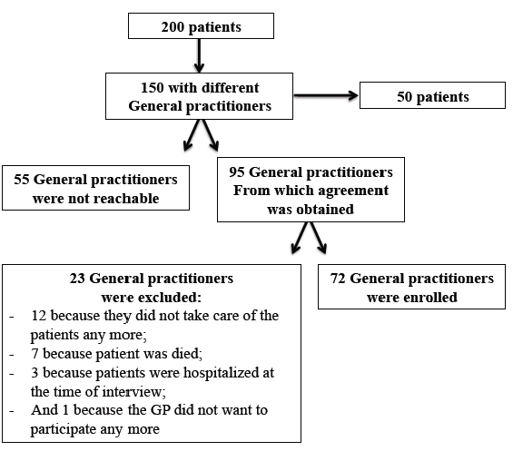
Data were prospectively collected from 200 consecutive patients aged 75 years or older discharged from the academic geriatric rehabilitation division of the University Hospital of Strasbourg (France) within a 4-month period (1st January to 31st April 2014). Patients were randomly selected throughout the hospitalisation list. Among them, we selected GPs who were taking care of only one patient in order to focus our attention on the factors influencing GPs’ prescriptions (patients whom GP were already candidates through another patient were not included). In addition, non-inclusion criteria for all patients were missing data for the medication list either at admission time, at discharge, or after 4 months, and patients for which the GP was not reachable. Criteria for secondary exclusion were withdrawal because of patient’s death before the end-point, patients for whom the GP was no longer in charge, patients who were hospitalized at the time of the interview with the GP, and GPs who withdrew from the study or who did not answer to the questionnaire despite three calls and one reminder. As depicted on the flow chart (figure 1), 150 GP/patient duos were identified; 95 were contacted by telephone (55 couldn’t be contacted); and 72 GPs consented to participate.

Figure 1. Flow chart of the population study

Figure 1. Flow chart of the population study

Appropriateness of prescribing medication during the hospital stay

In the present pilot study, all patients enrolled were admitted for rehabilitation following hospitalization either for acute medical conditions or orthopaedic surgery where interdisciplinary healthcare management was provided from admission to discharge. The interdisciplinary team consisted of the geriatric healthcare team with one full-time professor, one assistant senior physician and two fellows for the medical staff, supported by geriatric nurses, ancillary staff, physical therapists and psychologists. Specifically for psychiatric care, when necessary, an additional part-time senior geriatric-psychiatrist complemented the team. This interdisciplinary team designed, implemented and monitored comprehensive care and discharge plans for patients across a care continuum. This approach included a therapeutic plan with the aims of (i) limiting harmful effects through drug-drug or drug-disease interactions; (ii) ensuring the prescription of medications at the right doses and for the correct durations; (iii) systematically balancing the clinical benefit and the risk of adverse drug events (ADE) associated with any prescription with the patient’s needs, quality of life and expectations; and (iv) reducing the rate of omission of indicated medications with proven efficacy according to the patient’s level of functionality and life expectancy. [4] The medical team was present in the unit on a daily basis, participated in daily medical rounds and weekly interdisciplinary meetings and had direct contact with patients, care givers and patients’ families. For every patient admitted, a complete medication history was performed with the help of the patient’s GP if necessary. At their discharge, specific therapeutic recommendations were transmitted to their GP by telephone ( 1 day around the discharge day according to GP’s availability) and via the medical report usually sent by post within 3-4 weeks after discharge.

Elaboration of the questionnaire for General Practitioners (GPs)

The questionnaire was structured in 5 sections with 13 closed-ended questions (yes or no) and 1 open-ended question for personal comments. The average time to complete the survey was estimated to be 5 minutes.

The first section was dedicated to the list of medications prescribed 4 months after the hospital discharge. The GP or the referent nurse (if the patient was living in an institutional setting) filled it out. Changes in therapeutics were defined by the modification of at least one pharmaceutical molecule between the list at discharge and 4 months later. The remaining 4 sections were dedicated to GPs. Two sections focused on the prescriber’s characteristics (i.e. age, gender, year of installation), his continuing medical training and the type of activity (i.e. own practice, health clinic, and/or practitioner who practices in an institutional setting, training). The last two sections were dedicated to GPs’ expectations and more particularly the quality of therapeutic information transmitted (4 items); the issue of the complexity of therapeutic recommendations was also addressed (2 items).

Before starting the present study, the questionnaire was first tested on 3 GPs (not included in the present study) during the last quarter of 2013 in order to confirm that the questionnaire fitted both the study objectives and was acceptable according to GPs’ activity.

Complementary data collection

At the inclusion time, in addition to the treatment list upon admission, at discharge (M0) and 4 months later (M4), socio-demographic data (age, gender, living conditions) and health status were recorded by either the senior or attending physician. Thus, for each patient, the number co-morbidities was also recorded. For medications, the active pharmaceutical ingredient was considered as a statistical unit (for example, if in one pill two antihypertensive drugs were combined, two active pharmaceutical ingredients were recorded).

Statistical analysis

Results pertaining to numerical variables are presented as mean ± standard deviation (SD). For prescriptions of medicines, the median, maximum and minimum numbers of medications are also presented. For categorical variables, number and percentage are presented. Comparative analyses were computed with SAS software (version 9.1, SAS Institute, Cary, NC). Categorical outcomes were tested using the Chi2 (Χ2) test or Fisher’s exact test, and Student’s t-test or the Wilcoxon signed rank test for paired samples were used for numerical outcomes as appropriate. The level of significance was set at p = 0.05 for all analyses.

Results

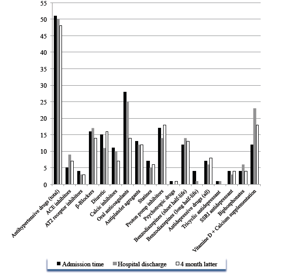
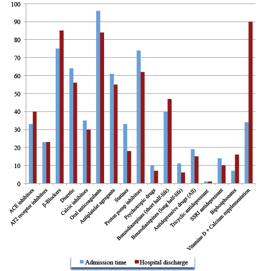
The sample of 150 patients was aged 84.8 ± 6.5 years on average (max-min: 94-74 years); 69.3% were women. They suffered, on average, from 4.9 ± 2.1 co-morbidities (max-min: 10-1) and took 8.4 ± 3.2 medications every day at admission time and 7.9 ± 3.0 at upon their discharge (p = 0.038) corresponding to 1257 and 1196 active pharmaceutical ingredients respectively. The details of medications prescribed at both these times are presented in figure 2.

imroj_103_figure2_1

Figure 2. Descriptive analysis of the number of medications prescribed at admission time and upon discharge among 150 geriatric patients. Results are given according to therapeutic class (ACE, angiotensin-converting enzyme inhibitor; AT2, Angiotensin-2 receptor inhibitor; SSR, specific serotonin-reuptake inhibitor)

Figure 2. Descriptive analysis of the number of medications prescribed at admission time and upon discharge among 150 geriatric patients. Results are given according to therapeutic class (ACE, angiotensin-converting enzyme inhibitor; AT2, Angiotensin-2 receptor inhibitor; SSR, specific serotonin-reuptake inhibitor)

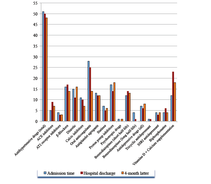
On the 72 GPs initially enrolled in the study, only 78.0% participated in the first part of the study concerning the medication list 4 months after discharge and 61.5% answered the sections about their attitudes toward therapeutic recommendations formulated in the final medical report. The corresponding 39 aged patients were not significantly different from the initial sample of 150 in terms of age, gender, co-morbidities and average number of medications at admission and upon discharge (p>0.05). These 39 patients accounted at admission time and discharge for 342 (8.8±3.7) and 306 (7.8±3.5) different medications respectively. During the hospital stay in the rehabilitation centre, 79 therapeutic modifications were done by the medical team with a 10.5% overall reduction of the number of treatment (p=0.03). Four months after discharge, a further 16.0% reduction of prescribed medication was also observed (6.6±3.5) corresponding to 257 molecules for a total reduction of 24.8% since admission time (p=0.02). For 17 patients, treatment was strictly similar (43.6%). As shown in figure 3, the most modified therapeutic class was antihypertensive drugs, which accounted for 33.0% of therapeutic modifications occurring during the hospital stay. With an overall reduction of 5.8%, 77.0% of antihypertensive treatments adapted during the hospital stay were maintained 4 months later. However, underuse of ACE inhibitors and β-blockers was observed during the stay. Conversely, the number of prescribed anti-calcics and AT2-inhibitors was dramatically reduced. One quarter of statins were stopped during the stay, and this recommendation was maintained 4 month latter in 97.4% of cases. Oral anticoagulants were dramatically reduced not only during the stay but also after discharge (by 25.6% in total). With respect to proton pomp inhibitors, the overall reduction during the stay was 18.2%. However, 4 months later, for proton pomp inhibitors, patients were back to initial values or even more (+4.5%). The main indication was the combination with anti-platelets.

Figure 3. Descriptive analysis of the number of medications prescribed at admission time, discharge and 4 months later among 39 geriatric patients whose GPs responded to the questionnaire. Results are given according to therapeutic class (ACE, angiotensin-converting enzyme inhibitor; AT2, Angiotensin-2 receptor inhibitor; SSRI, specific serotonin-reuptake inhibitor)

Figure 3. Descriptive analysis of the number of medications prescribed at admission time, discharge and 4 months later among 39 geriatric patients whose GPs responded to the questionnaire. Results are given according to therapeutic class (ACE, angiotensin-converting enzyme inhibitor; AT2, Angiotensin-2 receptor inhibitor; SSRI, specific serotonin-reuptake inhibitor)

Among the 39 GPs who answered the questionnaire, 87.5% were men and 2/3 were aged 50 years or over. On average 15.0% of their ambulatory patients were aged 70 or over and 91.0% of them were working at least part time, in institutional settings (e.g. nursing homes, long term care facilities).

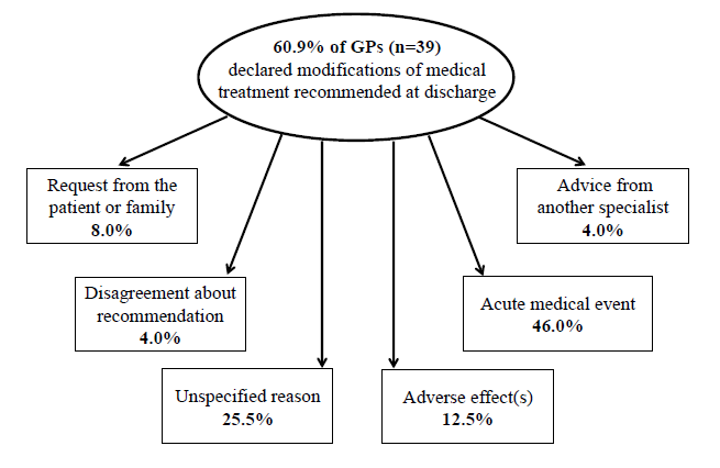
With respect to expectations for and the required level of medical report, GPs were (58.0%) satisfied with a report sent 3 to 4 weeks after hospital discharge. For 91.6% of them, GPs considered that the therapeutic changes were mentioned in the report and for 83.3%, had been properly explained. The usefulness of the report for the medical follow-up by GPs was highlighted by 83.0% of the GPs while 46.0% judged that the optimization of medications prescribed at discharge did not facilitate patient management. At 4 months, 60.9% of GPs had operated further modifications on the medication list prescribed at discharge; the reasons for are presented in figure 4.

Figure 4. Description of reasons motivating changes in medications prescribed by general practitioners during 4 months following discharge from the hospital

Figure 4. Description of reasons motivating changes in medications prescribed by general practitioners during 4 months following discharge from the hospital

The analysis of answers to the open-ended question revealed that most of GPs considered the hospital as expert for optimizing pharmacotherapy. They also underlined that some of the modifications operated were not always appropriate for ambulatory patients. One of the most frequent GP requests was that the hospital physicians keep the same active pharmaceutical ingredient when a treatment was unchanged during the stay. The second one was that a direct contact should be established between the hospital medical team and GPs before any treatment modifications or, at least, when the patient was discharged.

Discussion

This pilot study investigated GPs’ therapeutic adherence for patients discharged from a geriatric rehabilitation center. The first lesson provided is that, from an initial selected sample of 150 patients, the participation rate by GPs is very low (26.0%). This result was obtained despite an initial contact by telephone, with three successive attempts and one reminder by e-mail or post with an explicative letter. Moreover, the questionnaire sent was presented in a very short format and was composed by close-ended questions. The second observation is that the therapeutic optimization made during the hospital stay was maintained in only 43.6% patients. During the stay, the mean number of medications was reduced by 25.0%. Four months after discharge, it was reduced by 15.3%. While nearly 60% of secondary modifications after discharge were motivated by medication side effects or the occurrence of an acute medical event, 25.5% were unexplained, 8% were related to patient or patient’s family requests, or on basis of a secondary opinion given by another specialist. While most of the GPs considered the hospital setting as an expert environment for pharmacotherapy optimization, we did not use any tool or specific protocol to drive the optimization and assess its quality during the stay. Commonly, Evidence-Based Medicine (EBM) is used to standardize clinical practice and prevent errors. Based on EBM, guidelines are intended to help clinicians to prescribe appropriately. However, EBM does not often reflect age-specific differences, the high level of medical complexity of older patients, the presence of geriatric syndromes, and general geriatric vulnerability. [13] Guideline-driven prescribing does not help in achieving the goal of appropriate drug treatment and most of the time leads to substantial polypharmacy. [14, 15]

Thus, for patients with multiple conditions and polypharmacy, successful interventions included structured medication review, medication regimen simplification, administration aids and medication reminders, but no firm conclusion in favor of any particular intervention could be made. On average each patient considered in the present study suffered from 4.9 co-morbidities and took 8.4 different medications every day at admission time. Interventions to optimize geriatric pharmacotherapy focused most commonly on pharmacological outcomes (drug appropriateness, adverse drug events, adherence). [3] Systematic reviews, which have analyzed the effect of interventions to reduce inappropriate prescribing across healthcare settings [16-18], have shown that geriatric medicine services (involving geriatrician consultation, comprehensive geriatric assessment, multi-disciplinary geriatric team input into care or specialist case conferences) and multi-disciplinary team interventions (mostly pharmacist and physician collaboration or continuing education) reliably reduce inappropriate prescribing. [3]

At their discharge from the geriatric rehabilitation division, the number of prescribed medications for the 150 patients was significantly reduced, corresponding to 1196 active pharmaceutical ingredients compared to 1257 initially (figure 2). However prescribing appropriateness is not only reducing polypharmacy, but it also encompasses the use of medicines where the clinical benefits outweigh the risk of ADEs. It also includes the use of medicines that reduce the likelihood of drug-drug and drug-disease-interaction, the mis-prescribing of medicines (incorrect dose, frequency and duration) and the under-use of clinically indicated medicines. [16, 19]

With this in mind, the STOPP/START set of criteria can provide additional benefits to interdisciplinary geriatric team when it is necessary to design an intervention protocol. These criteria cover both common and important instances of potentially inappropriate prescribing and potentially serious errors of prescribing omission in older people. [20] Moreover, since the first iteration in 2008, it has been demonstrated that STOPP criteria medications are significantly associated with ADEs, and STOPP and START criteria applied as an intervention within 72 hours of admission significantly reduce adverse drug reactions and improve medication appropriateness [19, 21, 22]. Moreover, this effect was maintained 6 months post-intervention when subsequent recommendations to the attending clinicians to modify the prescription medications accordingly were notified into final medical reports. This is explained not only because this set of criteria represents the consensus views of a panel of experts in prescribing for older people but also because the clear wording of each criterion gives the opportunity to illustrate easily and precisely all medication changes operated during the stay. This reinforces comments obtained from the open-ended question of the questionnaire sent to GPs. Indeed, GPs had mentioned the quality of the medical report and how it has to be informative about the changes of the treatment regimen during the stay. This is of particular interest because among prescriber-associated barriers to optimal adherence are multi-professional communication and the transition and continuity of care. [3] A range of strategies has been implemented to increase adherence by targeting modifiable provider-related barriers. Although numerous systematic reviews have been published, no conclusion about the effectiveness of strategies to counteract non-adherence could be drawn. [23, 24] Furthermore, interventions were delivered by a single group of professionals and did not respect the geriatric medicine principles of teamwork and multi-professionality. However, when studies demonstrated statistically significant improvement in adherence, a significant reduction in mortality, better control of chronic conditions and reduced overall healthcare costs were observed. [3] There were many different classes of drugs for which GPs were non-adherent. Cardiovascular treatment was the most adjusted system during the stay, with introduction of β-blockers, ACE inhibitor and reduction of oral-anticoagulant treatment, cholesterol-lowering drugs and calcic-inhibitors. It was also the system that was the most readapted by GPs with proton pomp inhibitors (figure 3). No information in the present study concerned the appropriateness or not of these changes.

Thus, given that optimizing pharmacotherapy can be extraordinarily challenging and complex for physicians, in order to gain maximal benefit from pharmacotherapy and to achieve the best possible quality of life for the patient, one important step towards successful outcomes for complicated pharmacotherapeutic strategies is to favour the patient’s capacity to adhere to a complex treatment regimen. For that, it is necessary for GPs to set treatment priorities. However, this step is probably the most difficult because disease-specific guidelines do not capture the full clinical complexity of the patient’s pharmacotherapy. Thus, the GP’s judgement, experience, skills and attitudes are critical in determining whether or not the patient will have positive therapeutic outcomes from pharmacotherapy.

Despite limitations (e.g. small sample size, no measurement of appropriateness of prescribing recommendations), this pilot study emphasizes that the hospital discharge is an important time for non-adherence to prescribing medication according to geriatricians’ recommendations. It reinforces the need not only to enhance the quality of transition but also continuity of care between hospital and community. Thus, it could be tested in a future research protocol the impact of “faxmed” (i.e. a brief summary of the hospital stay with the complete list of medications sent by fax or e-mail the day before or the day of one patient’s discharge) or of systematic successive contacts by phone (i.e. at admission time, 48 to 72 hours before discharge, and at discharge) to discuss of medications with GP, and/or of the role that could play nurse practitioners. In that way, it could also be interesting to develop specific guidelines for writing prescriptions for GPs in which, for example, it should be systematically mentioned, in addition to medicines that have been introduced recently, those that have been stopped and those for which the dosage has been adapted with the reason for. This study also suggests the interest of specialized geriatric medical training to improve GPs’ skills and knowledge concerning pharmacotherapy in old age.

Acknowledgement

We would like to sincerely thank all the General Practitioners who made my project of medical thesis project a reality.

References

  • O’Mahony D, Cherubini A, Petrovic M (2012) Optimizing pharmacotherapy in older patients: a European perspective. Drugs Aging 29: 423-425. [crossref]
  • Ferner RE, Aronson JK (2010) Preventability of drug-related harms – part I: a systematic review. Drug Saf 33: 985-994. [crossref]
  • Topinková E, Baeyens JP, Michel JP, Lang PO (2012) Evidence-based strategies for the optimization of pharmacotherapy in older people. Drugs Aging 29: 477-494. [crossref]
  • Lang PO, Vogt-Ferrier N, Hasso Y, Le Saint L, Dramé M, et al. (2012) Interdisciplinary geriatric and psychiatric care reduces potentially inappropriate prescribing in the hospital: interventional study in 150 acutely ill elderly patients with mental and somatic comorbid conditions. J Am Med Dir Assoc 13: 406. [crossref]
  • Fallis BA, Dhalla IA, Klemensberg J, Bell CM (2013) Primary medication non-adherence after discharge from a general internal medicine service. PLoS One
  • Haynes R, Ackloo E, Sahota N, et al. (2008) Interventions for enhancing medication adherence. Cochrane Database Syst Rev.
  • Conn VS, Hafdahl AR, Cooper PS, et al. (2009) Interventions to improve medication adherence among older adults: metaanalysis of adherence outcomes among randomized controlled trials. Gerontologist 49: 447-62.
  • Hughes CM (2004) Medication non-adherence in the elderly: how big is the problem? Drugs Aging 21: 793-811. [crossref]
  • Cooper C, Carpenter I, Katona C, Schroll M, Wagner C, et al. (2005) The AdHOC Study of older adults’ adherence to medication in 11 countries. Am J Geriatr Psychiatry 13: 1067-1076. [crossref]
  • Gellad WF, Grenard JL, Marcum ZA (2011) A systematic review of barriers to medication adherence in the elderly: looking beyond cost and regimen complexity. Am J Geriatr Pharmacother 9: 11-23. [crossref]
  • Osterberg L, Blaschke T (2005) Adherence to medication. N Engl J Med 353: 487-497. [crossref]
  • McDonald HP, Garg AX, Haynes RB (2002) Interventions to enhance patient adherence to medication prescriptions: scientific review. JAMA 288: 2868-2879. [crossref]
  • Wehling M (2011) Guideline-driven polypharmacy in elderly, multimorbid patients is basically flawed: there are almost no guidelines for these patients. J Am Geriatr Soc 59: 376-7.
  • Van den Akker M, Buntinx F, Metsemakers JF, et al. (1998) Multimorbidity in general practice: prevalence, incidence, and determinants of co-occurring chronic and recurrent diseases. J Clin Epidemiol 51:367-75.
  • Boyd CM, Darer J, Boult C, Fried LP, Boult L, et al. (2005) Clinical practice guidelines and quality of care for older patients with multiple comorbid diseases: implications for pay for performance. JAMA 294: 716-724. [crossref]
  • Spinewine A, Schmader KE, Barber N, Hughes C, Lapane KL, et al. (2007) Appropriate prescribing in elderly people: how well can it be measured and optimised? Lancet 370: 173-184. [crossref]
  • Kaur S, Mitchell G, Vitetta L, et al. (2009) Interventions that can reduce inappropriate prescribing in the elderly: a systematic review. Drugs Aging 26:1013-28.
  • Steinman MA, Hanlon JT (2010) Managing medications in clinically complex elders: “There’s got to be a happy medium”. JAMA 304: 1592-1601. [crossref]
  • O’Mahony D, Gallagher P, Ryan C, Byrne S, Hamilton H, et al. (2010) STOPP & START criteria: A new approach to detecting potentially inappropriate prescribing in old age. Eur Geriatr Med 1: 45-51.
  • Lam MP, Cheung BM (2012) The use of STOPP/START criteria as a screening tool for assessing the appropriateness of medications in the elderly population. Expert Rev Clin Pharmacol 5: 187-197. [crossref]
  • Hamilton H, Gallagher P, Ryan C, Byrne S, O’Mahony D, et al. (2011) Potentially inappropriate medications defined by STOPP criteria and the risk of adverse drug events in older hospitalized patients. Arch Intern Med 171:1013-9.
  • Gallagher PF, O’Connor MN, O’Mahony D (2011) Prevention of potentially inappropriate prescribing for elderly patients: a randomized controlled trial using STOPP/START criteria. Clin Pharmacol Ther 89: p. 845-54.
  • Conn VS, Hafdahl AR, Cooper PS, et al, (2009) Interventions to improve medication adherence among older adults: meta- analysis of adherence outcomes among randomized controlled trials. Gerontologist 49:447-62.
  • Haynes R, Ackloo E, Sahota N, et al. (2008) Interventions for enhancing medication adherence. Cochrane Database Syst Rev 4: CD000011.

Discussing Why Hemochromatosis Is Ignored

DOI: 10.31038/IMROJ.2016114

Abstract

Hereditary Hemochromatosis is the most common, life threatening genetic disorder in the Western World. It is an autosomal recessive disorder, which means it is passed on to an individual by both parents who each have the HFE gene. When this gene mutates, it causes the small intestine to absorb high levels of iron from the diet into the bloodstream. The iron is commonly deposited in the liver, but it can also accumulate in the heart, lungs, brain, and the pancreas. This excessive accumulation of iron in the body can lead to toxicity and eventually to organ failure. Although, quite common, there is little public awareness of the disease with only 4 known cases diagnosed in the Grand Cayman. Some affected individuals have no symptoms. Those that have early signs and symptoms have nonspecific ones that resemble those of other common conditions. In a survey, conducted by the Centers for Disease Control and Prevention (CDC), 67% of those with the right symptoms had initially received various multiple diagnoses, including arthritis, liver disease, hormonal deficiencies, and diabetes (Centers for Disease Control and Prevention, 2008). Since, according to CDC, it takes approximately 9.5 years after the onset of symptoms for a patient to be diagnosed, early diagnosis and treatment is essential to prevent the development of severe complications such as diabetes mellitus, hepatocellular carcinoma, liver failure, cardiomyopathy, and even death. Consequently this raises the question, of whether the small number of known cases in Grand Cayman was due to lack of awareness or misdiagnosis of this common, but rarely diagnosed disease.

Key words

cirrhosis, Hemochromatosis, HFE = High Iron, Mutation, Phlebotomy, Transferrin

Introduction And Historical Background

In 1865, the first medical description of a patient with hemochromatosis was first described in a French pathology publication, by legendary diagnostician and educator Dr. Armand Trousseau [1]. He described it as an unrecognized illness involving the triad skin bronzing, cirrhosis, and diabetes. Two decades later in 1889, the German Pathologist Friedrich von Recklinghausen autopsied a series of patients dying of mysterious “bronze diabetes.” He noted that what Trousseau described was related to iron accumulation leading to the pigment changes in tissues, this prompted him to name the disorder hemochromatosis. Joseph Sheldon later explained the inherited nature of the disease in 1935. He organized a detailed written study of published cases of hemochromatosis, in which he concluded that the disease was not a complication of diabetes, cirrhosis, or excess copper but a familial disorder.

For more than 125 years, hemochromatosis was thought to be extremely rare [1]. Treatment for this disease was not effective until 1950 when Davis and Arrowsmith cleverly suggested phlebotomy as a treatment option. At the same time, progress was made when autopsy studies showed a much higher prevalence of the disease in the general population than previously known. Unfortunately, numerous supporting studies did not lead to changes in clinical practice. Nearly 150 years later in 1996, after Dr. Trosseau’s initial report, researchers identified the genetic culprit: a mutated HFE gene encoding a tyrosine molecule instead of the intended cysteine at the 282nd position of the protein chain (a mutation abbreviated as C282Y by biochemists). Since then, researchers have estimated that the C282Y mutation occurring on the HFE gene originated 60-70 generations ago. This means that the appearance of Hemochromatosis can be traced back between 600 to 1100 C.E. [2]

While hemochromatosis is prevalent worldwide, the likelyhood of carrying at least one copy of the HFE gene was most common in people of Western European descent. Specifically the chance of carrying this gene for people of Western European ancestry is about one in three individuals. Yet, only one in two hundred actually have hemochromatosis and its symptoms. This is particularly important since Caymanians not only have African ancestry and European ancestry as well.

In the United States, approximately 1 in 10 Caucasian population are heterozygous for the C282Y mutation while 4.4 per 1000 are homozygous for this same mutation [3]. Roughly, one million people have this disease in the USA. Since this is an autosomal-recessive condition, a difference in disease rates between the sexes would not be expected. Nonetheless, the clinical disease is more common in men, other than women because it is thought that women are protected from it by the loss of iron by their bodies during menses and pregnancy [3].

In the Grand Cayman, there were four confirmed cases of the disease 2 men and 2 women in 2013. One of the females was recently diagnosed and all four of them were light skin Caymanians from the district of West Bay [4]. Although the number of people with hemochromatosis is quite low, there could be cases out there that have yet to be diagnosed or that have been mistaken for other chronic diseases.

Iron is an essential part of our diet. Although it is mainly found in red meat, it is also found in green leafy vegetables and fortified foods such as cereal and breads. Iron is essential to the human body because it is required for vital functions. For example, it is crucial for the transportation of oxygen through the bloodstream, energy production, formation of the hemoglobin in the red blood cells and myoglobin in muscles, DNA synthesis, and numerous enzymatic metabolic processes. Normally, iron is absorbed in the duodenum located in the small intestine. Once absorbed, the iron is transported by a glycoprotein called Transferrin and is thereby distributed to sites of iron utilization and storage. This carrier protein plays a role in regulating the transport of iron from the site of absorption to virtually all tissues [5]. Depending on the need for iron, the body is able to increase or decrease the amount absorbed by the intestinal tract and thus maintain iron homeostasis. Hepcidin is the hormone responsible for controlling iron homeostasis. It controls how much iron is absorbed by the intestines, how iron is used in various body processes, and how it is stored in various organs [6].

Initially, iron is stored in ferritin molecules abundantly found in the heart and liver. A single one of these molecules can store up to 4000 iron atoms [5]. When excess iron from the diet is absorbed, the body responds by producing more ferritin to facilitate iron storage. On average, 3.5 g of iron is contained within the body and is maintained such that mucosal absorption of iron is equal to the iron loss. Men lose only about 1 mg of iron daily through the shedding of skin cells and secretions of the gut and skin. Women end up losing an additional milligram of iron due to menstrual bleeding and can lose approximately 500 mg when pregnant.

The daily intake of iron is about 10mg. Healthy people usually absorb about 10% of this iron, which is enough to meet normal requirements. In a person with hemochromatosis, 30% is absorbed because mucosal absorption is greater than body requirements. The mutated HFE gene causes a false signal that iron stores are low and thus, dietary iron is absorbed 2-4 times the normal rate amounting to approximately 4mg or more of iron per day [7] . This leads to accumulation of 0.5-1.0g of iron per year [5]. The progressive accumulation of iron increases plasma iron, saturation of transferrin, and results in a progressive increase of plasma ferretin. Although iron absorption is greater, the excretion rate remains the same. As there is no natural way for the body to excrete excess iron other than through blood loss, it is stored in synovial joints and various tissues, such as the liver, heart, pancreas, brain and lungs [8]. Over many years, this excess stored iron accumulates to toxic levels and can damage and bring about organ failure. The iron overload can cause many health problems, most frequently a form of diabetes that is often resistant to insulin treatment [9].

The gene responsible for regulating iron absorption from the diet is called the HFE gene which stands for High Fe (iron). Hemochromatosis is caused by the mutations in the HFE gene. As mentioned previously, hemochromatosis is an autosomal recessive disease and thus an individual must inherit two mutated genes for the disorder to manifest. This is termed homozygous. While researchers have identified more than 20 mutations in the HFE gene, only two in particular are responsible for this disorder. Each of these mutations changes one of the protein building blocks (amino acids) used to make the HFE protein [10]. The two main mutations that can occur on the HFE gene are the C282Y mutation, which accounts for 90 to 95% of cases, and the H63D mutation. C282Y is associated with a more severe form of iron absorption than the H63D mutation. Individuals who inherit one of the mutated genes and a normal gene are heterozygous for hemochromatosis. They are generally asymptomatic, but in rare cases they may also display signs and symptoms of the disease [9].

Signs and Symptoms

The signs and symptoms of this disease are so varied, non-specific and so vague that proper diagnosis is often difficult. Some of the early signs and symptoms include fatigue, weakness, weight loss, abdominal pain, and arthralgia. As iron accumulation progresses, patients may also experience arthritis, shortness of breath/ dyspnea or symptoms of gonadal failure such as amenorrhea, early menopause, loss of libido, and impotence. Iron accumulates in the parenchymal cells of several organs; the liver is a major site, followed by the heart and pancreas. Conditions associated with advanced stages of hemochromatosis include: arthritis, abnormal liver function such as elevated transaminase and clinical liver disease, glucose intolerance and diabetes, chronic abdominal pain, severe fatigue, hypopituitarism, hypogonadism, cardiomyopathy (enlargement of heart) and arrhythmia (abnormal heart beat), cirrhosis, liver cancer, heart failure, and gray or bronze skin pigmentation similar to a suntan [5].

Obviously, as seen by the extensive list of symptoms, hemochromatosis can be extremely difficult to diagnose. Most advanced hemochromatosis complications are also common primary disorders. Therefore, a hemochromatosis diagnosis can be missed even in advanced stages unless it is looked for specifically. Looking back at the low number of cases diagnosed in the Grand Cayman and some hospitals in the United States, one has to wonder if physicians in the Cayman Islands (Territory of Briatain) and their counter parts from all over the world are actively screening for hemochromatosis or mistaking its signs and symptoms for other common primary disorders. In speaking with a handful of physicians, most of them were vaguely aware of what hemochromatosis is. Most noted that it was “too much iron in the blood”. However, when asked about signs, symptoms, and treatments, none could recall any.

Some complications of hemochromatosis are not clearly related to excess iron, yet, when excess iron is removed, many individuals report feeling better [5]. Manifestations of iron accumulation can vary from person to person. The most common presenting symptom is chronic fatigue which occurs in about 50-75% of individuals. Over 70% of patients have liver function abnormalities, weakness, and lethargy at the time of diagnosis. Excessive skin pigmentation is present in more than 90% of symptomatic patients at diagnosis. The liver is usually the first organ affected. Hepatomegaly is one of the most frequent findings in clinical presentation, followed by cirrhosis. Primary hepatocellular carcinoma is more common in those with hemochromatosis than those in the general population. Diabetes mellitus occurs in 25%-75% of people and is more likely to develop in those with family histories of diabetes, suggesting that direct damage to pancreatic islets by iron absorption occurs in combination with genetic predisposition. Arthropathy develops in 25%-50% of people and usually occurs after age 50, but it may also occur as a first manifestation. Cardiac involvement is another presenting manifestation in about 15% of people, but the most common manifestation is palpitations as symptoms of arrhythmia [5].

The signs of hereditary hemochromatosis usually do not appear until about age between 40 and 60 years, when iron in the body has reached damaging levels. The reason for this is that it takes many years for iron to accumulate to the level at which clinical manifestations occur. Because women lose iron to a greater extent than men because of menses, pregnancy, and lactation, they tend to become symptomatic slightly later in life than men, often after menopause [8].

Penetrance is a term indicating the likelihood that a given gene will actually develop into disease. Thus, an individual with two mutated HFE genes does not necessarily mean have to exhibit symptoms and may actually remain symptom-free for life. Early studies that used both self-reported symptoms and clinical signs to define hemochromatosis, reported clinical penetrance estimates ranging from 40% to 70%. In contrast, more recent studies that used clinical signs or objective laboratory measures to define hemochromatosis have reported clinical penetrance estimates ranging from 1% to 50%. Inconsistencies regarding penetrance estimates persist and so further studies are needed to more fully understand the role of genetic and environmental factors that may affect penetrance [5]. Of people with the HFE mutations, only a subset will develop elevated transferrin saturation (TS). Of these, only a subset will develop an elevated serum ferritin (SF), only a further subset will develop hemochromatosis symptoms. Of those with symptoms, only a subset will develop clinical signs consistent with hemochromatosis. Thus, diagnosis is reserved for those whose signs and symptoms are clearly referable to documented iron overload [5].

Diagnosis

Currently, the clinical guidelines recommend that testing for hemochromatosis should be performed in individuals with any unexplained signs or symptoms associated with hemochromatosis, those with porphyria, hepatitis, or other liver diseases, and those with abnormal blood tests consistent with hemochromatosis. Individuals that have a family member with the condition should specially be examined for this inherited disorder because these individuals have an increased risk of developing iron overload and are an ideal group for targeted prevention efforts.

A number of laboratory tests are available to measure the amount of iron in the blood and diagnose iron overload.  Biochemical tests include: Serum iron (SI), total iron-binding capacity (TIBC), unsaturated iron-binding capacity (UIBC), transferrin saturation (TS), and serum ferritin (SF). The Centers for Disease Control and Prevention (CDC) has established a testing protocol involving 3 steps to determine a diagnosis of hereditary hemochromatosis. Involving a transferrin saturation test, a serum ferritin test, and a confirmation of the hemochromatosis diagnosis.

Transferrin is a blood protein that measures the amount of iron absorbed by the intestines and transports if from on location to another. When iron absorption is abnormally high, transferrin proteins become more saturated with iron. An elevated TS value therefore reflects an increase in iron absorption. This transferrin saturation test (TS) is a sensitive and relatively inexpensive biochemical measure of iron overloading [5]. When interpreting the results of a fasting transferrin saturation (TS), it is important to keep in mind that several factors can falsely elevate TS values, including the use of vitamin C, dietary supplements containing iron, medicinal iron, and estrogen preparations. Individuals should be advised to avoid these products for 24 hours prior to the fasting blood draw. On the other hand, colds, inflammation, liver disease, and malignancies can falsely lower TS values. Pathologic blood loss or a history of frequent blood donations should be considered reasons for normal iron status in those who have symptoms consistent with hemochromatosis [5].

Those with elevated TS values should proceed with serum ferritin testing and additional workup as needed. As mentioned before, ferritin is a protein that stores iron. The body increases serum ferritin production when excess iron is absorbed. Serum ferritin levels therefore reflect the body’s iron stores. It is important to note that because serum ferritin is also an acute phase reactant affected by cancer and inflammatory or infectious processes, SF values may increase if these underlying conditions are present.

The final test involves acquiring additional biochemical evidence of iron overload and is typically required before the hemochromatosis diagnosis can be made. This confirmation can be achieved in three ways: indirectly by quantitative phlebotomy, HFE genotyping, and directly by liver biopsy.

Quantitative phlebotomy is considered as a confirmatory test choice because the amount of mobilizable iron removed from the body by weekly or biweekly phlebotomy aids in measuring the degree of iron overload. This typically requires approximately 15 phlebotomies, each removing 500ml of blood. Each 500mL of blood extracted then removes approximately 200mg of iron. The goal is to reduce the ferritin level.

Genotyping for HFE mutations can provide additional confirmatory evidence though this information should be combined with clinical history, examination, and laboratory assessment. Identifying any HFE mutation alone is insufficient for diagnosing hereditary hemochromatosis [5]. Other genes involved in iron metabolism may also be responsible for iron overloading. Therefore, if a patient is negative for an HFE mutation yet has disease symptoms and iron overload, phlebotomy treatment and proper management of the patient’s iron overload are still important.

Another method of confirming hemochromatosis after getting elevated iron levels is liver biopsy. Since liver biopsy directly evaluates the amount of iron per gram of liver tissue, it is more commonly used for prognostic reasons to determine the level of damage [5].

Once used as the definitive confirmation test for hemochromatosis, liver biopsy is now recommended for those with high risk of liver involvement or liver damage. Most health care providers use liver biopsy in patients with elevated liver enzymes and serum ferritin levels greater than 1,000 ng/mL.

Screening

The screening process is the main thrust of this paper, since the disease is common and complications can be easily prevented with early diagnosis and treatment, the question of community screening has been raised and much debate has ensued. Concerns include: incomplete knowledge about disease penetrance, the potential for discrimination with insurance and employment, potential for increased anxiety in people who may never develop manifestations of the disease, the cost effectiveness of screening, compliance with clinical management, and whether screening should be by iron studies or genetic testing [11]. One major concern regarding screening is that people who test positive may never return for confirmation testing or may not take action to treat their disease. A study performed in Italy found that 67% of people who had elevated iron levels upon screening did not return for definitive testing [11]. Talking with the Genetic Councilor at Health Service Authority (HSA) of Cayman Islands, it was determined that HFE screening in the Grand Cayman was non-existent due to similar concerns.

Here in the USA a number of medical doctors (from Graves Gilbert hospital in Bowling green Kentucky, Wood county hospital in Bowling green Ohio, University of Toledo Medical center in Toledo Ohio, Toledo Hospital in Ohio) whom I interviewed for condition have indicated they don’t really “screen” people in the typical sense of the word, “screening” is usually reserved for asymptomatic individuals. One named hematologist said he usually checked for iron overload in patients with unexplained liver disease or in patients with underlying hematologic diseases that predispose to iron overload such as hemolytic anemia and transfusion dependent conditions. One cardiologists said, there was no current recommendation to screen for hemochromatosis unless they have symptoms or disease. Definitely there are several genotypes as well as accompanying phenotypes. Some patients are quite unlikely to have an end organ damage even with a quite elevated ferritin whereas other patients may have evidence of an end organ damage with moderate elevations in ferritin. The public health departments throughout USA and even in the Cayman Islands (territory of Britain) do conduct sporadic screening of certain diseases such as Hypertension, Diabetes, in public places (Myers stores here in USA, Foster’s food fair in Cayman Islands), this is good because people can be treated early once they have been diagnosed with these diseases and make informed decisions regarding their diets. May be the Department of public health in conjunctions with hospital hematologists, cardiologists could come up with a similar program that could implement the screening of people for hemochromatosis. People that have been diagnosed with diabetes, hypertension etc as a result of screening programs, take precaution regarding what they eat and they go back to the hospital for regular checkups to make sure the blood sugar levels or blood pressure is within normal range.

Individuals diagnosed with hemochromatosis can modify their diet accordingly; again each person is unique which must be taken into consideration before using the following suggested diet modifications in Table 1 below. This is exactly what happens with individuals diagnosed with hypertension (take low or no salt diet) or diabetes mellitus (on sugar substitutes or no sugar) do once they have known their status.

Table 1. Iron Fe (mg) content of selected foods per common measure

Food common measure content per measure
Alcohol beverage, beer 12 fl oz 0.11
Apples raw with skin 1 apple 0.17
Apple raw without skin 1 cup 0.08
Asparagus, canned drained solids 4 spears 1.32
Asparagus, cooked boiled drained 4 spears 0.55
Bananas, raw 1 banana 0.42
Beans, baked, canned plain 1 cup 3.00
Beans, baked, canned with pork/ tomato 1 cup 8.20
Beef, chuck 3 oz 3.13
Beef, ribs 3 oz 1.99
Beef, round 3 oz 2.09
Beef, ground 3 oz 2.21
Blackberries raw 1cup 0.89
Broccoli raw 1 spear 0.23
Broccoli raw 1 cup 0.64
Carrots raw 1 carrot 0.22
Carrot raw 1 cup 0.33
Chicken, broilers, cooked, roasted 3 oz 0.96
Chicken, broilers, cooked meat/ skin 1 thigh 1.25
Fish, salmon smoked 3 oz 0.72
Fish, salmon , pink, solids/bone 3 oz 0.71

Treatment and Management

Initial treatment and long-term management of hemochromatosis often depends on the level of iron in the body and associated symptoms at the time of diagnosis. In addition to treatment, diseases caused by hemochromatosis also need separate management, such as liver disease and diabetes mellitus [12]. Phlebotomy is the most common treatment and management method. Phlebotomy works by stimulating the bone marrow to make new red blood cells as old ones are extracted. Iron is moved out of iron stores in the body to make more hemoglobin. Therefore, phlebotomy reduces the patient’s iron level and can restore it to a healthy level.

In the initial de-ironing phase, normalization of iron stores involves weekly removal of blood by phlebotomy until mid hypoferritinemia occurs. That is, ferritin should equal 20ng/ml. This phase usually takes from 3 months to 1 year. The volume of blood to be removed varies among patients. Typically, 1 unit (500ml) of blood is removed per week but those who are smaller in size (less than 110lbs), elderly, and those with anemia, heart and lung problems can only manage 250ml of blood removal per week [5]. Careful monitoring of each patient throughout treatment is essential. If the treatment is too aggressive, anemia may result. Post-treatment monitoring is required and is key to appropriate patient management. Phlebotomy should be performed throughout a patient’s life to keep ferritin levels between 25 and 50ng/ml. Table 2: indicates the expected benefits from the pre-treatment state of individuals experiencing symptoms from hemochromatosis.

Table 2. Symptoms and benefits of Hemochromatosis management

Pretreatment State Expected Benefit
No symptoms prevention of complications of iron over­load; normal life expectancy.
Weakness, fatigue, lethargy Resolution or marked improvement if iron related.
Elevated serum concentrations of hepatic enzyme. Resolution or marked improvement
Diarrhea Cessation if iron related
Hepatomegaly Resolution often occurs
Hepatic cirrhosis No change or slower progression of liver failure
Right upper quadrant pain Resolution or marked improvement
Arthropathy Some improvements in arthralgia, change in joint deformity; progression sometimes seen.
Hypogonadotrophic hypogonadism Resolution is rare

Chelation therapy is an option for patients who are not allowed to bleed due to other heritable and acquired anemias [13]. Iron chelation is the pharmacological removal of metals by chemicals that bind metal so that it is excreted in urine. However, the only pharmacological iron-chelating agent approved by the FDA for use in humans is intravenous deferoxamine, or desferrioxamine or Desferal. This approach lacks the complete efficacy of phlebotomy and should be used only when absolutely necessary [5].

There are lifestyle and home remedies that may reduce the risk of complications from hemochromatosis. Those with hemochromatosis should avoid iron supplements and multivitamins containing iron because these can further increase the iron levels.  Individuals with the disease need to limit their intake of alcoholic beverages to lessen the effects of liver cirrhosis. They should also refrain from consuming Vitamin C which increases the absorption of iron from within the intestinal tract, and consume minimal amounts of red meat which is high in iron. They should avoid eating raw shellfish because individuals with hemochromatosis are susceptible to infections, especially those caused by certain bacteria in raw shellfish [6]. Also, increasing intake of substances that inhibit iron absorption, such as high tannin tea, and calcium may help slow the accumulation of iron in the body.

Conclusion

Hemochromatosis is a common yet rarely diagnosed genetically disorder. It is more common than the well-known sickle cell disease. Left untreated this disease leads to certain end organ damage and consequently death. Fortunately, if the condition is diagnosed and treated early, the damage from hereditary hemochromatosis is completely preventable. The HFE gene mutation responsible for hemochromatosis is found in a small but significant proportion of the general Caucasian population. Although it is rare to find HFE mutations in African Americans who have iron overload, these mutations have been found in a few individuals. It has been suggested that their appearance is due to admixture [5]. This might also hold true for Caymans. However, it is important to remember that only 4 known cases were in the Cayman Islands in 2013, thus, the disease might be under recognized by both physicians and individuals. It would thus be beneficial for Countries of the world to become more aware of the symptoms and management of this condition. The ongoing under diagnosis of hemochromatosis exhibited by screening individuals who have an end organ damage is not enough, or hematologists have a low sensitivity to the condition when symptoms compatible with the early stages of the disease are present and even sometimes when late complications are present. With early screening and diagnosis, preventive therapy can be instituted in the form of regular phlebotomy. If treatment is begun before end organ damage (cirrhosis or diabetes) has occurred, the prognosis is good. However, late and missed diagnoses lead to under- utilization of this readily accessible preventive treatment. This is worse in developing countries in Africa and elsewhere where the screening of hemochromatosis is never done and where most patients due to end organ damage die of the preventable diseases.

Acknowledgements

The author gratefully acknowledges support from Cynthia Powell and Jody Sims students at University college of Cayman Islands. Dr. David Kennedy University of Toledo for linking me up with Physicians from University of Toledo Medical Center, Federman Douglas MD University of Toledo medical Center Ohio, Robert Grande MD Toledo Hospital in Ohio and Jamie Jarboe MD Graves Gilbert Medical Center, Bowling green, KY.

References

  • Moalem, S. (2007) Survival of the sickest. New York, NY: HarperCollins Publishers.
  • Moalem S, Weinberg ED, Percy ME (2004) Hemochromatosis and the enigma of misplaced iron: implications for infectious disease and survival. Biometals 17: 135-139 [crossref]
  • Connor RF (2013) Haemochromatosis. Retrieved on November 10th, 2013, from Best Practice database http://bestpractice.bmj.com/bestpractice/monograph/134/basics/epidemiology.html
  • S.A (2013) Health Services Authority of Cayman Islands, Records 2013.
  • Centers for Disease Control and Prevention (2010) Training & Education- Epidemiology Retrieved on November 23rd, 2013 from the Centers for Disease Control and Prevention Web Site: http://www.cdc.gov/ncbddd/hemochromatosis/training/epidemiology/prevalence.html
  • Mayo Clinic (2008) Hemochromatosis. Retrieved November 17th, 2013 from Mayo Clinic Web Site: http://www.mayoclinic.com/health/hemochromatosis/DS00455
  • Colledge N, Ralston S, Walker B (2010) Hemochromatosis. Davidson’s Principles and Practice of Medicine. 21st ed. Textbook.
  • Adams P, Beaton M (2013) Hemochromatosis: A Guide to Diagnosis and Treatment. Retrieved on November 26th, 2013 from the British Medical Journal Database http://learning.bmj.com/learning/module-intro/haemochromatosis-diagnosis-treatment.html?moduleId=10012540&locale=en_GB
  • Plaut D, McLellan W(2009). Hereditary hemochromatosis. Journal of Continuing Education Topics & Issues 11(1): 18-21.
  • Genetics Home Reference (2009) HFE. Retrieved November l7, 2013th from the U.S. National Library of Medicine http://ghr.nlm.nih.gov/gene/HFE
  • Allen K, Nisselle A, Collins V, Williamson R, Delatycki M (2008) Asymptomatic Individuals at Genetic Risk of Haemochromatosis take appropriate steps to prevent disease related to iron overload. Liver International: Official Journal of the International Association for the Study of the Liver 28: 363-369.
  • National Heart Lung and Blood Institute (2009)
  • Dunet DO, Reyes M, Grossniklaus D, Volansky M, Blanck HM (2008) Using evaluation to guide successful development of an online training course for healthcare professionals. J Public Health Manag Pract 14: 66-75. [crossref]

Novel Agents for the Treatment of Hereditary Hemorrhagic Telengiectasia: Are They Effective Enough?

DOI: 10.31038/IMROJ.2016113

Editorial

Hereditary hemorrhagic telangiectasia (HHT) is a relatively rare (1/5000) autosomal dominant disorder characterised with arteriovenous malformations located in mucosal areas, gastrointestinal tract, pulmonary, hepatic and cerabral circulations. Several mutations (endoglin, activin A receptor type II-like 1, and less frequently SMAD4 gene) are determined, however the main cause of HHT remains unclear [1, 2]. Current guideliness recommend to take measures for the prevention of bleeding, avoidance of antiplatelet or anticoagulant agents (if possible), electrical or chemical coagulation techniques via endoscopic procedures, and treatment of iron-deficiency anemia [2].

Circulating vascular endothelial growth factor (VEGF) levels are elevated in HHT. Recently bevacizumab, a VEGF inhibitor, was administered parenterally to patients with HHT in order to investigate whether it has systemic anti-angiogenic effects in HHT. The bevacizumab dose administered was the same as its usual dose recommended for metastatic colorectal cancer patients (5 mg/kg, every 2 weeks, for 6 cycles). Among the evaluable subjects (n= 5), all patients had a decrease in transfusion requirements, and one of them gained transfusion independency. Improvement in hemoglobin levels were detected 2 months following bevacizumab administration. Some patients (n= 2) needed additional infusions of bevacizumab, but no serious (grade III or IV) side effects were reported. Bevacizumab was found to be more effective in patients suffering from epistaxis [3]. To avoid its systemic side-effects bevacizumab was also administered locally. In ELLIPSE phase-1 study which was performed on 40 HHT patients, nazal bevacizumab was well tolerated but showed no efficacy at the doses of 12.5, 25, 50, 75, and 100 mg/mL, respectively [4]. Subsequently, in a placebo-controlled and double-blind study with 15 HHT patients, a single intranasal submucosal bevacizumab injection (10 mL, 100 mg) reduced epistaxis severity and visual analog scores (27% vs 3%) non-significantly [5].

Beyond its immunomodulatory effects, thalidomide has anti-angiogenenic potency, and also suppresses tumor necrosis factor alpha  [1]. The efficacy of thalidomide in preventing gastrointestinal hemorrhages from angiodysplasias was proven in 2011. This prospective study included 2 arms, 100 mg/day thalidomide versus 400 mg/day iron (controls) were administered for 4 months. As expextedly, the subjects in thalidomide arm had significantly higher response rate which was described as a decrease by ≥50% in bleeding episodes. Thalidomide treatment was associated with a reduction in VEGF levels without any serious adverse events [6]. These promising results led clinicians to administer thalidomide for patients with HHT. Although no randomised clinical trial is reported, some small case series have emerged its clinical efficacy in HHT patients presenting with epistaxis at a dose of 50-250 mg/day (most commonly 100-200 mg/day). These patients have also showed less transfusion requirements and have described improvements in quality of life scores [7]. Special attention needs be paid while prescribing thalidomide. Teratogenicity, sedation, peripheral sensorial neuropathy, hematologic cytopenias, and venous thrombosis are the potential adverse effects of thalidomide. 100 mg/day thalidomide should be used to avoid from neuropathy instead of 300 mg/day. Interestingly, thromboembolic events were not reported in previous studies including patients with HHT [1].

Lenalidomide, a novel immunomodulatory drug which has less side effects than thalidomide, was successfully used in a 69-year-old women with chronic gastrointestinal bleeding due to HHT. Initially she was under thalidomide therapy (50-100 mg/day), but the drug was stopped because of thalidomide-induced grade-3 peripheral neuropathy. She has became free of gastrointestinal hemorrhage and drug side effects, with higher hemoglobin values (10.9 vs. 13.2 g/dL) and reduced iron and blood transfusion requirements after a 13-months period of lenalidomide treatment [8]. Both thalidomide and lenalidomide carry anti-angiogenic properties in a dose-dependent manner [9]. Additionally, the inhibitory potency of lenalidomide on growth factor-induced Akt phosphorylation and anti-migratory effects on endothelial cells were clearly shown in a rat mesenteric window assay by Dredge and colleagues [10]. Lenalidomide can suppress bone marrow activity, and therefore individual dosing should be monitored according to regular blood counts. However, high treatment costs, lack of endication in patients with HHT regarding health insurance, and most importantly limited evidence-based data about the efficacy of lenalidomide in HHT should be kept in mind.

In conclusion, novel agents such as bevacizumab or IMIDs should be used cautiously because of their potential side effects. Bevacizumab and thalidomide seems to be effective in HHT patients presenting with epistaxis. However, IMIDs could be preferred in gastrointestinal hemorrhages in HHT. These new treatment options will also provide improvements in quality of life in HHT. Although serious side effects are not reported in the recommended doses of these agents, there is insufficient evidence to recommend them as first line therapy. Further randomised and well-designed studies are needed to better identify the optimal treatment modality for the prevention and treatment of bleeding episodes in HHT.

References

  • Bauditz J (2016) Effective treatment of gastrointestinal bleeding with thalidomide–Chances and limitations.World J Gastroenterol 22: 3158-3164. [crossref]
  • Faughnan ME, Palda VA, Garcia-Tsao G, Geisthoff UW, McDonald J, et al. (2011) International guidelines for the diagnosis and management of hereditary haemorrhagic telangiectasia. J Med Genet 48: 73-87. [crossref]
  • Epperla N, Kapke JT, Karafin M, Friedman KD, Foy P (2016) Effect of systemic bevacizumab in severe hereditary hemorrhagic telangiectasia associated with bleeding. Am J Hematol 91:E313-4.
  • Dupuis-Girod S, Ambrun A, Decullier E, Samson G, Roux A, et al. (2014) ELLIPSE Study: a Phase 1 study evaluating the tolerance of bevacizumab nasal spray in the treatment of epistaxis in hereditary hemorrhagic telangiectasia. MAbs 6:794-9.
  • Riss D, Burian M, Wolf A, Kranebitter V, Kaider A, et al. (2015) Intranasal submucosal bevacizumab for epistaxis in hereditary hemorrhagic telangiectasia: a double-blind, randomized, placebo-controlled trial. Head Neck 37:783-7.
  • Ge ZZ, Chen HM, Gao YJ, Liu WZ, Xu CH, et al. (2011) Efficacy of thalidomide for refractory gastrointestinal bleeding from vascular malformation. Gastroenterology141:1629-37.e1-4.
  • Franchini M, Frattini F, Crestani S, Bonfanti C (2013) Novel treatments for epistaxis in hereditary hemorrhagic telangiectasia: a systematic review of the clinical experience with thalidomide. J Thromb Thrombolysis 36:355-7.
  • Bowcock SJ, Patrick HE (2009) Lenalidomide to control gastrointestinal bleeding in hereditary haemorrhagic telangiectasia: potential implications for angiodysplasias? Br J Haematol 146:220-2.
  • Dredge K, Marriott JB, Macdonald CD, Man HW, Chen R,et al. (2002) Novel thalidomide analogues display anti-angiogenic activity independently of immunomodulatory effects. Br J Cancer  87:1166-72.
  • Dredge K, Horsfall R, Robinson SP, Zhang LH, Lu L, et al. (2005) Orally administered lenalidomide (CC-5013) is anti-angiogenic in vivo and inhibits endothelial cell migration and Akt phosphorylation in vitro. Microvasc Res 69:56-63.

Ileum Ulcer Bleeding By Cytomegalovirus Infection In A Colon Cancer Patient

DOI: 10.31038/IMROJ.2016112

Abstract

We present an immunocompromised case of colon cancer who received a compound of tegafur and uracil as postoperative adjuvant chemotherapy. However, massive bloody stool passage was found and endoscopy revealed hemorrhagic erosions and ulcers at the ileum, which also caused partial obstruction of proximal small bowel. As persistent massive bleeding, segmental bowel resection was performed and the pathology of the resected lesion confirmed cytomegalovirus ileitis. Meanwhile, the polymerase chain reactions for cytomegalovirus DNA in the stool and peripheral blood samples were both positive. After surgical intervention, however, the intestinal bleeding still persisted and was stopped soon by ganciclovir therapy. In conclusion, intestinal bleeding due to cytomegalovirus ileitis may not be easily controlled without appropriate antiviral therapy. The polymerase chain reactions may be helpful in the earlier detection of cytomegalovirus intestinal disease and provide early initiation of pre-emptive therapy before the histopathological diagnosis is made.

Key words

cytomegalovirus, immunocompromised patients, ileitis

Introduction

Cytomegalovirus (CMV) is a virus that belongs to the family of Herpesviridae and usually causes an asymptomatic infection or produces mild flulike symptoms. Afterward, it remains latent throughout life and may reactivate in the immunocompromised status, thus causing a serious disease with significant morbidity and mortality of the patients [1]. CMV infection can affect any field of the gastrointestinal tract. The most common affected areas are the colon and the rectum, whereas other locations such as the duodenum and ileum are rarely reported [2-4]. Clinical symptoms of CMV ileitis include fever, abdominal pain, anorexia, nausea, vomiting, diffuse abdominal pain, lower abdominal pain, diarrhea, hematochezia or melena [2-4]. Herein we reported a rare case of CMV ileitis in a colon cancer patient presenting with partial small intestine obstruction and massive intestinal bleeding.

Case Report

This 75-year-old man had history of diabetes, gout and chronic hepatitis B virus infection. He was diagnosed with colon cancer and received laparoscopic radical right-sided hemicolectomy and subsequent adjuvant chemotherapy with tegafur/uracil (UFUR) 2 months prior to the admission. He suffered from nausea and vomiting with watery diarrhea since July 23, 2015. The laboratory data showed leukocytosis and the computed tomography (CT) of the abdomen showed segmental small bowel swelling at distal ileum (Figure 1A), causing partial obstruction of the proximal small bowel (Figure 1B). Then, he was admitted to the intensive care unit due to septic shock with acute respiratory failure. However, intermittent bloody stool passage was found. Colonoscopy through the right-sided colostomy showed diffuse hemorrhagic erosions and ulcers at distal ileum (Figure 1C). Thereafter, massive lower gastrointestinal bleeding persisted, thus requiring surgical intervention. Enterolysis and segmental bowel resection with end ileostomy were performed on August 17, 2015. Meanwhile, the polymerase chain reactions (PCRs) for CMV DNA in the stool and peripheral blood sample were both positive. The pathology of the biopsied ileum lesion confirmed CMV ileitis with evidence of some scattered stromal or endothelial cells with eosinophilic nuclear inclusion, which were highlighted by anti-CMV immunostain. Nonetheless, small bowel bleeding persisted even after surgery. Therefore, ganciclovir therapy was initiated and stopped the bleeding soon. Then the patient reached successful weaning from mechanical ventilation and he was uneventfully discharged.

Figure 1. The CT of the abdomen shows segmental small bowel swelling at distal ileum till anastomosis (arrow, A), causing proximal small bowel partial obstruction (arrow, B). Colonoscopy through the right-sided colostomy reveals diffuse hemorrhagic erosions and ulcers at the ileum (C).

Figure 1. The CT of the abdomen shows segmental small bowel swelling at distal ileum till anastomosis (arrow, A), causing proximal small bowel partial obstruction (arrow, B). Colonoscopy through the right-sided colostomy reveals diffuse hemorrhagic erosions and ulcers at the ileum (C).

Discussion

CMV enteritis with lower gastrointestinal (GI) haemorrhage is a rare presentation. As CMV infection in immunocompromised patients can be lethal, it is important to diagnose and initiate early treatment. In systemic CMV infections, ulceration of the GI tract is common, which could be a primary infection, a superimposed infection, reactivation of latent infection or re-infection with a new virus (1). CMV ileitis is rarely reported in the literature, but could still occur in the immunocompetent patients [2-4]. However, even in an immmunocompromised patient like our presented patient with colon cancer, CMV ileitis bleeding is usually not considered in the earlier course of the GI bleeding disease. Most gastrointestinal CMV infections respond well to ganciclovir treatment. Therefore, the patient should be offered an antiviral treatment as soon as possible. Early diagnosis of suspected CMV infection in immunosuppressed patients with gastrointestinal symptoms is of the utmost importance. Delayed management of CMV ileitis might cause stenosis of the ileum [4] and massive lower gastrointestinal bleeding [5, 6], just like our case; or could also result in small bowel perforation [7]. Nonetheless, CMV ileitis in an immunocompetent patient with mild symptom of epigastric pain may spontaneously recover without antiviral therapy [2].

Repeat endoscopy may be considered if previous study did not meet a concluding gastrointestinal CMV disease. The ileum ulcer is difficult to approach by endoscopy. In the current patient, we could easily assess the lesion site through the right-sided colostomy but endoscopic biopsy was not performed during bleeding episode. CMV-infected hemorrhagic ileitis was diagnosed by subsequent surgical biopsy and thereby antiviral therapy was rather delayed. In this scenario, we recommend pre-emptive antiviral therapy based on the positive results for stool and/or blood CMV-PCRs.

Authorship

Hui-Chun Chao contributed to acquisition of data and drafting the article. Khee-Siang Chan contributed to the patient care and analysis and interpretation of data. Wen-Liang Yu contributed to conception, design and critical revision of the article. All authors approved the final version to be published and agreed for all aspects of the work related.

Conflict of interests

We declare no funding and no conflict of interests. The above study has been granted exemption from review by the Institutional Review Board of Chi-Mei Medical Center (application no.10410-E02).

References

  • Ljungman P, Griffiths P, Paya C (2002) Definitions of cytomegalovirus infection and disease in transplant recipients. Clin Infect Dis 34: 1094-1097. [crossref]
  • Ryu KH, Yi SY (2006) Cytomegalovirus ileitis in an immunocompetent elderly adult. World J Gastroenterol 12: 5084-5086. [crossref]
  • Varma V, Perera MT, Olliff S, Taniere P, Isaac J, Mirza DF (2011) Cytomegalovirus ileitis causing massive gastrointestinal haemorrhage in a patient following hepatic resection. Tropical Gastroenterology 32:145-7.
  • Tejedor Cerdeña MA, Velasco Guardado A, Fernández Prodomingo A, Concepción Piñero Pérez MC, Calderón R, et al. (2011) Cytomegalovirus ileitis in an immunocompetent patient. Rev Esp Enferm Dig 103: 154-156. [crossref]
  • Meza AD, Bin-Sagheer S, Zuckerman MJ, Morales CA, Verghese A (1994) Ileal perforation due to cytomegalovirus infection. J Natl Med Assoc 86: 145-148. [crossref]
  • Lai IR, Chen KM, Shun CT, Chen MY (1996) Cytomegalovirus enteritis causing massive bleeding in a patient with AIDS. Hepatogastroenterology 43: 987-991. [crossref]
  • Vilaichone RK, Mahachai V, Eiam-Ong S, Kullavanuaya P, Wisedopas N, et al. (2001) Necrotizing ileitis caused by cytomegalovirus in patient with systemic lupus erythematosus: case report. J Med Assoc Thai 84 Suppl 1: S469-473. [crossref]

Congenital Hypercoagulable Disorders

DOI: 10.31038/IMROJ.2016111

Abstract

The blood coagulation in the body’s vessels represents a main cause of thrombosis arterial and venous acute and chronic events. The causes of hypercoagulation can be acquired or inherited. Acquired hypercoagulable disorders most frequently occurs. It can be present in several conditions, included advanged age, and must be reported to one or more factors of Virchow’s triad. On the contrary, hereditary thrombophilia is less frequent and little known too. Its pictures manifest with repeated vascular acute events in subjects in young age, often belonging to the same family, in the absence of factor of Virchow’s triad . Inherited hypercoagulation is due to some genetic anomaly of MTHFR, Factor V Leiden, Factor II. Nevertheless, these states can be caused by a deficiency of anticoagulant proteins (Protein C, Protein S, Antithrombin III) or by antiphospholipid antibodies syndrome. But the anticoagulant tendency , besides congenital, may be also dependent by some acquired conditions, such as liver disease, pregnancy, some infection and the use of drugs, as estrogen, heparin or warfarin. The genetic causes and the pathogenetic mechanisms of inherited thrombophilia were briefly illustrated.

Key words

Hypercoagulability, hereditary thrombophilia, MTHFR, Factor V Leiden, Prothrombin, Anticoagulant proteins, Lupus Anticoagulant

Introduction

Hemostasis is highly regulated by coagulant and anticoagulant factors to maintain a balance between bleeding and coagulation. On the contrary, the prevalence of pro-coagulant on anti-coagulant factors causes an impaired hemostasis balance, inducing thrombosis that blocks or reduces blood supply in tissues. That represents a most frequent and important life-threatening human disease. Hypercoagulable states can be acquired and congenital [1]. Both conditions likely to develop clots in venous and arterial vessels. Venous thrombosis is the most common cause of potentially life-threatening blood clots in the lungs, the deep leg veins, the arm veins, the kidney veins, or others. Conversely, arterial thrombosis consists in clot-formation in the arteries, that can cause some complications associated with significant morbidity and mortality, such as acute myocardial infarction, stroke or TIA [2].

Acquired Hypercoagulability

Acquired form can happen in some conditions, as advanced age, diabetes, inflammation, cancer, obesity, immobility, and others. These act favouring one or more factors of Virchow’s triad (stasis, endothelial dysfunction, hypercoagulability) causing procoagulant tendency [3].

Congenital Hypercoagulability

Also defined hereditary thrombophilia, it occurs lesser than acquired type. It is the result of abnormal gene deriving from one (heterozygous) of both parents (homozygous). It should be suspected in patients with individual and/or familiar history of recurrent ischemic-thrombotic events, in absence of any of the risk factors related to the conditions reported above (Virchow’s triad). But, hereditary thrombophilia generally is also present in subjects at a young age (40 years), with history of thrombosis in unusual sites (mesenteric, renal, hepatic, retinal veins) or cerebral thrombosis [4].

Genes mostly codifying for hereditary thrombophilia include the following:

  • MTHFR (Methylene-Tetra-Hydro-Folate-Reductase);
  • Factor V Leiden (factor V);
  • Prothrombin (factor II).

Reduced MTHRF activity may be responsible for increased homocysteine levels (HHcy) that is a risk factor for arterial and venous blood clots. HHcy exerts its thrombotic effect acting both directly on endothelial layer and indirectly through DNA-hypomethylation [5,6] . Factor V Leiden (FVL) is factor of hereditary thrombophilia in Caucasian populations. Heterozygous FVL is much more common than homozygous. FVLa causes the activation of prothrombin in thrombin [7]. It predisposes mainly to venous thrombosis (especially retinal vein occlusion). Finally, activated prothrombin induces fibrinogen in to fibrin conversion and favours deep venous thrombosis.

Other conditions causing a hypercoagulable state are induced by the deficiencies (inherited or acquired) of  the following proteins defined as natural anticoagulants: Protein C; Protein S; Antithrombin III.

Once the coagulation process begins, these act limiting the process in accordance with the scheme following: (figure.1)

Figure.1: Prevalence of coagulation on bleeding induced by reduction of anticoagulant proteins

Figure 1. Prevalence of coagulation on bleeding induced by reduction of anticoagulant proteins

Activated protein C and protein S act by inhibiting the action of the cofactors (factor Va and factor VIIIa). Antithrombin inhibits the serine proteases (factor III, X, XI, XII). Obviously, deficiencies of these proteins are associated with thromboembolic disease [8].

Deficiencies of anticoagulant factors are inherited for patient’s parents. People born with deficiencies of one of the abnormal gene from either mother or father is heterozygous for this gene (more frequent). Conversely, patients can inherit abnormal gene from both parents. These are homozygous and rarely occurs. Nevertheless,  deficiencies of  anticoagulant factors can also be acquired. Individuals with normal levels of anticoagulants may develop deficiencies in certain situations, such as pregnancy, liver disease, some infections or vitamin K deficiency and the use of certain medication, such as  estrogen, heparin, warfarin [9].

Finally, antiphospholipid antibodies syndrome (also called lupus anticoagulant) must be considered such as a cause of inherited thrombophilia. It occurs in about 20% of patients with systemic lupus erythematous (SLE) and may be also associated with other autoimmune diseases [10]. The inappropriate name for  antibodies is due to the initial discovery in patients with SLE, although they can also occur in individuals without lupus. The mechanisms of thrombosis in this syndrome are not yet defined. Nevertheless, activation of platelets to enhance endothelial adherence or production of antibodies against protein C or protein S must be considered [11].

Conclusions

Arterial and/or thromboembolic events deriving from acquired hypercoagulability represent the most frequent causes of morbidity and mortality of the whole population and were enough studied. On the contrary, disorders deriving from inherited thrombophilia less frequently occur and its physiopathology barely is known. Thus, the better knowledge of its numerous types is requested to successfully contrast its dreadful complications.

References

  • Kitchens CS (1985) Concept of hypercoagulability: a review of its development, clinical application, and recent progress. Semin Thromb Hemost 11: 293-315. [crossref]
  • Reiner AP, Siscovick DS, Rosendaal FR (2001) Hemostatic risk factors and arterial thrombotic disease.Thromb Haemost 85: 584-595. [crossref]
  • Franchini M, Mannucci PM (2008) The hemostatic balance revised through the lessons of markind evolution. Intern Emerg Med 3: 3-8
  • Khan S, Dickerman JD (2006) Hereditary thrombophilia. Thromb J 4: 15. [crossref]
  • Welch GN, Loscalzo J (1998) Homocysteine and atherothrombosis.N Engl J Med 338: 1042-1050. [crossref]
  • Cacciapuoti F (2011) Hyper-homocysteinemia: a novel risk factor or a powerful marker for cardiovascular diseases? Pathogenetic and therapeutical uncertainties. J Thromb Thrombolysis 32: 82-88. [crossref]
  • De Stefano V, Chiusolo P, Paciaroni K, Leone G (1998) Epidemiology of factor V Leiden: clinical implications. Semin Thromb Hemost 24: 367-379. [crossref]
  • High KA (1988) Antithrombin III, protein C, and protein S. Naturally occurring anticoagulant proteins. Arch Pathol Lab Med 112: 28-36. [crossref]
  • Lipe B, Ornstein DL (2011) Deficiencies of natural anticoagulants, protein C, protein S, and antithrombin. Circulation 124: e365-368. [crossref]
  • Lockshin MD (2008) Update on antiphospholipid syndrome. Bull NYU Hosp Jt Dis 66: 195-197. [crossref]
  • Negrini S, Pappalardo F, Murdaca G, Indiveri F, et al. (2016) The antiphospholipid syndrome: from pathophysiology to treatment. Clin Exp Med . [crossref]