Introduction
Chronic wounds pose a significant challenge in the United States, affecting approximately 2.5% of the general population. These persistent wounds not only have a detrimental impact on the quality of life for patients but also exert a considerable financial burden on the healthcare system. Given the intricate nature of the wound healing process, healthcare professionals often approach the management of wounds based on their underlying causes. In cases where a wound fails to heal, clinicians commonly resort to the utilization of wound dressings and various technologies [1,2]. However, a crucial aspect that is frequently overlooked is the reassessment of the wound’s etiology, particularly when the wounds appear superficially normal. Incorporating a comprehensive understanding of the underlying cause of a chronic wound is paramount in formulating an effective treatment plan.
One essential tool in the assessment of chronic wounds is wound culture. By conducting a wound culture, clinicians can identify the presence of specific pathogens and gain insights into the appropriate course of treatment. Conventional wound culture techniques involving plating, though commonly employed, may not always capture the causative pathogens accurately. This limitation can potentially mislead treatment decisions and inadvertently prolong the healing process. To illustrate this point, two non-typical cases are presented as examples, shedding light on the importance of accurate wound culture techniques and the subsequent implications on healing outcomes.
Case #1
A 27-year-old male presented with multiple plantar warts on toes and feet. Two months later, a lesion appeared on the left thumb. The lesion was presumed to be a wart transmitted from the feet. Debridement with topical antiviral cream, containing 5% Cimetidine, 5% 5-Fluoracil and 10% Salicylic acid, did not resolve the lesion. More lesions developed on the fingers. Patient was referred to a dermatologist who performed Candida injections without success. HIV test was negative. Patient was referred back to continue the treatment of debridement and topical antiviral cream. However, the etiology of the wounds is now being questioned. The lesions appeared on fingers on the left hand only and on four fingers except the middle one (Figure 1).
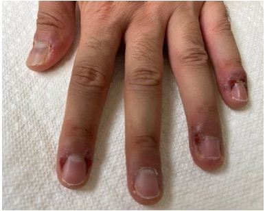
Figure 1: Suspected verrucae on the fingers
The lesions did not appear to follow the contact transmission logics. A wound culture by PCR (HealthTrackRx, Denton, TX) was performed. High volume of Peptostreptococcus anaerobius and Cutibacterium acnes were detected. Patient admitted to a habit of nail biting. All lesions on the fingers were resolved after seven days of Azithromycin and two weeks of topical antibiotic ointment (Figure 2).
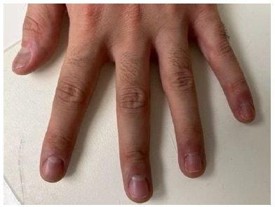
Figure 2: Fingers after the proper antibiotic treatment
Case #2
A 47-year-old female presented with an ingrown toenail and an incidental complaint of athlete’s foot. Patient indicated that the lesions were located in the left fourth interspace and had been resistant to over-the-counter antifungal for a few weeks. The webspace was erythematous with open skin and vesicles (Figure 3).
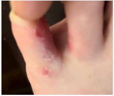
Figure 3: Suspected tinea pedis
Suspicion was immediately raised since she was a double-transplant recipient due to a rare desmin-related myopathy. A wound culture by PCR (HealthTrackRx, Denton, TX) was performed. High volume of Enterococcus faecalis, E. faecium, Klebsiella pneumoniae, K. Oxytoca, P. anaerobius, C. acnes, Staphylococcus spp, and Trichophyton mentagraphophytes were detected. Patient admitted to having issues with her gastric pouch. Patient immediately contacted her transplant team. Open wounds were resolved with seven days of Levofloxacin (Figures 4 and 5). Patient continued to apply topical antifungal for tinea pedis.
Six weeks later, patient returned with abscess and cellulitis in the same location. Wound culture by PCR was performed. The results showed a high amount of C. acnes and moderate amount of Trichophyton but no enterobacteria.
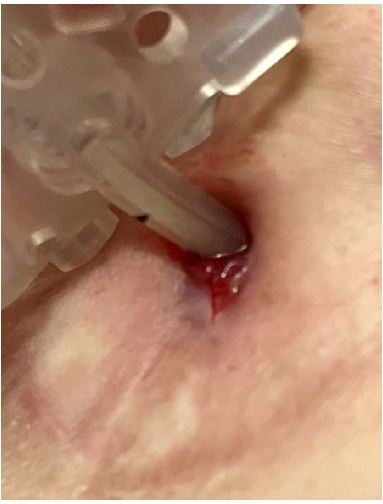
Figure 4: Gastrostomy tube
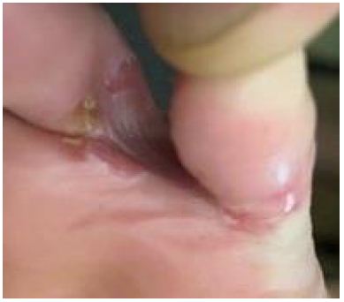
Figure 5: Resolved cellulitis
Conclusion
Verrucae and tinea pedis are prevalent skin conditions encountered frequently in podiatry and primary care practices [3-7]. As clinicians diagnose these conditions, they typically adhere to established treatment protocols. However, when wound healing fails to progress as expected, healthcare professionals often find themselves compelled to modify the course of action by altering wound dressings or incorporating additional therapeutic modalities. In such instances, it becomes imperative to consider the significance of conducting a proper wound culture to gain a comprehensive understanding of the microbial load. This timely and detailed assessment not only confirms the initial diagnosis but also plays a pivotal role in formulating an accurate and tailored treatment plan to promote optimal wound healing. By performing a wound culture, clinicians can identify the specific microorganisms present in the affected area. This information offers valuable insights into the nature of the infection, including its type, severity, and potential resistance patterns. Armed with this knowledge, healthcare professionals can make informed decisions regarding appropriate antimicrobial therapies, taking into account the specific pathogens involved. The implementation of targeted and effective treatments based on the wound culture results enhances the chances of successful wound healing.
In summary, when faced with persistent or non-progressing wounds, clinicians should recognize the significance of a wound culture. This diagnostic approach ensures a thorough assessment of the microbial load, enabling accurate diagnosis confirmation and the formulation of an individualized treatment plan. By leveraging the insights gained from a detailed wound culture, healthcare professionals can optimize their interventions and enhance the prospects of successful wound healing for their patients.
Acknowledgement
None
Funding
None
Conflict of Interest
None
References
- Childs DR, Murthy AS (2017) Overview of Wound Healing and Management. Surg Clin North Am 97: 189-207. [crossref]
- Korting HC, Schollmann C, White RJ (2011) Management of minor acute cutaneous wounds: importance of wound healing in a moist environment. J Eur Acad Dermatol Venereol 25: 130-137.
- Aldana-Caballero A, Marcos-Tejedor F, Mayordomo R (2021) Diagnostic techniques in HPV infections and the need to implement them in plantar lesions: A systematic review. Expert Rev Mol Diagn 21: 1341-1348. [crossref]
- Clebak KT, Malone MA (2018) Skin Infections. Prim Care 45: 433-454.
- Etcheverria E (2020) Recognizing and treating five common dermatologic conditions seen in primary care. JAAPA 33: 33-37. [crossref]
- Kaushik N, Pujalte GG, Reese ST (2015) Superficial Fungal Infections. Prim Care 42: 501-516. [crossref]
- Lai JY, Doyle RJ, Bluhm JM, Johnson JC (2006) Multiplexed PCR genotyping of HPVs from plantaris verrucae. J Clin Virol 35: 435-441. [crossref]