DOI: 10.31038/AFS.2021313
Abstract
Anomalous specimens of Tor tor and Tor putitora were noticed among fish collections made by fishermen from the river Chenab in Pargwal Wetland area, Akhnoor, over a period of three years, and are reported. Morphologically these deformed fishes were truncated and showed displacement of fins. Radiological analysis exhibited truncated vertebral column and compressed vertebrae with reduced vertebral thickness and intervertebral spaces. A possible cause of these aberrations is fast currents in various Himalayan tributaries of the torrential river Chenab in which Tor generally breeds.
Keywords
Deformed; Tor tor; Tor putitora; Truncated; The river Chenab; Currents
Introduction
Tor tor and Tor putitora, the important food and game fishes, are widely distributed in Himalayan lotic waters in India. In Jammu region of the Union territory of J&K, these fish species inhabit the river Chenab and its tributaries in Kishtwar, Doda, Banihal, Reasi, Udhampur, Rajouri and Jammu; the river Ravi and its tributaries in Kathua and Samba and Poonch river and its tributaries, including Mendhar nullah. Tor spp. migrate from plains, including Pakistan, for breeding in freshwater streams of Jammu region during monsoon and are netted in good number. Due to good water quality in streams and rivers of Jammu region, there are few reports of anomalous fishes in natural waters [1-10]. During hydrobiological studies of the river Chenab deformed specimens of Tor tor and Tor putitora were noticed along with normal fishes and have been described. The objective of the study is to find out the types and causes of various anomalies, though rare, in the Himalayan lotic water bodies.
Material and Methods
Deformed specimens of Tor spp. were purchased from fishermen collecting fishes from the river Chenab, in Pargwal wetland area, Akhnoor, and studied for morphological aberrations, parasitic infections and photographed. For detailed skeletal analysis these anomalous and normal fish specimens were radiographed with digital x-ray machine (Ray’s India).
For water quality characteristics, water samples were collected in plastic containers and analysed following standard methods [11].
Observations
During fish survey of the River Chenab six deformed specimens of Tor tor and five of Tor putitora were observed along with normal specimens and have been described as below.
Tor tor (Ham.-Buch.)
Head length is equal to body depth in a normal streamlined Tor tor Dorsal fin installation is midway between snout tip and caudal fin base and its longest fin ray is quite anterior to anal aperture. There is a wide space between longest pectoral fin ray and pelvic fin origin, pelvic fin ray and anal fin origin and anal fin ray and caudal fin base (Figure 1a).
Vertebral column is streamlined with normal uniform vertebral thickness and inter-vertebral spaces (Figure 1b).
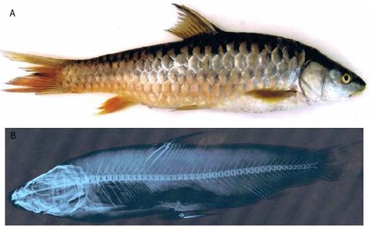
Figure 1a: Photograph of a Normal Specimen of Tor tor (Ham.-Buch.).
Figure 1b: X-ray Photograph of a normal specimen of Tor tor (Ham.-Buch.).
Morphological and vertebral deformities observed in six specimens of Tor tor collected from the river Chenab in Pargwal wetland area are shown in Table 1.
Table 1: Morphological and vertebral characteristics of abnormal Tor tor (Ham.-Buch.) collected from the river Chenab in Pargwal wetland, Akhnoor, Jammu
|
S.No. |
Size(Length cm/Wt. g) | Morphological characteristics | Fins placement |
Vertebral deformities |
|
1 |
23.5 cm/200 g |
Highly truncated body, abnormal height more than head length and curved caudal peduncle; (Figure 2a). |
Dorsal fin placement is towards caudal fin base and its longest dorsal fin ray extends beyond anal fin base. |
1st to 17th vertebrae irregularly compressed with reduced vertebral thickness and inter-vertebral spaces (Figure 2b). |
| 2 | 29 cm/500 g | Highly truncated globular body,
abnormal height more than head length and displacement of fins (Figure 3a). |
Dorsal fin installation is towards caudal fin base and its longest fin ray is short. Longest pectoral fin ray extends pelvic fin base, longest pelvic fin ray extends anal fin base and latter the caudal fin base. | 5th to 30nd vertebrae irregularly compressed and fused with variable vertebral thickness and intervertebral spaces. (Figure 3b). |
| 3 | 21 cm/155 g | Minor truncated body and displacement of fins (Figure 4a). | Dorsal fin placement is towards caudal fin base and its longest fin ray extend anal fin base. Longest pectoral fin ray extends pelvic fin base, pelvic fin ray anal fin base and anal fin ray caudal fin base. | 11th to 30th vertebrae, with variable vertebral thickness and inter-vertebral spaces, irregularly compressed. Compression is more marked between 11th to 16th vertebrae (Figure 4b). |
| 4 | 25 cm/185 g | Truncated body, displacement of fins, short caudal peduncle (Figure 5a). | Dorsal fin placement is towards caudal fin base and its longest fin ray extends beyond anal fin base. Space between longest pectoral fin ray and pelvic fin base, pelvic fin ray and anal fin base and anal fin ray and caudal fin base is short. | 10thto 29th vertebrae irregularly compressed with reduced vertebral thickness and inter- vertebral spaces (Figure 5b). |
| 5 | 22 cm/165 g | Dorsal dome (Figure 6a). | All fins like normal fish. | Vertebral column is dorsally curved in thoracic region (Figure 6b). |
| 6 | 20 cm/150 g | Highly truncated caudal peduncle and displacement of anal fin (Figure 7a). | Dorsal fin placement is towards caudal fin base and longest anal fin ray extends caudal fin base. | X-ray is not available. |
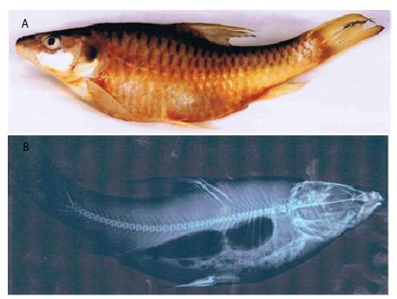
Figure 2a: Photograph of Tor tor (Ham.-Buch) showing highly truncated body, abnormal height and curved caudal peduncle.
Figure 2b: X-ray Photograph of Tor tor (Ham.-Buch) showing highly truncated body, abnormal height and curved caudal peduncle.
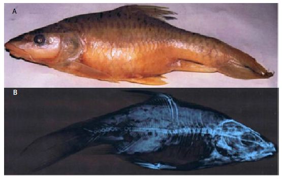
Figure 3a: Photograph of Tor tor (Ham.-Buch) showing highly truncated globular body, abnormal height and displacement of fins.
Figure 3b: X-ray Photograph of Tor tor (Ham.-Buch) with highly truncated globular body, abnormal height and displacement of fins.
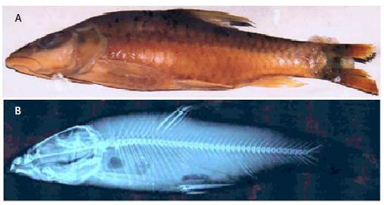
Figure 4a: Photograph of Tor tor (Ham.-Buch) showing minor truncated body and extension of the longest dorsal fin ray beyond the anal fin origin.
Figure 4b: X-ray Photograph of minor truncated Tor tor (Ham.-Buch) with extension of the longest dorsal fin ray beyond the anal fin origin.
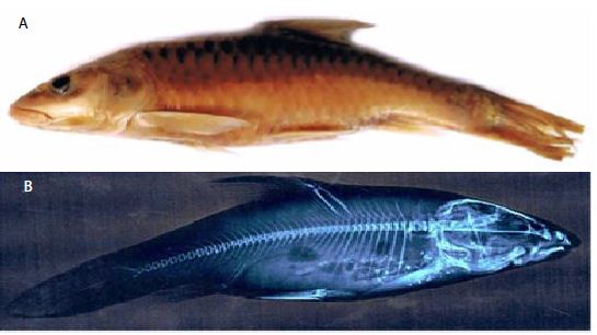
Figure 5a: Photograph of Tor tor (Ham.-Buch) showing truncated body, displacement of fins, short caudal peduncle and overlapping scales.
Figure 5b: X-ray Photograph of Tor tor (Ham.-Buch) with truncated body, displacement of fins, short caudal peduncle and overlapping scales.
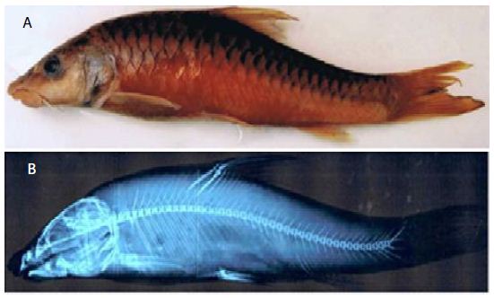
Figure 6a: Photograph of Tor tor (Ham.-Buch) showing a dorsal dome.
Figure 6b: X-ray Photograph of Tor tor (Ham.-Buch) with a dorsal dome.
Figure 7a: Photograph of Tor tor (Ham.-Buch) showing highly truncated caudal peduncle and extension of longest anal fin ray to the caudal fin base.
Tor putitora (Ham.-Buch.)
In a normal streamlined Tor putitora head length is greater than body depth. Dorsal fin insertion is midway between snout tip and caudal fin base. There is a wide space between longest dorsal fin ray and anal fin base, pectoral fin ray and pelvic fin base, pelvic fin ray and anal fin base and anal fin ray and caudal fin base (Figure 8a). Vertebral column is streamlined with normal vertebral thickness, inter-vertebral spaces, urostyle and caudal fin bones (Figure 8b).
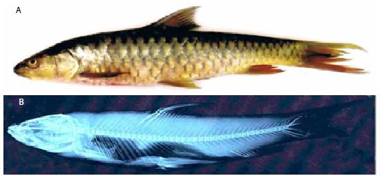
Figure 8a: Photograph of a normal specimen of Tor putitora (Ham.-Buch).
Figure 8b: X-ray Photograph of a normal specimen of Tor putitora (Ham.-Buch).
Various morphological and vertebral deformities observed in five specimens of Tor putitora are given in the Table 2.
Table 2: Morphological and vertebral column characteristics of abnormal Tor putitora (Ham.-Buch.) collected from the river Chenab in Pragwal wetland, Akhnoor, Jammu.
|
S.No. |
Size(Length cm/Wt. g) | Morphological characteristics | Fins placement |
Vertebral deformities |
|
1 |
25.4 cm/192 g |
Truncated body, short caudal peduncle and displacement of dorsal and anal fin. (Figure 9a). |
Dorsal fin placement is towards the caudal fin base and its longest fin ray reaches anal aperture. Longest anal fin ray extends caudal fin base. |
3rd – 9th and 17th – 23rd vertebrae are irregularly compressed and fused with variable vertebral thickness and inter vertebral spaces. 38th to 40th vertebrae highly compressed and fused (Figure 9b). |
| 2 | 55 cm/200 g | Mid truncated body, abnormal height and fins disposition.
(Figure 10a). |
Dorsal fin insertion is towards caudal fin base. Space between longest pectoral fin ray and pelvic fin base, pelvic fin ray and anal fin base and anal fin ray and caudal fin base reduced. | Vertebral column between 7th to 32nd vertebrae truncated and vertebrae irregularly compressed. 7th to 12th vertebrae are highly compressed and attenuated. Vertebral thickness and inter vertebral spaces reduced.
(Figure 10b and 10c). |
| 3 | 17.8cm/150 g | Mild truncated body and minor displacement of fins (Figure 11a). | Dorsal fin placement is towards caudal fin base and its longest fin ray extends opposite to the tip of longest anal fin ray. | 15th to 23th vertebrae compressed with irregular reduced vertebral thickness and inter vertebral spaces (Figure 11b). |
| 4 | 18.2 cm/96 g | Abnormal height more than head length, highly truncated caudal peduncle, fins displacement (Figure 12a). | Dorsal fin is located towards caudal fin base and its longest fin ray extends middle of anal fin. Longest pelvic fin ray reaches anal aperture and longest anal fin ray extends caudal fin base. | Vertebral column between 15th to 35th vertebrae truncated, vertebrae irregularly compressed with reduced vertebral thickness and inter-vertebral spaces (Figure 12b). |
| 5 | 23 cm/150 g | Highly truncated body, abnormal height more than head length, displacement of fins, short caudal peduncle (Figure 13a). | Dorsal fin insertion is towards caudal fin base and its longest fin ray extends beyond anal fin origin. Longest pelvic fin ray reaches anal aperture and anal fin ray extends caudal fin base. | First twenty nine vertebrae differently compressed and fused with variable vertebral thickness and inter-vertebral spaces (Figure 13b). |
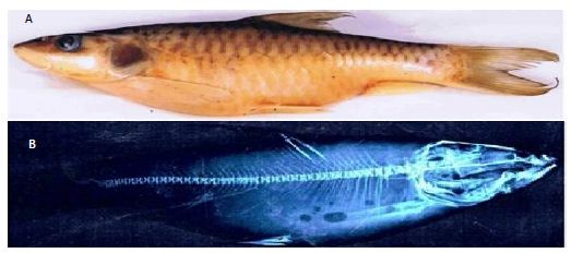
Figure 9a: Photograph of Tor putitora (Ham.-Buch) showing truncated body, short caudal peduncle and extension of the longest anal fin ray to the caudal fin base.
Figure 9b: X-ray Photograph of Tor putitora (Ham.-Buch) with truncated body, short caudal peduncle and extension of the longest anal fin ray to the caudal fin base.
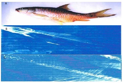
Figure 10a: Photograph of Tor putitora (Ham.-Buch) showing mid truncated body, abnormal height and disposition of fins.
Figure 10b: X-ray Photograph of deformed specimens of Tor putitora (Ham.-Buch) with mid truncated body, abnormal height and disposition of fins.
Figure 10c: Enlarged x-ray photograph of vertebral column of deformed Tor putitora (Ham.-Buch)
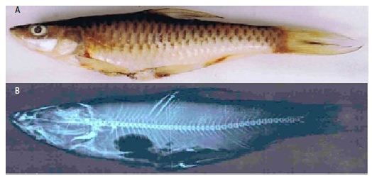
Figure 11a: Photograph of Tor putitora (Ham.-Buch) showing mild truncated body and minor displacement of fins.
Figure 11b: X-ray Photograph of Tor putitora (Ham.-Buch) with mild truncated body and minor displacement of fins.
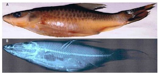
Figure 12a: Photograph of Tor putitora (Ham.-Buch) showing abnormal height, highly truncated caudal peduncle, displacement of fins and overlapping scales.
Figure 12b: X-ray Photograph of Tor putitora (Ham.-Buch) with abnormal height, highly truncated caudal peduncle, displacement of fins and overlapping scales.
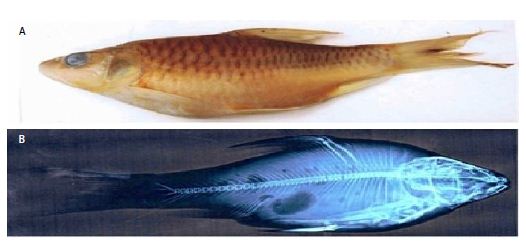
Figure 13a: Photograph of Tor putitora (Ham.-Buch) showing highly truncated body displacement of fins, short caudal peduncle and overlapping scales.
Figure 13b: X-ray Photograph of Tor putitora (Ham.-Buch) with highly truncated body displacement of fins, short caudal peduncle and overlapping scales.
Discussion
Records of only eleven adult deformed fishes, over a period of three years, in the river Chenab suggest their low percentage. This may be due to good water quality, inability of such fishes to resist against fast current or they easily fall prey to predators. Presence of these adult deformed fishes suggests that these aberrations are non fatal, feeding is normal and they are able to avoid predators.
Morphological aberrations observed among Tor species netted from the river Chenab are truncated body, abnormal height and displacement of various fins. Vertebral column deformities commonly reported among fishes include ankylosis, lordosis, kyphosis, scoliosis, irregular shape showing coiling and vertebral fusion and compression. Vertebral deformities noticed during the present study include truncated vertebral column, vertebrae compression and fusion, reduction in inter-vertebral spaces and vertebral thickness. Truncated body and displacement of fins observed in abnormal specimens of Tor is due to vertebral compressions and fusion. Vertebral fusion is known to alter the shape and length of the fish depending on the severity and number of structures affected [12].
Fish anomalies have been attributed to abiotic factors like temperature, light, low pH, salinity and low dissolved oxygen [13-23].
Water analysis in the river Chenab has revealed optimum range of water temperature (8-16°C), pH (8.23-8.46), conductivity (140.05-308.47 µs/cm-1), total dissolved solids (63.90-140.88 mg/l), salinity (0.2 ppt), DO (6.24-13.08 mg/l), BOD (1.07-5.56 mg/l), free CO2 (nil), carbonate (1.16-3.97 mg/l), bicarbonate (54.27-116.25 mg/l), chloride (2.33-9.28 mg/l), calcium (14.79-32.49 mg/l), magnesium (3.87-10.13 mg/l), total hardness (58.32-119.56 mg/l), sodium (1.11-1.69 mg/l), potassium (1.23-2.54 mg/l), phosphate (0.040-0.075 mg/l), nitrate (0.145-0.323 mg/l), silicate (4.08-9.33 mg/l) and sulphate (11.35-19.30 mg/l). Moreover, heavy metal analysis of lead, copper, nickel, zinc and iron is below detectable limits of instrument. This clearly suggests that abnormalities in Tor species, under discussion, are not due to fluctuations in abiotic characteristics of water. These optimum levels of water quality also suggest absence of any type of water pollution in the river Chenab. Therefore, fish aberrations caused by water quality degradation resulting from pollutants and suggested by earlier workers [24-27] are ruled out in the present case.
Among biological factors, fish aberrations have been attributed to parasitic infestations [3,27-32]. Absence of any parasite and sign of disease among the presently collected deformed specimens of Tor species from the river Chenab suggests that these aberrations are not due to this biological factor.
Aberrations in Tor tor and Tor putitora netted from the river Chenab are most probably induced by fast currents faced by larvae and young fishes in various tributaries (fish breeding grounds) of the river Chenab. Young fishes migrating from the Himalayan streams (breeding grounds) into the river Chenab are also exposed to torrential waters inducing various aberrations. Fish anomalies due to currents are well documented [2,5,33-36].
A detailed study on fish larvae and young fishes in their breeding grounds in various Himalayan streams and young fishes in the river Chenab is suggested to understand the role of currents in inducing various fish aberrations in torrential lotic waters.
Acknowledgements
This paper is a part of the project supported by the UGC, New Delhi and is gratefully acknowledged. HOD, Environmental sciences, University of Jammu, Jammu, is acknowledged for providing necessary facilities in the department.
References
- Dutta SPS (1989-90) Vertebral-spinal deformity in Barilius vargra (Ham.-Buch.) from Jammu. Matsya 15-16: 166-168.
- Dutta SPS (2016) Record of an abnormal Schizothoraichthys esocinus (Heckel) from the Himalayan River Chenab draining Jammu region of the J&K State, India. Journal of Aquaculture Research & Development 7: 1-3.
- Dutta SPS (2016) Some deformed specimens of Mystus bleekeri (Day) and Labeo bata (Ham.-Buch.) from the river Chenab in Pargwal wetland, Akhnoor, Jammu. Journal of Applied and Natural Science, 8: 481-484.
- Dutta SPS, Kour H (1994) Pelvic fin deformity in Schizothorax richardsonii (Gray and Hard) inhabiting Rajouri river, (J&K). J Freshwater Biol 6: 195-196.
- Dutta SPS, Sheikh A (2017) Skeletal deformities in Bagarius bagarius (Ham.-Buch.) and Crossocheilus latius diplocheilus (Ham.-Buch.) from river Tawi, a Himalayan stream, in Udhampur area, Jammu region, J&K, India. International Journal of Fisheries and Aquatic Studies 5: 247-251.
- Dutta SPS, Sharma J, Koul V (1993) A truncated specimen of Garra lamta (Ham.). J Nature Conservators 5: 115-116.
- Dutta SPS, Kour H, Sharma J (1996) On the occurrence of malformed specimen of Labeo bata (Ham.-Buch.) in river Tawi, Jammu. J Nature Conservators 8: 147-149.
- Gupta SC, Dutta SPS, Verma M (1998) A report on abnormal specimen of Puntius sarana (Ham.-Buch.) from river Basantar, Samba, Jammu (J&K). J Freshwater Biol 10: 137-140.
- Kour H, Kaul V, Dutta SPS (1997) Deformities in some freshwater fishes of Jammu. J Freshwater Biol 8: 213-216.
- Shekhar C, Dutta SPS (1993) An abnormal specimen of Shizothorax richadsonii (Gary and Hard) with vertebral deformities. Him J Env Zool 7: 101-102.
- APHA (1998) Standard methods for the examination of water and waste water.20th American Public Health Association, New York.
- Gavia PJ, Dinis MT, Cancela ML (2002) Osteological development and abnormalities of the vertebral column and caudal skeleton in larval and juvenile stages of hatchery reared Senegal sole (Solea senegalensis) Aquaculture 211: 305-323.
- Alderdice DF, Wickett WP, Brett JR (1958) Some effects of temporary exposure to low dissolved oxygen levels in Pacific salmon eggs. J Fish Res Bd Canada 15: 229-249.
- Al–Hassan LAJ (1982) Vertebral deformities in fishes from Iraq and United Arab Emirates, Arabian Gulf. Iraqi Jour Mar Sci 1: 13-23.
- Beamish RJ (1972) Lethal pH for the white sucker (Catostomus commersonii) (Scapede).Trans. American Fish Society 101: 355-358.
- Bolla S, HolmefJord I (1988) Effect of temperature and light on development of Atlantic halibut (Hippoglossus hippoglossus) larvae Aquaculture 74: 355-358.
- Hubbs C (1959) High incidence of vertebral deformity in two natural populations of fishes inhabiting warm springs. Ecology 40: 154-155.
- Jawad LA, Oktener A (2007) Incidence of lordosis in the freshwater mullet, Liza abu (Heckel, 1843) collected from Ataturk Dam Lake, Turkey Anales de Biologia 29: 105-113.
- Lee CS, Williams WD (1970) Abnormality in fishes due to salinity fluctuations. J Fish Biol 2: 55.
- Lee CS, Menu B (1981) Effects of salinity on egg development and hatching in grey mullet, Mugil cephalus J Fish Biol 19: 179-188.
- Steingraeber MT, Gingerich WH (1991) Hatching, growth, ion accumulation and ossification of brook trout (Salvelinus alpinus) alevins in acidic soft waters. Can J Zool 69: 2266-2276.
- Wang LH, Tsai CL (2000) Effects of temperature on the deformity and sex differentiation of Tilapia, Oreochromis mossambicus. J Expt Zool 286: 534-537.
- Antunes M & Lopes Da-Cunha P (2002) Skeltal anomalies in Gobius niger (Gobiidae) from Saddo estuary, Portugal. Cybium 26: 179-184.
- Dulcic J (2004) Incidence of spinal deformities in natural populations of grass-goby, Zosterisessor ophiocephalus, from the Karin Sea, Eastern middle Adriatic. Cybium 28: 7-11.
- Subba BR (2004) Anomalies in bighead carp Aristichthys nobilis and African catfish Clarias gariepinus in Birat nagar, Nepal. Our Nature 2: 41-44.
- Sloof W (1982) Skeletal anomalies in fish from polluted surface waters. Aquat Toxicol 5: 157-173.
- Sun PL, Hawkins WE Overstreet, RM, Brown-Peterson NJ (2009) Morphological deformities as biomarkers in fish from contaminated rivers in Taiwan. J. Environ. Res. Public Hlth 6: 2307-2331.
- Kent ML, Watral VG, Whipps CM, Cunningham ME, Criscione CD et al., (2004) A digenean metacercaria (Apophallus sp.) and a Myxozoan (Myxobolus sp.) associated with vertebral deformities in cyprinid fishes from the Willamette River, Oregon. Journal of Aquatic Animal Health 16: 116-129.
- Schwartz FJ (1973) Spinal and cranail deformities in the elasmobranchs Carcharhinus leucas, Squalus acanthias and Carcharhinus milberti. J. Elisha Mitchell Sci. Soc 89: 74-77.
- Taylor LH, Hall BK, Miyake T, Cone DK (1994) Ectopic ossicles associated with metacercariae of Apophalluus brevis (Trematoda) in yellow perch, Perca flavescens (Teleostei) development and identification of bone and chondroid bone. Embryol (Berl.) 190: 29-46.
- Villeneuve DL, Curtis LR, Jenkins JJ, Warner KE, Tilton et al., (2005) Environmental stresses and skeletal deformities in fish from the Willamette River, Oregen. Environmental Scienece and Technology 39: 3495-3506.
- Yokoyama H, Freeman MA, Itoh N, Fukuda Y (2005) Spinal curvature of cultured Japanese mackerel, Scomber japanicus associated with brain myxosporean, Myxobolus acanthogobii. Dis. Aquat. Org 66: 1-7.
- Backiel T, Kokurewicz B, Ogorzalek A (1984) High incidence of skeletal anomalies in carp, Cyprinus carpio, reared in cages in flowing water. Aquaculture 43: 369-380.
- Chatain B (1994) Abnormal swimbladder development and lordosis in sea bass (Dicentrarchus labrax) and sea bream (Sparus auratus). Aquaculture 119: 371-379.
- Divanach P, Papandroulakis N, Anastasiadis P, Koumoundouros G, Kentouri M (1997) Effect of water currents on the development of skeletal deformities in sea bass (Dicentrarchus labrax L.) with functional swimbladder during postlarval and nursery phase. Aquaculture 156: 145-155.
- KitaJima C, Watanabe T, Tsukashima Y, Fujita S (1994) Lordotic deformition and abnormal development of swim bladders in some hatchery-bred marine physoclistous fish in Japan. World Aquaculture Soc 25: 64-77.