Abstract
Frozen shoulder (FS) remains one of the most debated and ill-understood conditions causing painful shoulder. It is an extremely painful condition that can be treated in primary care facilities with a lengthy natural history resulting in resolution over 12-18 months. Codman, who coined the word “frozen shoulder” in 1934 had defined it as a painful condition of slow onset in the shoulder coupled with stiffness and sleeping discomfort on the affected side. He had described a pronounced decrease in forward elevation and outward rotation which are the disease’s hallmarks even today. The prevalence of frozen shoulder is estimated to affect 2-5% of the general population, from 10.8 to 30% of diabetic patients and much higher prevalence (27.2%) and incidence (10.9%) of hypothyroidism patients. FS has higher prevalence among men (70:30) than women. It usually affects the non-dominant shoulder although it can occur in either shoulder. Bilateral frozen shoulder occurs in around 14% of patients. Though the single most effective treatment is uncertain, response to a combination of conservative treatment results in gradual resolution of symptoms in 12-18 months.
Frozen shoulder once fully resolved rarely recurs among healthy population but not rare among the diabetes and Thyroid patients. Recurrence is known to occur usually in 5-7 years after the first episode. It’s unusual for frozen shoulder to recur on the affected side earlier. I present here an autobiographic case of rare recurrence of frozen shoulder. It is a rare case because it affected the non-dominant and same side (left shoulder) and after nearly 24 years in contradiction to recurrence in about 5-7 years known in the literature. Though the second and current episode is milder than the first with roughly one third pain and half restriction of range of movement and is being managed conservatively by physiotherapy without analgesics. Resistant cases that do not respond to conservative treatment for 6-9 months are offered surgical treatments called as 1) an arthroscopic capsular release (ACR) or 2) a manipulation under anaesthesia (MUA), both being equally effective. Manipulation process could result in unwarranted complications like fractures of humerus or rotator cuff tear, there for less preferred. Surgical procedures are out of the scope of present case report.
Keywords
Frozen shoulder, Adhesive capsulitis, Conservative treatment, Manipulation, Arthroscopic capsular release, AIIMS, Rehabilitation exercises
Introduction
Adhesive Capsulitis of the Shoulder (ACOS) commonly known as Frozen shoulder (FS) is a condition of uncertain aetiology characterized by significant restriction of both active and passive shoulder movements that occur in the absence of a known intrinsic shoulder disorder [1]. It was first described by Duplay in 1872 as ‘periarthritis’ scapulohumeral, later described as Frozen shoulder by Codman in 1934 [2], Adhesive capsulitis (Naviaser 1945) and Fibrotic capsulitis (Hsu 2011). International Society of Arthroscopy, Knee Surgery & Orthopaedic Sports Medicine (ISAKOS) prefers the term _ ‘frozen shoulder’_ and discourages adhesive capsulitis as there are no adhesions in the shoulder joint [3]. The ISAKOS Upper Limb Committee has classified a stiff shoulder into1. primary idiopathic stiff shoulder (frozen shoulder) and secondary stiff shoulder. Primary idiopathic stiff shoulder develops without any specific trauma or any underlying disease process, however a patient can have a condition that is known to have a link to stiffness (diabetes, thyroid disorders) but not necessarily known to cause stiffness. Secondary FS is shoulder stiffness following an underlying cause such as trauma, infection, or inflammatory disorder. The prevalence of frozen shoulder is estimated to affect 2-5% of the population, and affects men more than women. The peak incidence is observed between 40 and 60 years 20% of patients develop similar symptoms in the opposite shoulder in later years. Bilateral simultaneous involvement is reported in 14% of the patients.
Two conditions namely diabetes mellitus (DM) and thyroid dysfunction are associated with FS. The incidence of frozen shoulder in diabetic patients could vary from 10.8 to 30% [4,5] with a tendency of more severe symptoms and resistance to treatment. The prevalence of DM is ten folds in patients with frozen shoulder, and higher HbA1C and poorly controlled diabetes is associated with the development of FS [6]. Several studies have confirmed higher prevalence (27.2%) and incidence (10.9%) of hypothyroidism in patients with FS [7,8]. Other associated conditions with FS are smoking, cardiac disease, Parkinson’s disease, stroke, neck and cardiac surgery, hyperlipidaemia and Dupuytren’s contracture.
The diagnosis is most often by clinical examination and sometime supported by Xray, MRI or Scanning. Treatment options address reduction in pain, improved function, with high level of patient satisfaction. Minimum of 6 months of supervised conservative treatment should be attempted before surgical treatments are considered. Standard conservative therapy include i) Self-limited disease course: “supervised neglect” with analgesia ii) Non-steroidal anti-inflammatory medications (NSAIDS for 2-3 weeks, iii) Modalities – like Heat/Ice application iv) Oral corticosteroids to reduce night pain v) One or two Corticosteroid injections for pain relief – Glenohumeral/Intra-articular either Landmark-guided or Image-guided, Hydro dilatation and Suprascapular nerve blockade. The surgical procedures like Manipulation under anaesthesia (MUA) and b) Arthroscopic capsular release (ACR) are beyond the scope of this article. The modern evidence of frozen shoulder pathogenesis involves fibrosis and/or contracture of the tendons, joint capsule, and other soft tissues surrounding the glenohumeral joint specifically the rotator cuff interval. In severe cases, the RC interval is “obliterated,” and the coracohumeral ligament is “transformed into a tough contracted band,” like arthritis of soft tissues.
My Case Report
I started to have insidious onset of pain and stiffness in my left shoulder without any preceding traumatic, infective, or inflammatory event in mid May 2022. Pain is around the shoulder over deltoid muscle. Sleeping on my left side was a bit troublesome in the night. My left shoulder examination revealed restriction of both active and passive range of movements (ROM). The loss of external rotation with arm by the side of the chest and folding my left hand behind my back and raising hands overhead were the key symptoms. The range of movement is more than 100° in forward flexion, less than 30° in external rotation, and more than L5 vertebral level in internal rotation. The strength of the rotator cuff is relatively unaffected.
History of Significance
I am a known diabetic since August 1991, managing well on oral anti-diabetics, diet, and regular exercises. I had Left sided Frozen shoulder in 1998 (in the 8th year after diabetes diagnosis). I was treated conservatively at All India Institute of Medical Sciences (AIIMS) New Delhi. The treatment then included NSAIDS, Physiotherapy and Extracorporeal shock wave therapy. After about 3 months of intensive therapy and follow-up of another 12 months I had fully recovered with less than 5% restriction of the movements. I had some complications of Diabetes like Coronary artery disease undergoing By-pass surgery (CABG) in August 2005, Bilateral early cataract surgeries in 2010 and 2014, and Benign enlargement of the prostate. I am under annual check-ups for Diabetic retinopathy,Kidney function tests. My diabetes was fairly well managed until 2018. Since 2018 following relocation to Bengaluru, I am on Carbohydrates restricted diet, walking and muscle strengthening exercises for about 90 minutes per day for 6 days week and oral anti-diabetes drugs. This led to reduction of oral diabetes drugs requirements by one third of what I used to take in early 2018 and Hb1Ac has been around 6.5-7. The FS was almost forgotten.
Two events 1) A head injury due to a fall in the bathroom in August 2021 (Brain scan was normal) 2) the precautionary dose of Covid 19 vaccination on 25 January 2022 had disrupted my diet and exercise routine.
Current Clinical presentation
The current Frozen shoulder pain started insidiously around 15th of May 202 and in a week’s time range of movements of the shoulder got restricted. The pain this time in much less, not needing analgesics but with the restriction of movements (50% of what it was in the first episode). The first sign observed was of painful usual exercise of joining both hands behind the back around L5 level. I had to do this for left shoulder with passive pulling from right hand. Over last 4 weeks it is improved, but the first few attempts of all shoulder exercises in the morning are painful. The worst effected movement is folding (90°) at elbow keeping the shoulders and arms parallel to the ground and extending the left upper limb to touch the ground in supine position or stretching above head standing with my back to wall. Fortunately, there is no disturbance in the sleep. I intensified my shoulder exercises and in 4 weeks I see 25% improvement of ROM.
Physical Examination
Current examination findings include Painful and restricted active and passive ROM, especially of Elevation – forward flexion/abduction, Rotation – external/internal rotation. Difficult active and passive internal and external rotation with shoulder abducted 90° and tenderness of anterior and posterior capsule is felt. Has not affected my sleep.
Basic investigations on 26 May 2022, indicated a Fasting BS= 159 mg/Dl, Hb1Ac level of 7.0, Hb% of 12.2 g/Dl, B12= 150 mg/ml which were a matter of concern. Other biochemical parameters like serum calcium, creatinine, Iron, Vitamin D (25 Hydroxy) = 50 ng/ml and serum Cholesterol = 106 mg/Dl were well within acceptable limits. No Imaging is done this time.
My Management Approach in Current Episode
I am a 76 years aged male, diabetic for 32 years, FS diagnosis based on previous experience. The clinical stages are indistinguishable, ROM and pain are limited. Have been doing all activities as much as tolerated. I have not used either heat, or analgesic medications. I have been cautiously doing physiotherapy, including shoulder exercises as recommended in the first episode,listed later with illustrations, that were interrupted for about 3 months including stretching, strengthening and Pendulum stretch with weights, inwards and outwards rotations, and other Yoga asanas.
Prognosis
I intensified my shoulder exercises and in over last 4weeks ROM has improved,but the first few attampts of all shoulder exercises in the morning are painful.
Discussions
Two Episodes of My FS Episodes in Comparison with National Recommendations
Pathogenesis
A major role of inflammatory mediators (interleukins, cytokines, B- and T-lymphocytes, growth factors, matrix metalloproteinases, tumour necrosis factors and fibroblast activation markers) is postulated in FS characterized by intense inflammatory changes in capsule indicating a role of and disturbance in local collagen translation, which result in global fibroplasia. The capsule of the FS appears thick, congested, and inflamed, particularly around the rotator interval and anteroinferior capsule along with thickened coracohumeral ligament (CHL) and superior-middle-inferior glenohumeral ligaments to the naked eyes during surgery. This results in loss of flexion, abduction, and rotations. Microscopic tissue samples reveal dense collagen matrix and high population of fibroblasts and contractile myofibroblasts, a process like Dupuytren’s contracture, with the fibrotic process predominantly limited to anterior capsule.
Epidemiology
Exact cause is not known. Evolution of synovial inflammation to capsular fibrosis and a Combination/progression of inflammation and fibrosis like Dupuytren’s chronic inflammatory response with immunomodulated fibroblastic proliferation (Hand, JBJS, 2007). Contracture of the rotator interval, coracohumeral ligament, and anterior /inferior capsule is observed.
Literature review indicates that mostly men (60-70 %) are affected and Med Sport data indicated 67% (Housner, 2017). Majority (70-75%) of the affected are between 40-60 years old (peak age 50). My first episode was at the age of 46 year and the current at 76 years. While the first episode was well within expected age frame the second episode is well beyond known upper age limit.
Risk of recurrent in contralateral shoulder 15-20% usually within 5 years (range of 6 months to 7 years (Bridgman 1972, Reeves 1975, Shaffer 1992, Hand 2008) and Risk of recurrence in same shoulder essentially 0% (Codman 1934, Lippman 1944, Hsu 2011). My case is very rare as both these conditions are contradicted as the recurrence has occurred after 24 years and has affected same shoulder.
Natural History
Total duration may be 12 to 40 months; recovery is always sure and can be confidently expected but there is a Controversy over residual pain and/or loss of motion of 50% patients at 7 years with no functional limitation. My first episode had similar experience as I had recovered within 15 months fully, with a functional loss of less than 5% and absolutely no pain. The second episode now is insidious and much less painful and ROM. The different stages described below were evident and distinct in the first episode but not now.
Stage 1 (Painful or “Freezing”). Usually lasts up to 9 months from onset of symptoms, pain precedes the restriction in motion. Sharp pain at end range of motion and gradual loss of motion. The earliest to get affected and latest to return finding is usually loss of external rotation. May affect sleep if there is aggressive synovitis or angiogenesis. In my case in the first episode, my sleep was disturbed for nearly 2 months but not affected at all now.
Stage 2 (“Frozen”). 6 to 15 months from onset leading to loss of motion in all planes and throbbing pain worse with motion and usually disturbs sleep. MRI shows Capsuloligamentous fibrosis. My first episode had bothered me for about 6 months in this stage. The image inferences were like the ones described in the literature. In the current episode I have not gone for any imaging as the condition is evident and mild. I need to observe the long-term resolution.
Stage 3 (“thawing”). Lasts for 12 to 24 months from onset. It is gradual spontaneous improvement of shoulder mobility and function. Pain starts decreasing, range of motion improves, mild mobility deficits and pain may persist. Most patients report minimal to no disability. Pathologically Synovial involvement recedes. Poor function of reaching overhead (getting dressed, putting on deodorant), reaching behind back (putting on shirt/coat) and reaching out to the side (getting mail, using ATM). Patient finds it difficult to explain onset of pain to attributes to a trivial injury. My first episode had taken about 6 months in this stage. Pain had receded slowly over 6 months and after a total of about 15 months neither there was a residual pain or no was there any residual restriction of movements. In the current episode I need to observe this stage. Looking at the current progress in first 4 weeks, I strongly feel that the entire course of returning to normalcy may take around 6 months.
Treatment
By and large, conservative treatment of frozen shoulder is successful in up to 90% patients. Latest recommendation of the treatment is ‘use it or lose it’ by movement therapy [Jun 2022-11]. The age old conservative treatment incudes:
NSAIDs and Other Analgesics
NSAIDs remain one of the most common medical interventions, a short course of NSAIDs for 2-3 weeks minimises the intense pain of in treating frozen. In my first episode this was adopted for about 3 weeks. The current episode pain is tolerable and hence have not taken any analgesics.
Corticosteroids
Both oral steroid and local steroid injections are widely used. They are beneficial only in early stages to control inflammation and ensuing pain and may not be useful in late stages with established fibrosis without much inflammation. Oral steroids for improving pain, ROM and function when prescribed for ‘short term’ of up to 6 weeks in early stage is helpful. Systematic reviews and metanalysis have confirmed usefulness of steroid injections in improving pain and ROM in the short term, and moderate evidence in the midterm. Fortunately, neither in the first nor the current episodes this intervention was either advised or tried.
Hydro-dilatation (HD)
A single HD of the glenohumeral joint using saline, steroid, local anaesthetic agent is supposed to distend the capsule by breaking the ‘early intracapsular fibrosis’ helping in improving ROM In early/late frozen stage. However, more than one repeated HD after 2 weeks have no added effect over FS. There was no need of such an intervention in my case.
Calcitonin
Calcitonin is supposed to decrease the systemic inflammatory response and stimulate the release of endorphins, improving mRNA expression of fibrosis-related mole, but further research is required in this area to validate.
Extracorporeal Shock Wave Therapy (ECSWT)
ECSWT significantly improves the functional outcome and ROM without any adverse events. ECSWT was used for about 4 weeks twice a week in the first episode and was instrumental in remission of pain in 6 months as against expected time frame of 9-12 month, that was considered needed then.
Acupuncture
A few studies have reported reasonable relief in pain and improved forward flexion by using acupuncture in the treatment of FS.
Nerve Block
A single or multiple injections to block Suprascapular nerve in the treatment of FS have shown improved pain score and ROM Operative Management of Frozen Shoulder involves a) Manipulation under anaesthesia (MUA) and b) Arthroscopic capsular release (ACR). Both are equally effective, but ACR is ‘preferred’ surgical option for the treatment of refractory FS as it allows controlled and precise release of fibrosed capsule-ligament complex under vision, avoiding complications of MUA like as Humerus shaft fracture, rotator cuff tear, shoulder dislocation, labral tear, and nerve injury.
Rehabilitation
A Shoulder Range of Motions (Flexion, abduction, internal rotation, and external rotation) as detailed below was advised and done with the help of a physiotherapist of AIIMS for two weeks along with ECSWT and continued at home for another 4 months without ECSWT in my first episode of FS in 1998.
Pendulum Stretch
I was to relax my shoulders, stand and lean over slightly, allowing the affected arm to hang down. Swing the arm in a small circle, about a foot in diameter. Perform 11 revolutions in each direction, once a day. As my symptoms improved, increased the diameter of my swing, without forcing it. Slowly I increased the stretch by holding a light weight (1-5 kg) in the swinging arm even now [9] (Figure 1).
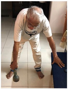
Figure 1: Stretch by holding a light weight (1-5 kg) in the swinging arm
Towel/Rubber Band Stretch
I hold one end of a three-foot-long towel (of late a rubber stretch band) behind my back and grab the opposite end with your other hand. Held the towel in a horizontal position and used my good (right) arm to pull the affected arm upward to stretch it. I held the bottom of the towel with the affected arm and pull it toward the lower back with the unaffected arm for 11 times a day (Figure 2).
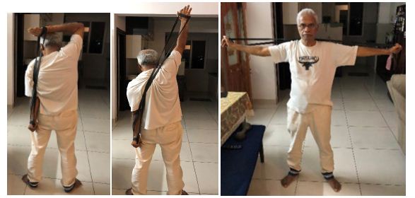
Figure 2: Held the bottom of the towel with the affected arm and pull it toward the lower back with the unaffected arm for 11 times a day
Finger Walk
I faced a wall three-quarters of an arm’s length away. Reach out and touch the wall at waist level with the fingertips of the affected arm. With my elbow slightly bent, slowly walk my fingers up the wall, spider-like, until I had raised my arm as far as I was comfortable. Only my fingers worked and not my shoulder muscles. Slowly lower the arm (with the help of the good arm, if necessary) and repeat. Perform this exercise 11 times a day.
Cross-body Reach
While standing, I used my good arm to lift my affected arm at the elbow, and bring it up and across my body, exerting gentle pressure to stretch the shoulder and hold the stretch for 15 to 20 seconds. Repeating these 11 times per day.
Outward Rotation
Holding a rubber exercise band between my hands with my elbows at a 90-degree angle close to my sides, rotated the lower part of the affected arm outward two or three inches and hold for five seconds. Repeated these movements for 11 times, once a day (Figure 3).
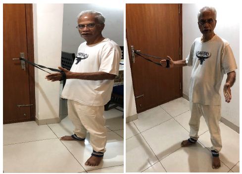
Figure 3: Repeated these movements for 11 times, once a day
Inward Rotation
Standing next to a closed door, hooked one end of a rubber exercise band around the doorknob. Holding the other end with the hand of the affected arm, and my elbow at a 90-degree angle, I pulled the band toward my body two or three inches and held for five seconds. Repeated this exercise for 11 times, daily at least 5 days a week. I had continued doing most of these exercises at least 3-4 days week since then. After relocating to Bengaluru in 2018 my apartment complex Gymnasium had equipment that provided for the above exercises – internal and external rotation exercises, that I did. In 2020 the gym was closed, and I continued to the similar exercising using the elastic stretch bands (Figure 4).
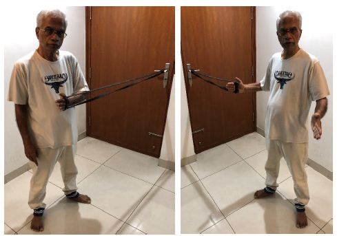
Figure 4: Similar exercising using the elastic stretch bands
The international recommendations are for 10-15/20 movements each time. Keeping in Indian tradition of the odd number sanctity I advocate for 11 or 21 movements for each shoulder.
These exercises are field tested under the name of Pune Shoulder Rehabilitation Program (PSRP) in 2013. PSRP an exercise program involving exercises with low resistance, high repetition performed in sub impingement region for strengthening of scapular and rotator cuff muscles to normalize scapular muscle strength, normalize scapula humeral rhythm, pain relief, rotator cuff muscle strengthening, restoration of range of motion (ROM), restoration of function, and maintaining posture and core. The Pune study findings showed that over the 6th-week protocol, statistically significant improvements were found in pain reduction and shoulder range of motion, concluding that such exercise protocol is effective in increasing the range of motion and decreasing the pain in the shoulder caused by frozen shoulder [10].
Prevention: A few easy steps to help prevent FS are:
- Stretching your shoulder and back muscles daily.
- Stretching your tendons (by rotating hands and palms to stretch different tendons).
- Practicing good ergonomics while sitting at a desk and using a computer.
- Maintaining a healthy immune system.
Conclusion
These days, adhesive capsulitis is the precise descriptive jargon for the condition, but no better than “periarthritis. Frozen shoulder (FS) continues to be an extremely painful condition since it was first identified in 1934, that can be treated in primary care. It has a lengthy natural history resulting in resolution over 12-18 months. The shoulder is the only joint that often “freezes” like this and is a common biological puzzle. It’s hard to define precisely, diagnose accurately, or treat effectively. In fact, frozen shoulder treatment is one of the best examples of how musculoskeletal medicine is surprisingly primitive still. Of late there is a hypothesis that FS is more of Functional freezing than adhesive freezing. Three main ways that a functional limitation of shoulder ROM would probably work, are put forth as a) The brain can “shut down” a joint with neurological inhibition, b) because it has become sensitized or c) The muscles may have gotten rotten with trigger points.
Frozen shoulder once fully resolved does recur, though in small proportion among the diabetes and Hypothyroid patients. Usually, recurrence occurs in 5-7 years after the first episode and affects contralateral side. It’s extremely rare for frozen shoulder to recur on the same side affected earlier. Second episode is much milder, hardly disturbs sleep and can be managed with exercises only [11-13].
References
- Duplay S (1892) Archives Générales de Médecine.
- Codman E (1984) The Shoulder Rupture of the Supraspinatus Tendon and Other Lesions in or about the Subacromial Bursa. Medicine.
- Vivek Pandey et al. (2021) Clinical Guidelines in the Management of Frozen Shoulder. Journal List Indian J Orthop. 55: 2. [crossref]
- Jeffrey A, Housner. Adhesive Capsulitis of the Shoulder. Departments of Family Medicine, and Orthopaedic Surgery, University of Michigan.
- Future Sci OA (2020) Frozen shoulder: Overview of clinical presentation and review of the current evidence base for management strategies[crossref]
- J Zreik NH et al. (2016) Adhesive capsulitis of the shoulder and diabetes: A meta-analysis of prevalence. Muscles Ligaments Tendons. 6: 26-34. [crossref]
- Bridgman JF (1972) Periarthritis of the shoulder and diabetes mellitus, Annals of the Rheumatic Diseases. 31: 69-71. [crossref]
- Chan JH et al. (2017) The relationship between the incidence of adhesive capsulitis and haemoglobin A(1c). Journal of Shoulder and Elbow Surgery. 26: 1834-1837. [crossref]
- Schiefer M et al. (2017) Prevalence of hypothyroidism in patients with frozen shoulder, Journal of Shoulder and Elbow Surgery. 26: 49-55. [crossref]
- Cakir M et al. (2003) Musculoskeletal manifestations in patients with thyroid disease. Clinical Endocrinology – Oxford. 59: 162-167. [crossref]
- Shoulders stretching exercises for frozen shoulder.
- Seema Saini et al. (2022) Effectiveness of Pune shoulder rehab protocol on patients with frozen shoulder.
- Complete Guide to Frozen Shoulder.