DOI: 10.31038/CST.2017262
Abstract
Primary Cardiac Lymphoma (PCL) is a very rare condition accounts for 2% of all primary cardiac tumours. This is a case report of a 76 year old male patient with shortness of breath, cough and pyrexia of unknown origin for 2 months. In the initial diagnostic workup, echocardiography revealed aortic and mitral regurgitation with no pericardial effusion. With the development of right sided pleural effusion about one month after the initial presentation, ultrasound scan guided pleural fluid aspiration performed. Pleural fluid culture revealed no bacterial growth. Second echocardiography showed no flow in the Superior Vena Cava (SVC) and suspected of a thrombus. Contrast enhanced Computed Tomography scan of the chest demonstrated a mass lesion in the right heart and distal inferior vena cava (IVC). The lesion was compressing the SVC without complete obstruction. The pericardium was intact with no pericardial effusion. No mediastinal lymphadenopathy identified. Initial differential diagnosis was leiomyosarcoma of the distal IVC extending in to the right heart. As there was no pericardial involvement, possibility of lymphoma was made as a remote cause. Subsequently digital subtraction angiography guided biopsy performed from the mass in the distal IVC and right atrium. Diffuse lymphoma was confirmed on histology. As there were no Hodgkin cells in the sample, probable diagnosis of Non-Hodgkin’s lymphoma was made. Subsequently patient died due to cardiovascular compromise before starting specific therapy.
Keywords
Cardiac lymphoma, Inferior vena cava, Radiological imaging, Histological diagnosis
Introduction
Primary Cardiac Lymphoma (PCL) is a very rare condition usually of B-cell non-Hodgkin’s type. They account for 2% of all primary cardiac tumours and 1% of extranodal lymphomas [1, 2]. They are usually diffuse large cell lymphomas manifest as an ill-defined, infiltrative mass [3]. PCL are more commonly seen in immunocompromised states, either iatrogenic including those associated with solid organ or bone marrow transplantation or HIV related and these are usually associated with EBV infection. Lymphomas in immunocompetent individuals are extremely rare [4]. As there are no specific clinical symptoms, the clinical diagnosis of the condition is difficult. Typically when B symptoms develop, (fever, weight loss, fatigue common in lymphoid malignancies), progressive heart failure will ensue in these patients [5]. However advanced cross sectional imaging modalities allowed early and accurate detection of primary cardiac malignancies [6]. The definitive diagnosis of the condition needs histological evaluation of the tumour. One of the accessible diagnostic method is transoesophageal echocargiography-guided transjugular biopsy, which has a relatively low risk of complications [7]. Endomyocardial biopsy via percutaneous cardiac approach is also another approach for histological diagnosis [8].
We present a case of lymphoma arising in the heart with extension in to the superior and inferior vena cava.
Case Report
A 76 year old male was initially investigated for shortness of breath and cough of 2 months duration in a specialized hospital for respiratory disease. On initial presentation the patient was not dyspnoeic but found to have 94% of saturation of oxygen in blood. Bilateral ankle oedema was evident at that time. The blood pressure was normal on admission to the hospital. During hospital stay patient develops mild fever episodes intermittently. Sputum culture and blood culture were negative during the febrile illness. Subsequently patient underwent trans oesophageal echocardiography suspecting infective endocarditis, and it revealed grade ii aortic regurgitation and mitral regurgitation. There was no evidence of pericardial effusion or features of congestive cardiac failure Figure 1.
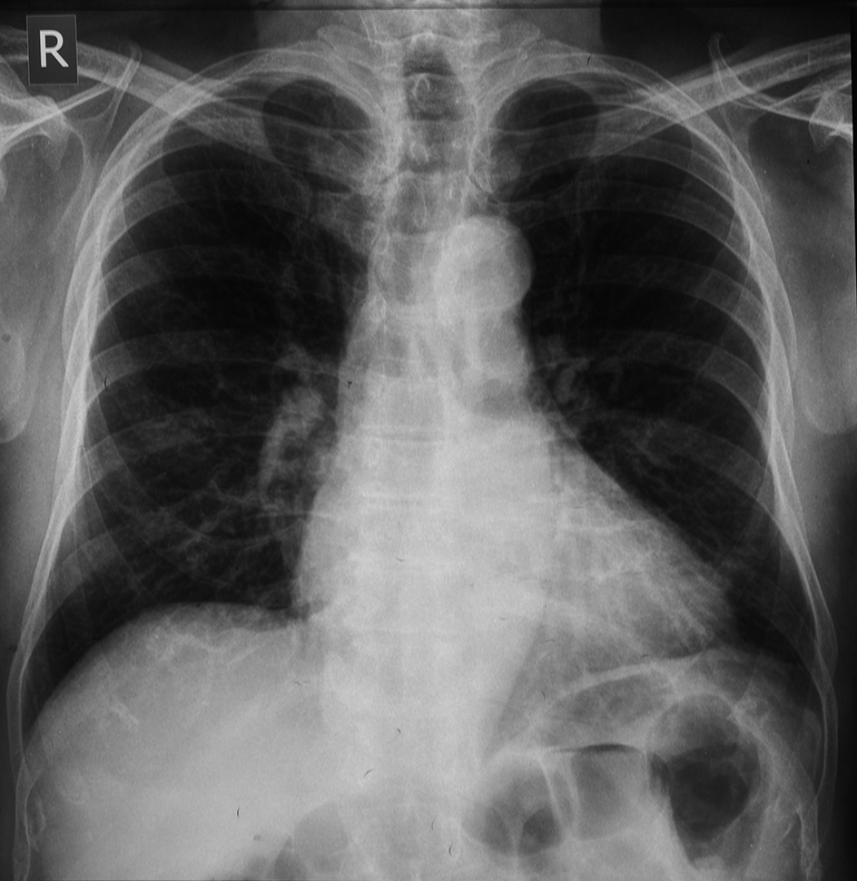
Figure 1.
About a month following initial presentation the patient develops right sided pleural effusion and then ultrasound scan guided pleural fluid aspiration was performed. There were 3.8g/dl of protein and 80% of lymphocytes in the pleural fluid. However there was no evidence of bacterial growth or acid fast bacilli in the pleural fluid.
Again patient was send for cardiology opinion and underwent 2 dimensional Echocardiogram which revealed no flow in the Superior Vena Cava (SVC) with suspicion of a thrombus in the SVC extending in to the right ventricle. Right atrium was dilated at that time. Then the patient was transferred to the National Hospital of Sri Lanka (NHSL) for further evaluation and management Figure 2.
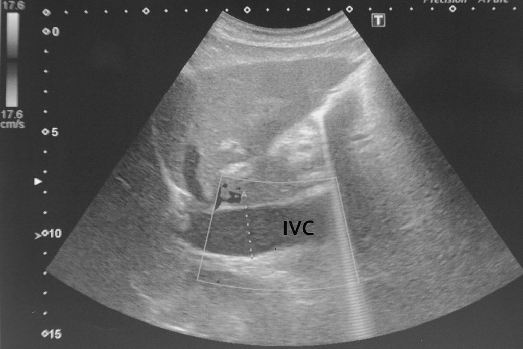
Figure 2.
A contrast enhanced Computed Tomography (CT) scan of the chest performed in the NHSL and detected a contrast enhancing mass lesion with few hypodence areas within the lesion involving the distal inferior vena cava (IVC), right atrium and right ventricle. The lesion was appear to compress the distal SVC without complete obstruction of the lumen. Right main pulmonary artery was pushed superiorly by the mass lesion without significant luminal narrowing. The pericardium was not involved by the lesion and there was no pericardial effusion. There was bilateral pleural effusion on CT scan. There was no significant hilar or mediastinal lymphadenopathy. In conclusion differential diagnosis of leiomyosarcoma of the distal IVC extending in to the right atrium and right ventricle was made. As there was no pericardial effusion or pericardial involvement by the mass lesion, the possibility of lymphoma was made as a remote possibility Figure 3.
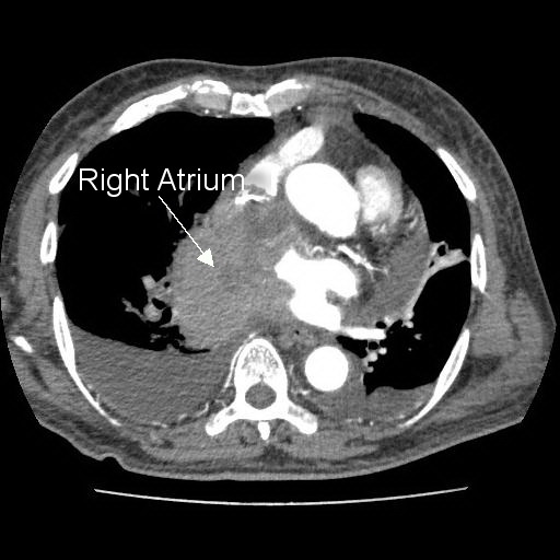
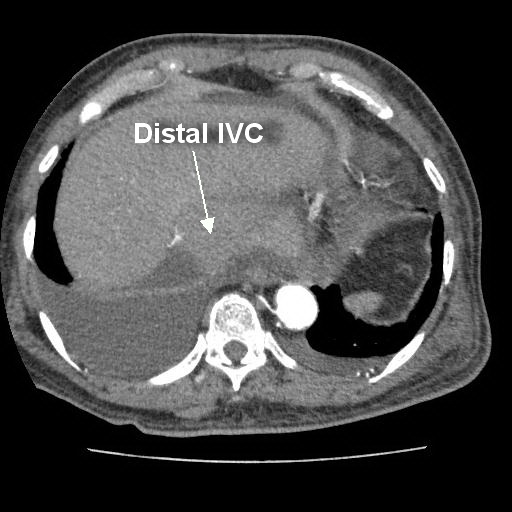
Figure 3.
Subsequently the patient referred for digital subtraction angiography guided biopsy of the mass lesion in the distal IVC and right atrium. With right femoral vein puncture the venous access gained and IVC catheterization was performed. Five biopsy samples were obtained using 10 F guiding catheter and an Alligator forceps Figure 4.
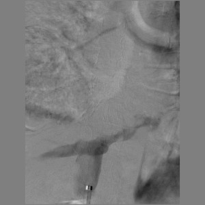
Figure 4.
On histological assessment with haematoxylin and eosin staining there were sheets of discohesive medium to large lymphoid cells with enlarged hyper chromatic nuclei and narrow rim of cytoplasm. Mitoses were frequently found. Immunohistochemistry (IHC) with Leukocyte Common Antigen (LCA) revealed strong membranous positivity in all lymphocytes. Diffuse lymphoma was confirmed by histology. As there were no Hodgkin cells in the biopsy, probable diagnosis of Non-Hodgkin type lymphoma was made [Figure 5 & 6].
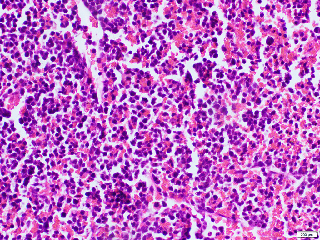
Figure 5.
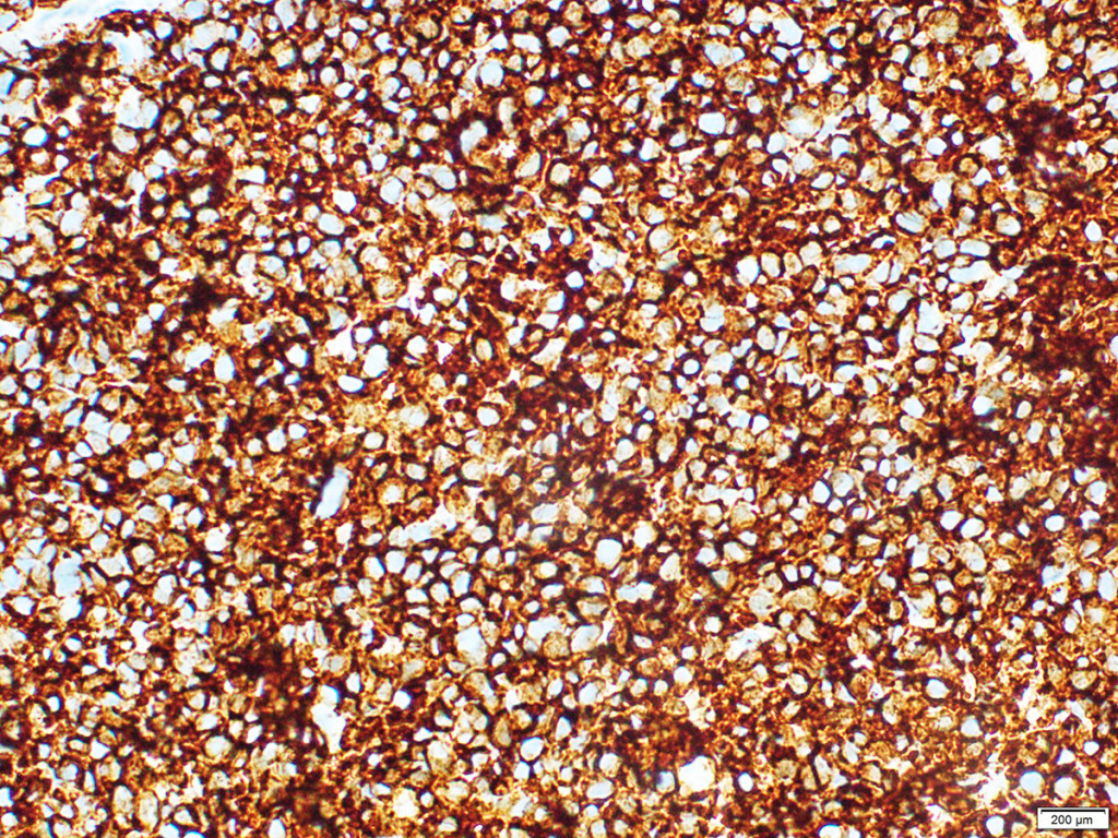
Figure 6.
Subsequently the patient was transferred to The National Cancer Institute, Sri Lanka for further management. However the patient died from cardiac arrest before starting specific therapy.
Discussion
Primary cardiac lymphoma (PCL) is a very rare condition. On histology they are usually of B-cell non-Hodgkin’s type lymphoma. The most frequent location of PCL is the right atrium, followed by the pericardium. Signs and symptoms are non-specific and depend on the location and extent of the tumour; these include right-sided heart failure, precordial pain, arrhythmias, conduction disorders and cardiac tamponade [7]. In literature there is a case of atypical presentation of ischemic hepatitis in a young adult with PCL, which highlights the wide variety of clinical manifestations of this rare tumour [9].
In our patient the initial presentation was nonspecific and the disease progressed rapidly. Radiological imaging played major role in the identification and sampling of the tumour. The diagnosis of diffuse lymphoma was confirmed by histology with probable diagnosis of Non-Hodgkin lymphoma.
The most effective treatment method for cardiac lymphoma is chemotherapy. Palliative surgery may be necessary to correct haemodynamics when venous blood flow to the lungs is disturbed [10]. Guilherme HO, et al. concluded in their study that cardiac lymphomas had worse survival compared with extra-cardiac lymphomas [11].
Unfortunately this patient died due to cardiovascular compromise before starting chemotherapy or palliative surgery.
Take home message: Primary cardiac lymphoma is a rare entity with nonspecific clinical presentation which may mislead the early diagnosis and prompt treatment. Radiological and histological evaluation plays major role in the diagnosis of this condition.
Limitation: In our local setting immuno-histochemical analysis was not available freely and therefore the diagnosis of definite subtype of the lymphoma was limited.
Ethical Consideration: Informed consent was taken from the relatives of the patient for publication of the case report.
References
- Fernandes F, Soufen HN, Ianni BM, Arteaga E, Ramires FJ, et al. (2001) Primary neoplasms of the heart. Clinical and histological presentation of 50 cases. Arq Bras Cardiol 76: 231–237. [crossref]
- Lam KY, Dickens P, Chan AC (1993) Tumors of the heart. A 20-year experience with a review of 12,485 consecutive autopsies. Arch Pathol Lab Med 117: 1027–1031. [crossref]
- Jean Jeudy, et al. (2012) From the Radiologic Pathology Archives, Cardiac Lymphoma: Radiologic Pathologic Correlation. RadioGraphics 32:1369–1380
- Bagwan IN, et al. (2009) Unusual presentation of primary cardiac lymphoma. Interactive CardioVascular and Thoracic Surgery 9: 127–129
- Shah K, Shemisa KA (2014) “low and slow” approach to successful medical treatment of primary cardiac lymphoma. Cardiovasc Diagn Ther 4:270–273
- Seok JR, Byoung WC, Kyu OC (2001) CT and MR findings of primary cardiac lymphoma: Report upon 2 cases and review. Yonsei Medical Journal 42: 451–456
- Angela FC, et al. (2003) Primary Cardiac Lymphoma: Diagnosis by transjugular biopsy. Rev Esp Cardiol 56:1141–4
- Jung YC, et al. (2009) Extensive primary cardiac lymphoma diagnosed by percutaneous endomyocardial biopsy. J Cardiovasc Ultrasound 17:141–144
- Yuan JS, et al. (2012) Acute ischemia hepatitis as a major clinical presentation of the primary cardiac lymphoma. J Emerg Crit Care Med 3: 118–23.
- Jonavicius K, et al. (2015) Primary cardiac lymphoma: two cases and a review of literature. Journal of Cardiothoracic Surgery 10:138
- Guilherme HO, et al. (2017) Characteristics and Survival of Malignant Cardiac Tumors: A 40-Year Analysis of Over 500 Patients.