DOI: 10.31038/CST.2018334
Case Report
A 40-year-old man with a 20-years history of hepatitis B virus infection was diagnosed as post-hepatitis cirrhosis a month ago. He sought to take Chinese Herbs (CH), aiming to softening the cirrhotic nodules. Unexpectedly, he experienced fever and diarrhea for 1 week and abdominal pain for 2 days after taking CH of 7 days. Blood investigations revealed severe leucocytosis and raised liver enzymes and bilirubinemia, elevated Alpha Fetal Protein (AFP) and incredible severe coagulation disorders that coincided with the diagnosis of Disseminated Intravascular Coagulation (DIC). Computed tomography revealed some cirrhotic liver with partial enhancement. Magnetic Resonance Imaging (MRI) for the first time unveiled multiple low-signal vaculoses throughout the liver (honeycomb sign) in its diffused weighted imaging phase (Figure 1), which were potentially made up of necrotic nodules of varying sizes but less than 1 centimeter. Unlike melioid liver abscesses, his inflammatory was soon cured after antibiotic (Meropenem) using of 10 days. In this case, DIC was a confusing issue about its definitive cause that was thought to be associated with the advanced liver cirrhosis or nodules carcinomarization or drug-related liver injury or other blood malignancy. In any way, CH may play a key role in inducing these changes. Strangingly, DIC didn’t significantly improved following improved liver function and cured infection. Further evaluation of petron emission tomography / computed tomography confirmed hepatic nodules carcinoma transformation and extensive abdominal peritoneum metastasis, which also clarified the initial conundrum of DIC cause, fluctuating value of AFP and honeycomb sign. On 3-month following-up visit, he seemed to obtain benefit from CH performance of promoting hard nodules-resolving because AFP began to go better and clotting disorder also improved gradually.
Consent statement: The patient has given his written informed consent for his data to be submitted or published.
Conflicts of Interest: All authors declare that they have no conflicts of interest concerning on this manuscript.
Authors’ Contributions: Zhong Jia and Jia-Qing Huang were involved in compilation of the data and drafting of the article. Both authors read and approved the final manuscript.
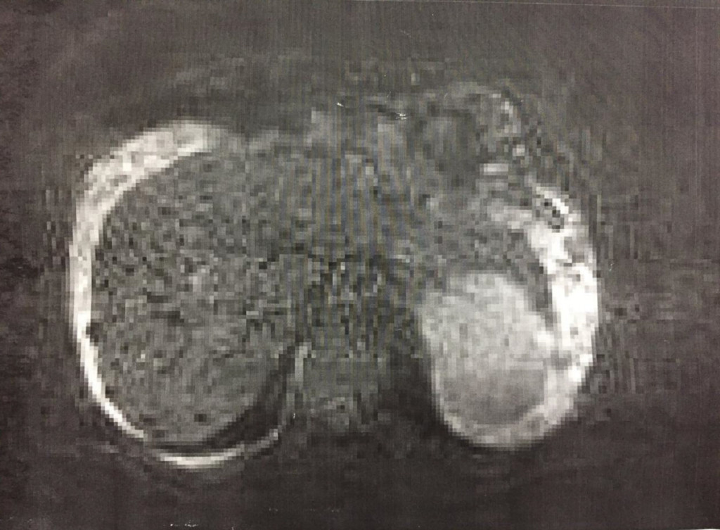
Figure 1. Magnetic Resonance Imaging (MRI)