Summary
A 41-year-old female patient, who developed trigger finger after she repeated cervical sprain several times, underwent myofascial evaluation and treatment, which were based on Fascial Manipulation®, in addition to evaluation by physical therapy, and favorable results were obtained. She showed symptoms of trigger finger, which suddenly occurred, for 5 months. Palliative treatment was the only method for the symptoms, and they did not reach improvement. The site and tissue, which are responsible for the condition, were specified first on evaluation by physical therapy. Subsequently addition of exercise test and palpation test for fascia in view of a past history and timeline may appropriately approach to problems with high-density sites and force transmission of the fascia related to pain.
Introduction
Many cases of trigger finger have been believed to be idiopathic, and the symptoms occur spontaneously. Some reports have indicated the relevance to occupational or repetitive activities [1]. For the pathogeneses, much attention was paid to specification of sites of myofascial dysfunction in view of a past history and living activity level and routes of force transmission, as well as conventional tests and evaluation. This article describes favorable results of the myofascial evaluation and treatment, which were based on Fascial Manipulation® (FM®) and conducted in addition to evaluation by physical therapy, in a patient who developed trigger finger after she repeated cervical sprain several times.
Roles of fasciae
Fascia has been explained at the International Conference of Scientific Research of Fascia, as follows: “Fascia, a soft tissue constituent of the connective tissue system all over the human body, forms a 3-D matrix in the whole body to support structures “[2]. Fascia spread over all organs, muscles, bones, and nerve fibers to cover them. The definition of fascia includes aponeurosis, ligament, tendon, retinaculum, articular capsule, capsules of organs and vessels, epineurium, meninges, periosteum, and intra- and intermuscular fibers of all fasciae. All of them indicate that fasciae have important roles in muscular biomechanics, coordination of muscular peripheral movements, and maintenance of proprioceptor and postures.
Some muscle fibers of epimysium enter the deep fascia; i.e., 37% of muscle origins and insertions enter the deep fascia and intermuscular septum (myofascial expansion) without insertion and the inserted tendon [3]. When fascial dysfunction occurs, muscle spindle and nociceptor are stimulated, spreading along the fascial arrangement via the deep fascia. For instance, aponeurotic fasciae cover the whole muscles of the upper limb, and collagen fiber bundles are arranged in different directions in the aponeurotic fasciae. The thoracic muscles receive tension at a proximal site by insertion of various fasciae, enabling to slide between them and inferior muscles. The brachial fasciae are connected at a proximal site to axillary fascia, greater pectoral fascia, deltoid fascia, and dorsolateral fascia, while they are connected to antebrachial fascia at a distal site. The mediolateral intermuscular septum is originated from the brachial fascia, by which the upper arm is divided into the front and the rear parts as segments. At the elbow the brachial fascia is reinforced by the anterior and rear retinacula, and the anterior retinaculum is composed of the brachial biceps aponeurosis. The brachial biceps aponeurotic expansion branches off two directions. In one direction a fiber bundle extends like a bow obliquely and medially below and binds at the antebrachial fascia. Inside the elbow many muscle fibers of the round pronator muscle and radial carpal flexor are inserted into the antebrachial fascia from the inside. In the other direction collagen fiber bundles are running in parallel to a median line of the forearm in a longitudinal direction. This fibrous expansion reaches the antebrachial fascia between the radial carpal flexor and the humeroradial muscle. Therefore, when the brachial biceps (muscle) tendon is extended at a proximal site, two force lines appear in a medial direction corresponding to the coved fibers and in a direction longitudinally running along the central part of the forearm.
On movements of the upper limbs in various directions, fascial expansion activates fascial proprioceptor with a specific moving pattern and extends a specific site of the brachial fascia, connecting different sites to transmit force. Owing to the relationship between the muscle and fascia coordinative movement to the periphery is realized by performance of movement in a correct direction and correct recognition [2, 4].
Fascia has a role in enhancement of muscle sliding. There is hyaluronic acid in loose connective tissue between deep fascial layers and between epimysium and endomysium, which acts as a lubricant [5]. When hyaluronic acid aggregation is induced by trauma, overuse, etc., fascial layer sliding is restricted. Contraction of the epimysium causes tendon extension due to high density of epimysium, and the extension stimulates the articular receptor to lead to its excitement and pain around the joint. When the body has pain, it responds to the pain with secondary compensation by a posture to avoid the pain. The change in base tension due to the compensation will be controlled by up-and-down tension on the competitive or ipsilateral side, leading to increased complication of the symptoms [6].
FM®
The subjects of FM® treatment include the point [referred to by Centre of Coordination (CC)] on the epimysium, to which one-way muscle strength converges, and the point [referred to by Centre of Fusion (CF)], to which force of adjacent two deep fascia (aponeurotic fascia) units converges. The body is divided into 14 segments, and a functional unit related to movement in each direction is called as fascial unit. All the segments include 6 CC points. There are 6 directions of movement: Sagittal plane (antemotion: AN and retromotion: RE); frontal plane (lateromotion: LA and mediomotion: ME); and horizontal plane (extrarotation: ER and intrarotation: IR). Each fascial unit is designated from the movement direction and segment. For example, the fascial unit of anterior movement of CU (elbow) is designated as AN-CU. There are 4 directions of movement of CF: Anterio-lateral (Ante-Latero: AN-LA); anterior-medial (Ante-Medio: AN-ME); retrolateral (Retro-Latero: RE-LA); and retromedial (Retro-Medio: RE-ME) [6,7]. CC forms the one-way continuous arrangement (called as fascial arrangement) associated with movement direction along the sagittal, frontal, and horizontal planes [8]. For example, the facial arrangement of anterior movement of the upper limbs is composed of 5 fascial units, i.e., AN-SC (scapula), AN-HU (upper arm), AN-CU, AN-CA (carpus), and AN-DI (finger), and anterior movement of the upper limbs is induced by the arrangement. CF has a fascial diagonal line, which appears on a diagonal line of fascial arrangement, and a fascial spiral, which integrates articular elements with retinaculum to influence wide-range pain. It comes to be important for treatment which arrangement has a problem [6, 7].
Pathogeneses of Trigger Finger (Snapping Finger)
Trigger finger is tenosynovitis of the flexor tendon due to imbalance between the tendon sheath and the flexor tendon passing the sheath. Transmission disorder occurs in the tendon sheath to lead to pain, swelling, and heat sensation of fingers. It has been reported that thickening of the tendon sheath, ligamentous intrathecal narrowing, edematous enlargement of the tendon itself, and so on are responsible for these conditions [9]. The augmentation of the symptoms early in the morning and amelioration of the symptoms by day are frequently observed. When they advance, a snapping phenomenon develops, and it may ultimately result in secondary contracture of the Proximal Inter Phalangeal (PIP) joint. The symptoms also frequently appear in a plurality of fingers. It has been believed that the phenomenon is one of the most common causes of hand pain in adults. It has also been reported that the prevalence is ca. 2% of the general population, and tends to be high in women in their fifties or sixties. The prevalence in women in the later stages of pregnancy is also high, and the condition is also characterized by its frequent occurrence due to overuse of hand and as sports injury. It frequently occurs in patients with diabetes, rheumatoid arthritis, or those under the conditions such as amyloidosis, in which systemic accumulation of proteins occurs. As for the treatment, some investigators have reported evidence of moderate efficacy of conservative intervention including the short-term use of Non-Steroidal Anti-Inflammatory drug (NSAID) [10]. The orthotic treatment has also been widespread [11]. In case of conservative therapy, surgical treatment may also be selected when any improvement is not achieved by conservative therapy.
Treatment case
1. General information
A patient is a 41-year-old woman, housewife.
The chief complaints included flexion contracture of and resting pain in the left thumb MP joint and thumb movement pain, and she grasped with difficulty because of pain.
Approximately 5 months ago she suddenly had left thumb pain and restricted Range of Motion (ROM) at the time of rising from her bed. In the early stage the pain was gradually reduced by day, but in the next morning she repeated pain and restricted ROM. The pain gradually augmented even by day. She visited a hospital, and received intrathecal steroid injection with a diagnosis of trigger finger. Even so, the symptoms were not improved. Subsequently she had an attempt to receive acupuncture as well, but the pain augmented. Her left thenar swelling also appeared, and she could do daily living activities (ADL), including clothespin pinching, taking out coins, grasping dish and mobile phone, etc., with difficulty. Thus, she came to interfere with the whole range of housework.
Diagnosis: Trigger finger (snapping finger)
Past history:
20 years of age: Cervical sprain-due to numbness in the region ranging from neck to left hand (traffic accident)
25 years of age: Surgery for right inguinal hernia
30 years of age: Cervical sprain-due to numbness in the region ranging from neck to left hand (traffic accident). Treated by cervical collar fixation
36 years of age: Cervical sprain-due to numbness in the region ranging from neck to both hands (traffic accident)
38 years of age: Right 5th metatarsal fracture
39 years of age: Cervical sprain (a violent fall)
In her late thirties she repeated pain around the scapula once or twice a year.
41 years of age: Lt Thumb trigger finger (snapping finger)
2. Evaluation by physical therapy
Pain: Resting pain (+), movement pain (+), numbness (-)
Active movement: ROM restriction in all directions of left thumb CM joint, in association with pain with score 8 based on the Numerical Rating Scale (NRS).
Passive movement: ROM restriction in all directions of left thumb CM joint, in association with NRS 8 pain.
Neurological test: Various neurological tests (-)
End feel: Empty
Joint play test: Hypermobility of C4th/5th and 5th/6th; low mobility of TH1st/2nd.
Muscle tightness: Brachioradial muscle, long palmar muscle, round pronator muscle, biceps muscle of arm, scalenus, and smaller pectoral muscle.
Observation of posture: Round back and forward head.
Observation of motion: Difficult movements of grasping and pinching.
3. Hypothesis
This patient developed numbness in her both upper limbs after she sustained cervical sprain several times. Since she had a past history of cervical collar fixation, her cervical vertebrae were examined. Various neurological tests are negative at present. She repeated cervical sprain, and had the imbalanced trunk because of coexistence of hypermobility of the cervical vertebrae with low mobility of the thoracic vertebrae. The patient may have been forced to have compensation for various postures under the situation with her unstable neck. It was further considered that the poor alignment accelerated excessive tension of her neck and chest to have led to tension stress to her left thumb through fascial arrangement and expansion from the trunk to the upper limbs. On the hypothesis that the stress spread from the cervicothoracic vertebrae downward, fascia was evaluated.
4. Palpation test [High density level (*~***)]: Comparative palpation test was conducted in TH (thorax), SC, and CA. LT LA-CA***, Bi LA-TH***, Lt LA-SC**, Rt LA-CL (neck)**, Lt ME-CA**, and Lt ME-CU**.
Palpation test revealed high density at the above-described sites, indicating remarkable areas of high density on the frontal plane arrangement.
5. Treatment
First treatment: Lt LA-CA (Figure 1), Lt LA-CU, Lt LA-DI (Figure 2), Lt ME-DI (Figure 3), and Lt ME-CA (Figure 4) (++).
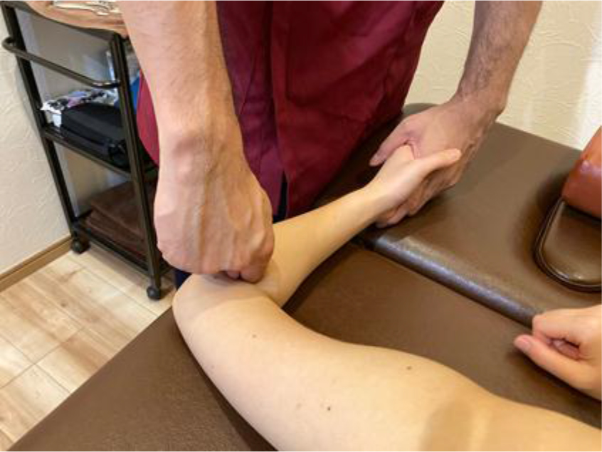
Figure 1. Lt LA-CA.
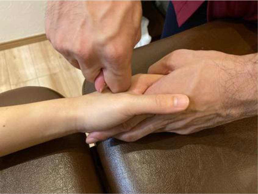
Figure 2. Lt LA-DI.
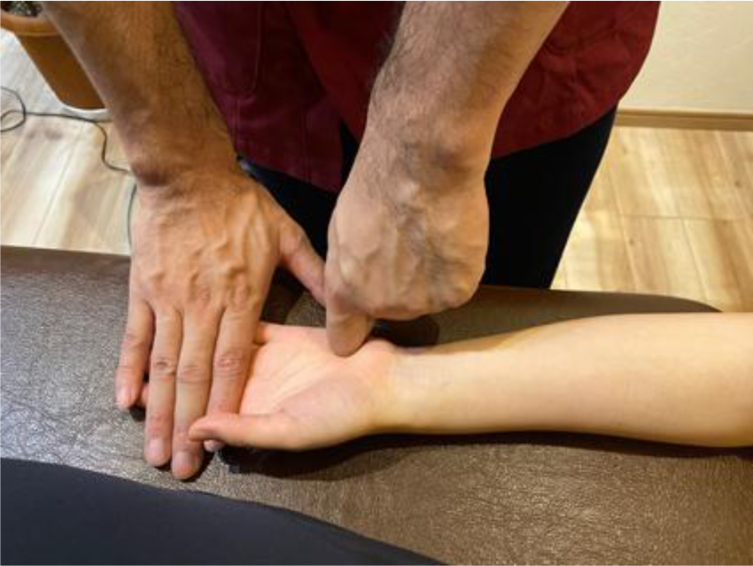
Figure 3. Lt ME-DI.
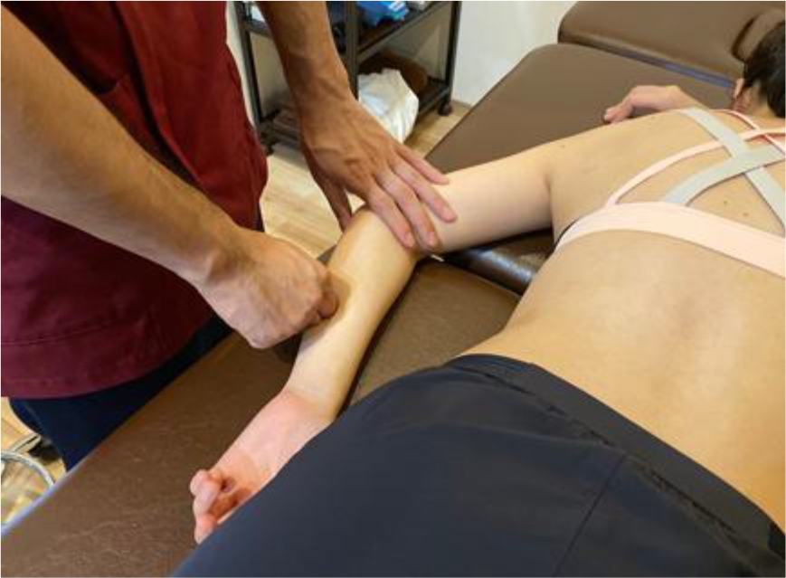
Figure 4. Lt ME-CA.
Second treatment: Lt LA-CA, Lt LA-SC (Figure 5), Rt LA-CL, Lt ME-CU, and Bi LA-TH (Figure 6) (+++).
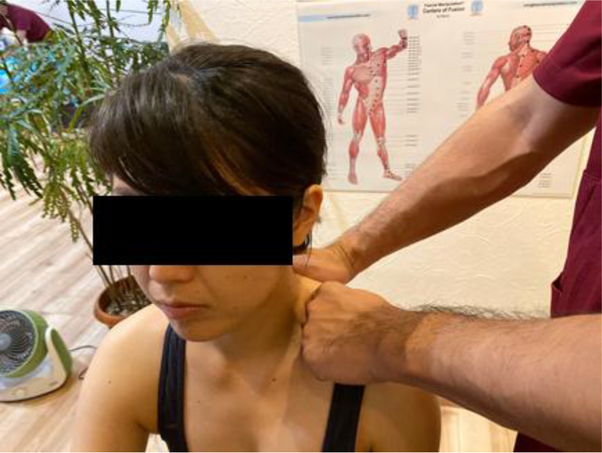
Figure 5. Lt LA-SC.
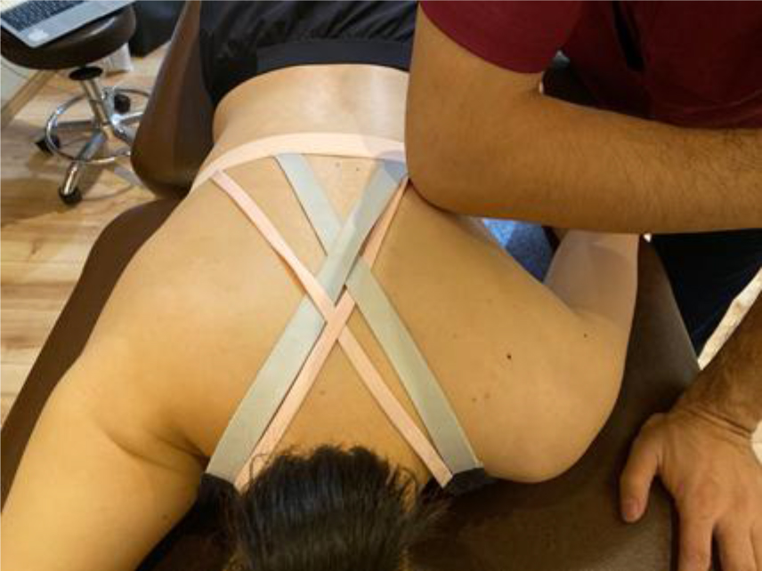
Figure 6. Lt LA-TH.
6. Treatment results
Although pain on grasping movement was reduced and ROM was improved after the first treatment, she complained of tension on her neck and upper back. One week later she received the second treatment, and pain on pinching movement, restricted ROM, and a sensation of tension on her upper back disappeared.
Discussion
The patient repeated cervical sprain, and had numbness in the region ranging from her neck to fingers. However, the region was not consistent with the dermatome of the C4th/5thor 5th/6th, which showed cervical hypermobility, and the site of numbness varied day by day. The patient’s condition repeated remission and exacerbation. Her life in such a situation is presumed to have caused various compensatory postures and activities. One of these causes is fascial compensation. Excessive tension of her trunk, which is originated from cervical instability, is considered to have received stress through fascial expansion to the hand along the arrangement of fascial connection. For this reason arrangement of fascial balance between the proximal and distal positions may have led to reduction in symptoms by intervention along the arrangement.
Conclusion
This article described the importance of evaluation and treatment with FM® from a musculofascial viewpoint for a patient who developed trigger finger after she repeated cervical sprain several times.
It has been believed that the occurrence of trigger finger is idiopathic. The symptoms appear spontaneously and have correlation with occupational or repetitive activities. The condition shows compensatory movements due to fascial dysfunction very frequently. Not only palliative treatment of the present symptoms but also specification of the causative sites and tissue by physical therapeutic evaluation may make an appropriate approach possible to resolve problems with the high-density site of pain and force transmission by addition of movement test and precise palpation test for fascia in view of a past history and the timeline. It is further important for trigger finger patient to do exercise for correction of compensatory movements after improvement in fascial sliding and the high-density site and to advance the movement to functional movement and to ADL movement.
References
- ……………………………
- Atsushi Yoshida (2019) The interaction between muscle and fascia (myofascial chain). Spine & Spinal Cord. 32: 301–306.
- …………….
- Stecco C (2018) Atlas of Functional Anatomy of Fascial System. Hitoshi Takei (Tr.). Ishiyaku Publishers, Inc. : 234–291.
- Hitoshi Takei (2014) Expansion of Systemic and Therapeutic Technique. H. Takei, et al. (Ed.). KYODO ISHO SHUPPAN CO., LTD., Tokyo.
- Atsushi Yoshida (2015) Practical approach to muscle and fascia (Special Issue: Manipulative physical therapy for sports injury). The Journal of Clinical Sports Medicine. 32: 1000–1004.
- Stecco L et al (2011) Fascial Manipulation—Theory Edition. H. Takei (Tr.) Ishiyaku Publishers, Inc., Tokyo.
- Schleip et al (2015) Membrane and Fascia. Up-to-date knowledges and therapeutical approach. H. Takei (Tr.), Ishiyaku Publishers, Inc., Tokyo, 343–349.
- Misako Nishimori: Pathological characteristics of tenosynovitis of the flexor tendon (trigger finger) on ultrasonic images of the joint. Japanese Journal of Medical Ultrasound Technology. 38: 2013.
- ……………….
- ………………..