DOI: 10.31038/JMG.2020311
Abstract
The Aim of this study is to focus at the evaluation of the potential of the antimicrobial activity of the synthesized Zinc chloride nanoparticles using Leptadenia hastata leaf extracts on selected bacterial; Escherichia coli (Gram–ve), Staphylococcus aureus, Gram +ve), Klebsielia Pneumonia (Gram +ve). The synthesized zinc oxide (ZnO) nanoparticles were characterized using UV, SEM and FTIR. The particles were further subjected to evaluation of their antimicrobial activity. The FTIR studies was observed to have shown the several chemical functional groups, while the SEM results showed the synthesized nanoparticles were in the range below 100 nm and the shape of the particles was slightly ellipsoidal as well as spherical. From the result it was observed that the synthesised nanoparticles have showed significant antibacterial activity against the gram-ve and gram +ve bacteria’s (Escherichia coli, Staphylococcus aureus and Klebsielia Pneumonia. This study being the first using Leptadenia hastata leaf extract should provide a new agent toward the fight of resistance bacterial and effective therapies in the field of medicine.
Keywords
Biosynthesis, Zinc oxide, nanoparticles, Leptadenia hastata, leaf, antimicrobial, characterization
Introduction
Green synthesis has been considered as another remedy in the field of medicine. Aside toxic chemical and physical method, biological method is considered use of medicinal plants extract for synthesis of nanoparticles. The surface and fraction of the atoms are responsible for the activity of the nanoparticles. This invention of green nanotechnology is considered eco-friendly and cost effective when compared to the others. The technology utilizes proteins as natural capping agents and its synthesis from plants utilize various secondary metabolites, enzymes, proteins and or other reducing agents which makes it suitable to use in various biomedical and clinical applications [1]. The advantage of using ZnO nanoparticles is that they have a strong potential against the pathogenic microbes when used in small concentration [2]. It was also reported that ZnO demonstrates significant growth inhibition of a broad spectrum of bacteria [3]. Thus, impaired wound healing in ulcer patients is a major complication. The elevated infections by bacterial such as Helicobacter pylori and many more resistant bacterial may interfere in the proper healing process. If left untreated will lead to delay in the healing process, as such introducing nanoparticle is an answer to such disturbing ailment. Many antibacterial agents have limitations in the clinical applications, because of complications and resistance towards pathogenic microorganisms. [4,5]. It was reported in some cases where few metal nanoparticles are known to have potential antimicrobial and consequential wound rejuvenating activity, but they also have some disadvantages related to the toxicity level [6].
This prompted the synthesize of nanoparticles by bio-assisted pathway to reduce their toxicity effect and provide better duodenal or stomach ulcer healing therapy to avoid and treat the resistance related pathogens. Thus, this study may provide a remedy with the green synthesis nanoparticles and its high potential towards antibacterial activity to control the resistance effect of Helicobacter phylori as well as other ulcer causing bacteria. Since this area is less explored, there is a need to consider the potential of this nanoparticles as to cutile the menace and the pain attached to this wound
Material and Method
Plant collection: The leafs of Leptadenia hastata (pers) Decne was collected in Michika local government in Adamawa state, Nigeria. It is a medicinally important plant. Fresh green leaves were harvested during the months of August to September.
Preparation of the plant extract: 5 g of fresh leaves were washed with distilled water and then cut, soaked in a 250 mL Erlenmeyer flask containing 100 mL of distilled water. The solution was boiled at 70 °C for 8 min. The leaf extract was allowed to cool to room temperature, filtered through Whatman number-1 filter paper, and the filtrate was stored for further experimental use.
Synthesis of ZnO Nanoparticles (NPs): 1 mM Zinc acetate [Zn (O 2CCH3)2 (H2O) 2] was dissolved in 50 ml distilled water and kept in stirrer for 1 hr, respectively as reported by [7]. Then 20 mL of NaOH solution was slowly added into the Zinc acetate solution and 25 mL of plant extract was added to the same. The colour of the reaction mixture was changed after 1 hr of incubation time. The solution was left in stirrer for 3 hrs. A light yellow colour was observed after the incubation time. This confirmed the synthesis of ZnO Nanoparticles (NPs). The precipitate was separated from the reaction solution by centrifugation at 8000 rpm at 60 °C for 15 min and pellet was collected from the filtered. The Pellet was dried using a hot air oven operating at 80 °C for 2 hrs and preserved in air-tight bottles for further studies.
Characterization of biosynthesized ZnO Nanoparticles (NPs): The Leptadenia hastata Optical properties of ZnO Nanoparticles (NPs) were characterized based on UV absorption spectra with the wavelength range of 300–500 nm. The FTIR characterization was obtained by taking 2 mg of ZnO Nanoparticle (NPs) and mixed with 200 mg of potassium bromide (FTIR grade) and pressed into a pellet for the characterization. The sample pellet was placed into the sample holder and FTIR spectra were recorded in FTIR spectroscopy at a resolution of 4 cm-1 [8]. The shape and size of the Nanoparticles were analysed by using SEM.
Antibacterial activity of synthesized ZnO Nanoparticles (NPs):
Preparation of Test Samples
The Leptadenia hastata zinc oxide (ZnO) nanoparticle from Leptadenia hastata leaf water extract was tested by disc diffusion method on nutrient agar medium as described by [9]. The extract exactly 5mg was dissolved homogeneity in 5mL of methanol giving a stock solution of 1000 μg/mL. Lower concentration of 10, 50, 100, 250, 500, and 1000ppm, i.e. six different volumes from the stock solution were taken for the studies.
Preparation of Bacteria Broth
The selected bacteria were used to evaluate the antibacterial activity of the Leptadenia hastata zinc oxide (ZnO) nanoparticle from Leptadenia hastata leaf water extract, Staphylococcus aureus, (+ve), Escherichia coli (–ve) and Klebsielia Pneumonia (+ve) were obtained from the stock culture provided by Virology Laboratory, Universiti Malaysia Sarawak. The nutrient broth was prepared according to manufacturer’s instruction, with 2.6 g of the dried broth dissolved in 200 mL distilled water followed by sterilization in autoclave at 121°C. The bacterial was sub-cultured in a 10 mL of broth, each in universal glass vail bottle for 16 hours inside an incubator equipped with shaker at 37°C [10]. After 16 hours’ incubation, turbidity (optical density/OD) of the bacterial broth was measured by using UV mini spectrophotometer (model 1240 of Shimadzu brand), comparable to that of nutrient broth standard tube for further use [11]. The measurement of the optical density was performed at wavelength 575nm and the bacterial broth was ready to be used when its turbidity was between OD 0.6 to 0.9. Nutrient broth was used to adjust the turbidity until the desired value was obtained.
Plate Inoculation
Inoculation of the bacteria was carried out in a biohazard cabinet and the procedure was based on method described by [12]. Approximately 1mL of the ready bacterial broth were transferred into mini centrifuge tubes. A sterile cotton swap was dipped into the mini centrifuge tube containing bacteria broth and streaked over entire of the agar plate surface, performed in four different directions. The agar plate was then left for 5–10 minutes before applying the test samples. The disc used was 6 mm diameter. A volume of 10μL of the test extract (Leptadenia hastata zinc oxide (ZnO) nanoparticle) of concentration 10, 50, 100, 250, 500 and 1000μg/mL were each pupated onto the discs and placed onto the agar plate by using sterile forceps and gently pressed to ensure contact. Next to be placed on the agar plate was the disc pupated with methanol as negative control, followed by 30μg of tetracycline as standard antibacterial agent (positive control). The plates were left at room temperature for 10 minutes to allow the diffusion of the test samples and the standards into the agar. Each of the test essential oil was tested in triplicate for the bacterium used. The plate samples were then incubated at 37°C for 24 hours before the inhibition zone around every sample disc being examined. The inhibition zone was measured in diameter (mm) to indicate the presence of antibacterial activity for each sample compared to the positive control.
Result and Discussion
Scanning Electron Microscope (SEM): SEM analysis is done to visualize shape and size of nanoparticle. JSM6510LV Scanning electron microscope was used to determine the shape of Leptadenia hastata capped ZnO NPs (Fig. 3). SEM images were seen in different magnification ranges which clearly demonstrated the presence of spherical shaped nanoparticle with mean average diameter of 70 nm [13].
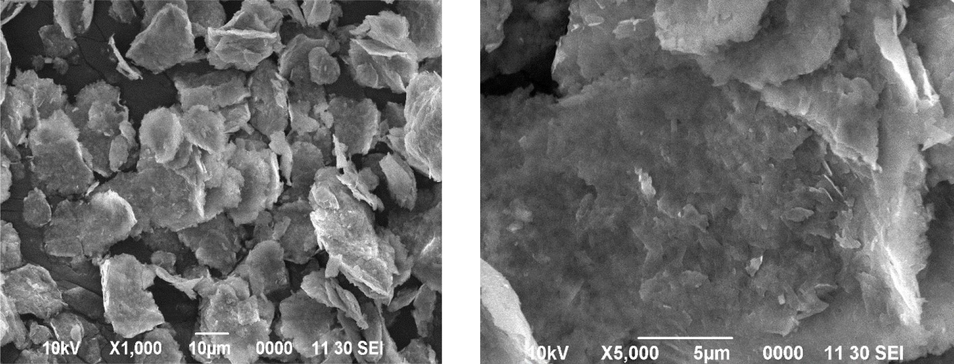
Figure 3. SEM images of ZnO Nanoparticles (NPs) of Leptadenia hastata leaf in different magnification ranges
Fourier Transform Infra-Red Spectrometry (FTIR): Substance-specific vibrations of the molecules lead to the specific signals obtained by IR spectroscopy. FT-IR spectra and functional group involved in Leptadenia hastata ZnO Nanoparticle synthesis illustrated peak in the range of 1000–4000 cm −1 (Fig 4). Absorption band of ZnO Nanoparticle IR spectrum showed the presence of the functional group (O-H) which appeared at 3363.66 cm-1 bond. The IR spectrum (Figure 4) also shows an absorption band which indicated a C-H bond at 2970.49 cm-1 which suggested the presence of methyl carbon in the chemical structure. However, a double bond C=C stretching at 1772.35 cm-1 and 1566.86 cm-1 was also observed in the spectrum of ZnO Nanoparticle. The peak in the range of 1502.35 and 1409.81 corresponded to C=C stretch and in aromatic ring and C=O stretch in polyphenols and C–N stretch of Amide-I in protein. Weak peaks obtained at 1089.98 cm-1, 1010.10 cm-1 and 886.75 cm-1, 830.25 cm-1, 764.32 cm-1, demonstrated the presence of C-O stretching in amino acid, C-N stretching, C-F, C-l, C-Br strong stretching, and C-H bending respectively. A very ignorable peak obtained at 686.62 cm-1 demonstrated the probable presence of C- Alkyl chloride and Hexagonal phase ZnO thus supported by the report of [14].
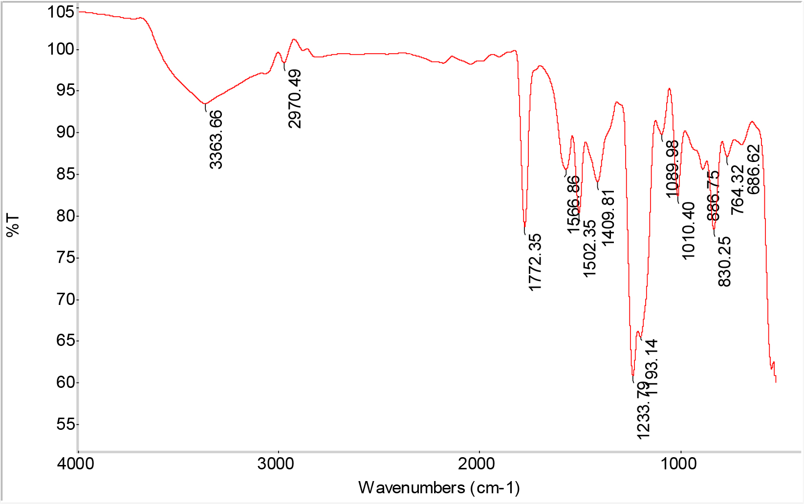
Figure 4. FT-IR Spectrum of ZnO Nanoparticles (NPs) of Leptadenia hastata fresh leaf
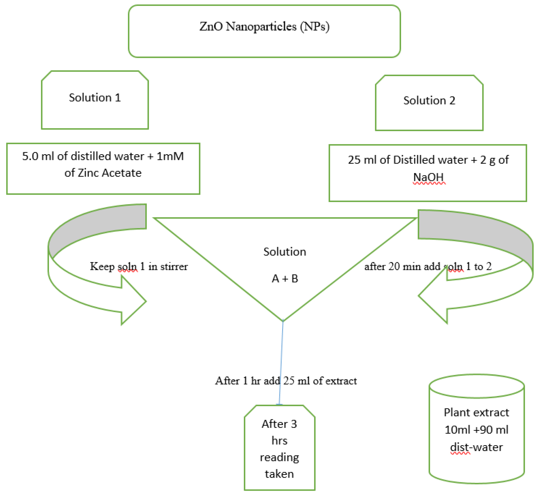
Figure 1. Schematic representation of ZnO Nanoparticles (NPs) synthesis.
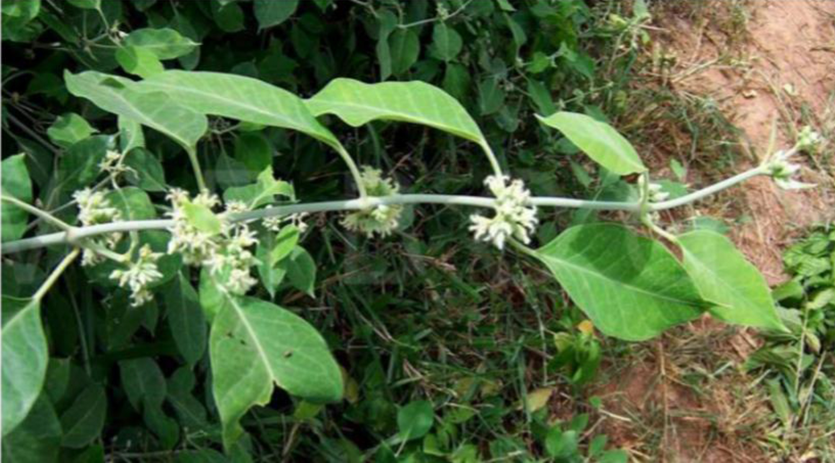
Figure 2. Leaves of Leptadenia hastata.
UV-visible Analysis: The UV-visible spectroscopy is usually conducted to confirm the synthesis of ZnO Nanoparticles. Conducting electrons start oscillating at a certain wavelength range due to surface Plasmon resonance (SPR) effect. Figure 5 represents the UV-visible spectra of freshly prepared Leptadenia hastata ZnO Nanoparticles. Peak observed at 380 nm clearly demonstrates the presence of ZnO Nanoparticles in the reaction mixture. Initial peak obtained at range of 420 nm got further raised due to oscillation of more electrons after 5 hrs which reports the continuous synthesis of Leptadenia hastata ZnO Nanoparticles.
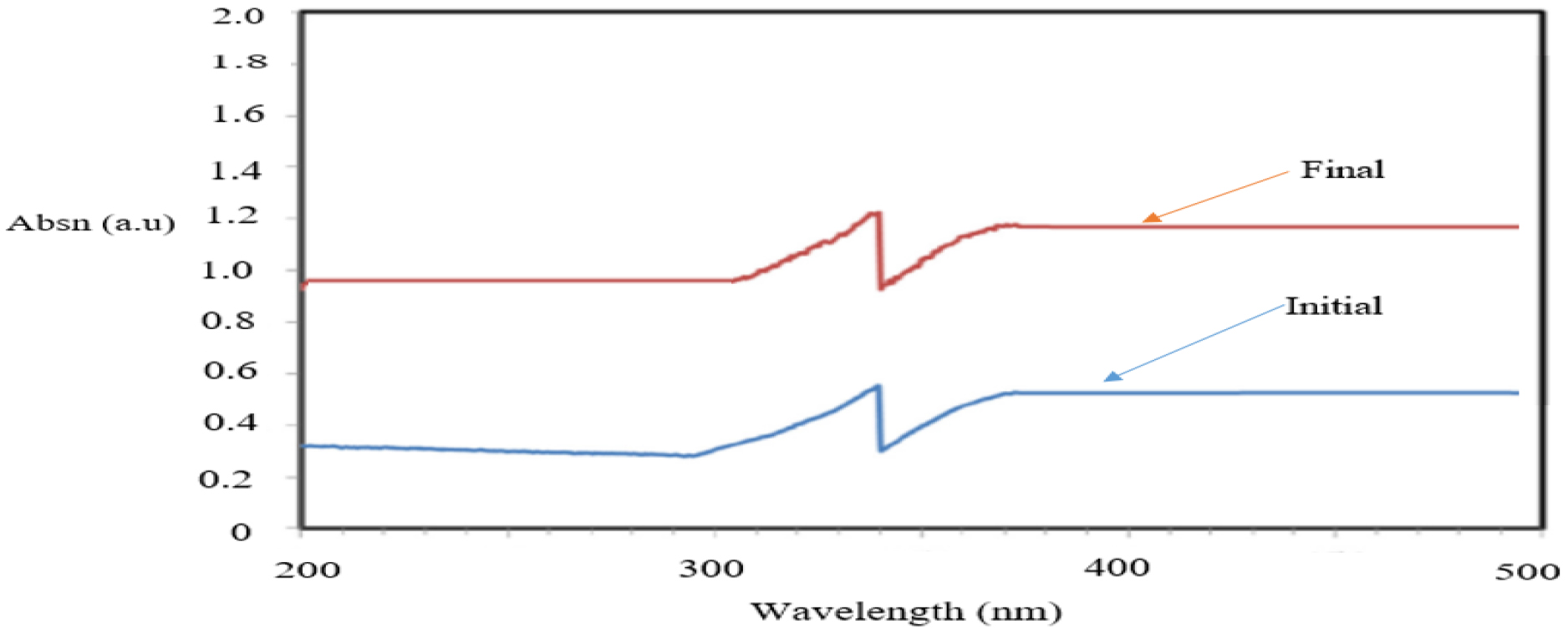
Figure 5. UV-vs spectrum of Leptadenia hastata leaf ZnO Nanoparticles.
Antibacterial Activity of ZnO Nanoparticles (NPs) of Leptadenia hastata fresh leaf: Anti-bacterial effect of Leptadenia hastata fresh leaf ZnO Nanoparticles was tested against Staphylococcus aureus, Escherichia coli, Klebsielia Pneumonia. Tetracycline disc was used as a standard, from the table 1 the result clearly demonstrated that the nanoparticles showed antibacterial activity in a dose dependant manner. Figure 6 and Table 1. A significant inhibition was observed at 25 ppm to 1000 ppm, maximum zone of inhibition was observed against all the bacteria at 1000 ppm with increase in concentration of the ZnO nanoparticles in all the gram-ve and gram +ve bacterium (Staphylococcus aureus, Escherichia coli, Klebsielia Pneumonia) of12.53 ± 0.12 mm, 12.34 ± 0.10 mm and 12.13 ± 0.12 mm, respectively. Minimum zone of inhibition was observed against all the bacterium at 25 ppm. However, the zone of growth Inhibition obtained using the nanoparticle was much significant when compared to the standard disc (Tetracycline) used which depicts the need of considering nanoparticle as an agent to compliment in the fight of resistance bacteria in the field of medical science. The entire tests were done in triplicate. The zinc oxide nanoparticles are inhibiting the microbial growth in in-vitro antimicrobial activities.
Table 1. Effect of Leptadenia hastata leaf zinc oxide (ZnO) nanoparticle on Staphylococcus aureus, (+ve), Escherichia coli (–ve) and Klebsielia Pneumonia (+ve)
|
Concentration (ppm) |
Escherichia coli (Gram–ve), |
Staphylococcus aureus, (Gram +ve) |
Klebsielia Pneumonia (Gram +ve) |
|
Control(tetracycline) |
13.15 ± 0.10 |
13.12 ± 0.81 |
13.10 ± 1.10 |
|
25 |
9.97 ± 0.21b |
7.75 ± 0.07 |
10.77 ± 0.23 |
|
50 |
11.20 ± 0.20 |
11.60 ± 0.10b |
11.55 ± 0.07 |
|
100 |
11.85 ± 0.07 |
11.00 ± 0.10 |
11.65 ± 0.07 |
|
250 |
11.90 ± 0.14 |
11.67 ± 0.14 |
11.95 ± 0.07b |
|
500 |
11.99 ± 0.10b |
12.17 ± 0.25 |
11.97 ± 0.35 |
|
1000 |
12.53 ± 0.12a |
12.34 ± 0.10a |
12.13 ± 0.12a |
Values are Mean ± SD
aSignificantly (p< 0.05) higher compared to different concentration in each column
bSignificantly (p< 0.05) higher compared at the same concentration in each row
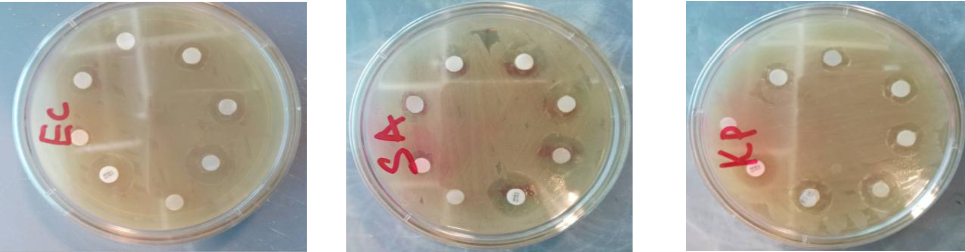
Figure 6. Showing the growth inhibition of Leptadenia hastata ZnO Nanoparticles
Conclusion
The study on the biosynthesis of ZnO nanoparticles using fresh leaf extract from Leptadenia hastata was carried out. This green synthesis was found to be eco-friendly, non-toxic and less usage of chemicals compared to the physical and chemical method. The presence of phytochemical constituents in the leaf extract helps in the synthesis of the ZnO nanoparticles by inducing redox reaction. The functional groups of phytochemicals such as amine, alkane and hydroxyl groups induced the formation of nanoparticles which are widely seen in the secondary metabolites, such as terpenoids, flavonoids, alkaloids. The preliminary confirmation of the ZnO nanoparticles through was measured using the Uv-visible spectroscopy at 380 nm, the SEM analysis demonstrate the size of the nanoparticles and the antibacterial activity of the Leptadenia hastata ZnO nanoparticles has confirmed the potential of the nanoparticles as an agent against bacterial.
Acknowledgement
The authors wish to acknowledge the research grant 07(ZRC05/1238/2015(2) Provided by Universiti Malaysia Sarawak which has resulted to this article.
References
- Parthasarathy G, Saroja M, Venkatachalam M, Shankar S, Evanjelene VK (2019) Green synthesis of zinc oxide nanoparticles- review paper. World Journal of Pharmacy and Pharmaceutical Sciences 5: 922–931.
- Dobrucka, R, Długaszewska J (2016) Biosynthesis and antibacterial activity of ZnO nanoparticles using Trifolium pratense flower extract. Saudi journal of biological sciences 23: 517–523. [Crossref]
- Kalpana VN, Kataru BAS, Sravani N, Vigneshwari T, Panneerselvam A (2018) Biosynthesis of Zinc oxide nanoparticles using culture filtrates of Aspergillus niger: Antimicrobial textiles and dye degradation studies. Open Nano 3: 48–55.
- Tacconelli E, Müller NF, Lemmen S, Mutters NT, Hagel S, et al. (2017) Infection Risk in Sterile Operative Procedures a Systematic Review and Meta-analysis. Dtsch Arztebl Int 113: 271–278.
- Ozgenç O (2016) Methodology in improving antibiotic implementation policies. World J Methodol 6: 143–153. [Crossref]
- Chauhan PV, Shrivastava V, Tomar RS (2019) Biosynthesis of zinc oxide nanoparticles using Cassia siamea leaves extracts and their efficacy evaluation as potential antimicrobial agent. Journal of Pharmacognosy and Phytochemistry 8: 162–166.
- Jamdagni P, Khatri P, Rana JS (2016) Green synthesis of zinc oxide nanoparticles using flower extract of Nyctanthes arbor-tristis and their antifungal activity.
- Mishra V, Sharma R (2015) Green synthesis of zinc oxide nanoparticles using fresh peels extract of Punica granatum and its antimicrobial activities. 143: 158–164.
- Isaac John Umaru, Fasihuddin A Badruddin, Hauwa Aduwamai Umaru (2018) Phytochemical, antifungal and antibacterial potential of Leptadenia hastata stem-bark extract. Toxicology 4: 263–268.
- Isaac John Umaru, Fasihuddin Ahmad Badruddin, Zaini B Assim, Hauwa Aduwamai Umaru (2018) Antibacterial and cytotoxic actions of chloroform crude extract of Leptadenia hastata (pers) Decnee. Clin Med Biochemistry 4:1–4.
- Umaru IJ, Badruddin FA, Assim ZB, Umaru HA (2018) Antimicrobial properties of Leptadenia hastata (pers) decne leaves extract. International Journal of Pharmacy and Pharmaceutical Sciences 10: 149–152.
- Isaac John Umaru, Fasihuddin A Badruddin, Hauwa A Umaru (2019) Phytochemical Screening of Essential Oils and Antibacterial Activity and Antioxidant Properties of Barringtonia asiatica (L) Leaf Extract.
- Raut S, Thorat PV, Thakre R (2013) Green synthesis of zinc oxide (ZnO) nanoparticles using Ocimum tenuiflorum leaves. Int.J.Sci.Res 14: 2319–7064.
- Yedurkar S, Maurya C, Mahanwar P (2016) Biosynthesis of zinc oxide nanoparticles using Ixora coccinea leaf extract, a green approach. Open J. Synth. Theory Appl 5: 1–14