DOI: 10.31038/AFS.2022423
Abstract
This study has been so far the first attempt of biomolecular investigation on three water mite species, namely, Hydrachna globosa, Hydryphantes dispar and Limnesia fulgida. UV-Vis spectroscopy was used to determine vibration frequencies of different biomolecules, and FTIR spectrum ranges were used to characterize the functional groups of different biochemical substances. A comparative study was performed to understand similarities and/or differences between these three species, based on the qualitative and quantitative analyses of various biocompounds. Despite having similar functional groups in all of them, significant similarities between H. dispar and L. fulgida were found in terms of UV-Vis absorbance peaks, IR graphical patterns and absorption percentage (%). On the other hand, although these three species comprise distinctive morphological features, H. dispar and L. fulgida were observed to be closer considering their body lengths, whereas H. globosa was identified to be the largest among these mites. In addition to the noble findings, this investigation will contribute to the identification and characterization of other water mites too.
Short Summary
In this study, functional structural groups of three different water mite species were examined by spectrophotometric analysis method and these values were compared between species and their systematic similarities were discussed. This method is a new molecular systematic approach to controversial systematic problems in this group. It is clear that the results obtained in the study will contribute to classical taxonomy since they are species-specific.
Keywords
Acari, Hydracnidia, Hydrachna globosa, Hydryphantes dispar, Limnesia fulgida, UV-Vis spectroscopy, FTIR analysis
Introduction
Water mites composed of more than 6000 species, and are the most abundant, diverse inland water invertebrates which play a vital role in fresh water ecosystem [1]. The life cycle of water mites is very complex and their eggs can be found attached with many aquatic plants. Besides, throughout their larval phase, they co-habit with different insects, and live as the ectoparasites on the bodies and wings of these insects. Nymphs and adults have four pairs of legs while larvae have three. Adults also comprise oval or oblong or elliptical bodies with abdomen and a flattened dorsum [2]. However, depending on the type of habitats and mobility, water mites posses a wide range of morphological variations, such as variable shapes (from rounded to elongate), diverse external morphology with different colours etc. Different species of Hydrachnidia can be used as bioindicators to determine the ecological quality of freshwater habitation [3]. Therefore, genetical, biochemical and ecological analysis on water mites is very essential, since they signify the most essential level of life in fresh water ecosystem. On the other hand, most classical taxonomic techniques for studying water mites can often have many systematic problems due to insufficient in the detection and diagnosis [4]. Hence, biomolecular analysis could be a novel approach for the characterization of water mites.
Various spectrophotometric approaches have been utilized for evaluating different microorganisms, planktons, microalgae, mammals etc. [5-10]. In recent years, use of different spectrophotometric (e.g. UV, FTIR) techniques as a new molecular taxonomic method for studying invertebrates especially water mite species have gained huge attention since it is quite new aspect for characterization. For instance, [4] applied UV-Vis spectrophotometry for the structural analysis of six different water mite species (Acari, Hydrachnidia). In the Acaridae family, the IR technique was first used in a study [11] for isolating Rosefuran and Perillene derivatives of Tyrophagus neiswanderi (Acariformes, Acaridae). This study explained the structure of these natural compounds by using IR and other spectroscopic techniques and compared them with synthetic ones.
The primarily aim of this study was to determine the similarities and/or differences between three species of water mites, respectively Hydrachna globosa, Hydryphantes dispar, and Limnesia fulgida, based on the quantitative analyses of different biomolecules. Aiming this, UV-Vis spectrophotometry and Fourier-transform Infrared spectroscopy (FTIR) were applied on these water mites. Being so far the first attempt of biomolecular investigation on these species, this study will also open up new scopes in identification and characterization of other water mites.
Materials and Methods
The samples of this study were collected from the Lake Karamık in Afyonkarahisar province of Turkey, between May and August 2017. Based on species type, the samples were isolated from each other with the help of a microscope, and collected in different glass bottles. For photographing, the microscopic images were taken and transferred to the computer with the help of Nikon SMZ445 microscope and Argenit Kameram 12 CCD camera using the Kameram GEN3 program. The isolated samples were then washed several times by distilled water, and analyzed separately using UV (Ultraviolet) Spectrophotometer and FT-IR (Fourier Transform Infrared) Spectrophotometer techniques.
Preparation of PBS (Phosphate Buffered Saline) Solution
The PBS solution was prepared in desired amount for using in ultraviolet spectrophotometric analysis. For 1 L of 1X PBS, 800 mL of distilled water (dH2O) was filled into a sufficiently large glass flask and 8 g NaCl, 0.2 g KCl, 1.44 g Na2HPO4 and 0.24 g KH2PO4 salts were weighed using a precision balance (SHIMADZU ATX224), and added into the distilled water. The resulting solution was shaken well until homogeneous appearance. The pH was adjusted to 7.4 with HCl, and the solution was adjusted to 1 L with dH2O. Sterilization was then carried out in an autoclave (JEIO TECH ST-65G) at 121°C for 20 minutes, and stored in a 4°C for further experiments.
Ultraviolet (UV-VIS) Spectrophotometric Analysis
Species used in the study (Hydrachna globosa, Limnesia fulgida, Hydryphantes dispar) were weighed separately with a precision scales, and then, approximately 10 mg of each water tick sample was added into 0.5 mL of concentrated PBS (Phosphate Buffered Saline) solution, and dissolve properly with homogenator. The solutions were then taken into Eppendorf tubes and centrifuged (AWEL MF 20-R) for 20 minutes at 14000 rpm at 4°C. Afterward, the supernatant was removed with the help of micropipette into different eppendorf tubes, and 1 mL of PBS solution was added to form the dilute solution. The diluted solution was taken into micro quartz cuvette and UV (SHIMADZU UV-1700 Pharma) was measured against concentrated PBS solution. Freshly prepared PBS (Phosphate Buffered Saline) solution was used as blank.
FTIR (Fourier Transform Infrared) Spectrophotometric Analysis
The identified washed species were dried in sterile containers at room temperature. For each species (Hydrachna globosa, Limnesia fulgida and Hydryphantes dispar), 100 mg anhydrous KBr (Potassium Bromide) was added to a sample of about 5 mg of water tick by weighing with a precision balance. The mixture was taken into agate mortar and thoroughly crushed to obtain a homogeneous mixture. Afterwards, this powder mixture was pressed for 2 minutes to form a thin transparent disc like pellet which was used for FTIR analysis via FTIR (SHIMADZU IRAffinity-1S) spectrometer.
Results and Discussion
Three water mite species i.e., H. globosa, H. dispar and L. fulgida used in the study belong to the families Hydrachnidae, Hydryphantidae and Limnesiidae, respectively. Initially, a microscopic examination was conducted for imaging and observing the morphological features of these three water mites.
Adult Hydrachna globosa is reddish-brown in colour with rounded body and red paw placed at equal distances (Figure 1A). Males (2300 to 2110 μm) are smaller in size than females (2500 to 2250 μm) [12]. On the other hand, Hydryphantes dispar is a medium-sized dark red colored water mite with flat, thin and papilled body (Figure 1B). The body length of male H. dispar varies from 1368 to 1180 μm whereas females were found to be 1380 to 1250 μm [12,13]. Besides, Limnesia fulgida is a medium-sized blackish coloured mite with long, thick, bluish palps (Figure 1C). The body length ranges from 1370 to 1125 μm for meals and 1480 to 1200 μm for females [12].
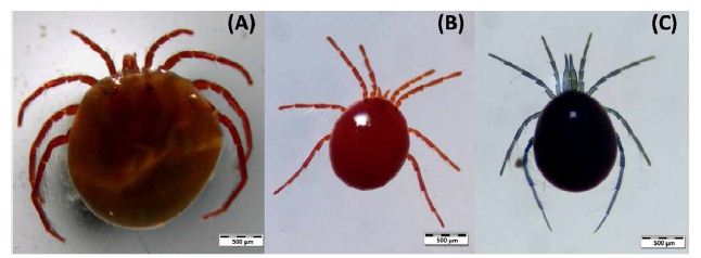
Figure 1: Microscopic dorsal views of Hydrachna globosa (A), Hydryphantes dispar (B), and Limnesia fulgida (C) at 500 µm scale
From this microscopic observation, Hydrachna globosa was identified to be the largest mites among all these three species. Besides, despite having differences in various morphological features i.e., body colour, palp length etc., Hydryphantes dispar and Limnesia fulgida were observed to be almost similar in their body lengths or size.
However, this study mainly focused on the UV-Vis spectrophotometric and Fourier-transform Infrared spectroscopic (FTIR) approaches for analyzing the chemical composition which can be used to compare these three Hydrachnidia species. This is due to the fact that, UV-Vis spectrophotometry and Fourier-transform Infrared spectroscopy (FTIR) are the most widely used methods applied for the qualitative and quantitative analyses of different biomolecules available in living organisms. In the UV-Vis spectrophotometry, ultraviolet and visible lights can transmit through a prepared dilution and determine the amount of light absorbed by that dilution by giving significant peaks. The higher contain of desirable agents in the dilution confirm the higher rate of absorption [4]. On the other hand, FTIR spectroscopy with a significant infrared region between 4000 – 670 cm-1 are utilized for determining and evaluating chemical information i.e., functional groups, side chains of different biomolecules such as nucleic acids, proteins, carbohydrates, lipids, and lipopolysaccharides [10].
Ultraviolet (UV-VIS) Spectrophotometric Analysis
The spectroscopic values of this study were calculated as absorbance versus wavelength graph. While the vertical axis in the graphs shows % absorbance, the horizontal axis shows the wavelength of the material in nm. The UV-vis spectrophotometric graphs of the studied species (Hydrachna globosa, Hydryphantes dispar and Limnesia fulgida) showed that the graphical values of each species are specific to that species. Moreover, the maximum wavelengths for both Hydryphantes dispar and Limnesia fulgida were 200 nm at the absorbance of 3.2 and 4.0, respectively (Figures 2-3). On the other hand, the third species, Hydrachna globosa confirmed the maximum wavelength of 230 nm at the absorbance of 3.9 (Figure 4). Based on the maximum wavelengths, Hydryphantes dispar and Limnesia fulgida were found to be very close to each other compared to Hydrachna globosa.
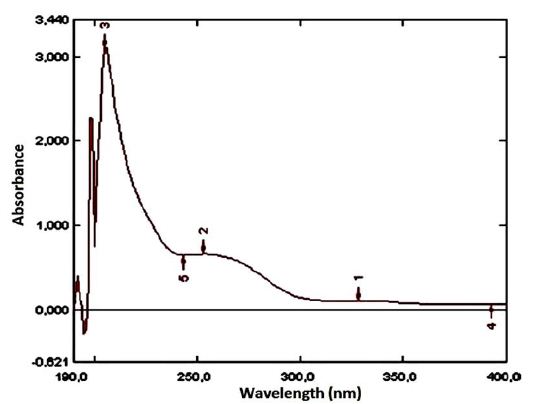
Figure 2: UV spectrum of Hydryphantes dispar
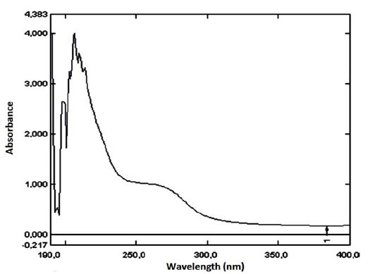
Figure 3: UV spectrum of Limnesia fulgida
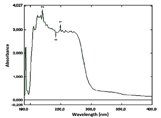
Figure 4: UV spectrum of Hydrachna globosa
A comparative study of water mites by using UV-Vis spectrophotometry analysis was conducted by [4]. In that study, the researchers performed UV-Vis analysis for Hydrodroma despiciens, Hydryphantes flexiosus, Eylais infundibulifera, Georgella helvatica, Torrenticola brevirostris and Hygrobates nigromacutlatus. According to the results, it was stated that all of these species are systematically very close to each other.
The optical properties as UV-vis spectra are able to provide quantitative information, like internal structure, abundance and chemical composition of microorganisms [14]. In a study, UV-Vis spectroscopy was applied on Bacillus globigii and Escherichia coli cell spores as model microbes to detect and identify microorganisms [14]. Furthermore, UV-Vis spectroscopy was utilized as a rapid method for identifying and quantifying some pathogenic protozoa in water, especially Cryptosporidium and Giardia responsible for causing serious waterborne diseases [15]. The outcomes of this study suggested that distinctive UV-vis spectra of these protozoa in the short wavelength region might be species specific for each protozoon, which can be applicable for water-control management.
FTIR (Fourier Transform Infrared) Spectrophotometric Analysis
The FTIR analysis was carried out for the three water mite species, and the result showed that the peaks of L. fulgida and H. dispar species were sharper and stronger, while the peaks of H. globosa species were weaker. Moreover, the graphical patterns as well as absorption percentage (%) of Hydryphantes dispar and Limnesia fulgida have strong resemblance in all aspects whereas Hydrachna globosa was slightly different from others regarding the infrared spectra at 3400-3200 cm-1 and 1200-1000 cm-1 (Figure 5). Thus, Infrared analysis also suggested that Hydryphantes dispar and Limnesia fulgida are closer to each other than Hydrachna globosa.
However, similar functional groups were observed in all species (Figure 5). The infrared spectra ranged from 3400 to 3200 cm-1 represent the intermolecular bonded alcohol (-OH) stretching and aliphatic primary amine (N-H) stretching. The IR absorbances from 2950 to 2800 cm-1 stand for various alkane and aldehyde based compounds (C-H stretching) which can be found in almost all organic compounds [16]. The graphs of this region are very similar in terms of the three species discussed in this study.
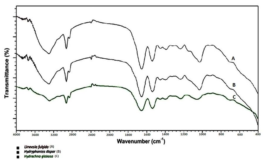
Figure 5: FTIR Analysis of Limnesia fulgida, Hydryphantes dispar, Hydrachna globosa
Besides, the peaks from 1670 to 1550 cm-1 are the consequences of nitro compound (N-O stretching), trisubstituted and tetrasubstituted alkene (C=C stretching), imine / oxime (C=N stretching) and secondary amide (C=O Stretch). These bonds are found in amino acids, the building blocks of proteins. On the other hand, the spectra from 1450 to 700 cm-1 is also referred to as the fingerprint region. This region designate basic building blocks of both carbohydrates and proteins considering different bonds including methylated alkane group (C-H bending), carboxylic acid (O-H bending), sulfate, sulfonyl chloride, sulfonic acid (S=O stretching), fluoro compound (C-F stretching), aromatic amine (C-N stretching), aromatic ester (C-O stretching) and C-X stretching for halo compound [17,18].
Attenuated total reflection-fourier transform infrared spectroscopy (ATR-FTIR) was utilized in a study for a fast and non-destructive identification of species variation of maggots and the developmental stage of their larvae and adult samples as entomological evidences, which could be useful in forensic investigation [19]. Similarly, six species of flesh flies (Diptera: Sarcophagidae) native to Neotropical regions were categorized and differentiated by applying infra-red spectroscopy, i.e., ATR-FTIR, NIRS [20]. On the other hand, Infrared spectra are used to determine the origin of edible insect powder with the aim of quality control of food products. A rapid chemical fingerprinting of seven edible insect powders from different species, i.e., Locusta migratoria, Acheta domesticus, Gryllodes sigillatus, Alphitobius diaperinus, and Tenebrio molitor was prepared in study by applying ATR-FTIR spectroscopy [21].
Conclusion
This study has been so far the first attempt of biomolecular investigation by using UV-Vis spectrophotometry and Fourier-transform Infrared spectroscopy (FTIR) on three water mite species, respectively Hydrachna globosa, Hydryphantes dispar and Limnesia fulgida. The three water mite species have various distinctive morphological characteristics since they belong to three different families. However, the species H. dispar and L. fulgida only showed significant resemblance in their body length, while the species Hydrachna globosa was found to be larger. Nevertheless, it is interesting that spectrophotometric analyses ravelled considerable similarities between Hydryphantes dispar and Limnesia fulgida in terms of UV-Vis absorbance peaks, IR graphical patterns as well as absorption percentage (%). Furthermore, despite having similar functional groups in all species, the obtained differences indicate that the quantity of some biomolecules might be different in Hydrachna globosa. Therefore, it can be concluded that Hydryphantes dispar and Limnesia fulgida are closer than Hydrachna globosa, particularly regarding their chemical constitution.
On the other hand, most classical taxonomic techniques for studying water mites encountered many systemic problems since the conventional methods can often be insufficient for the detection and diagnosis of this group. However, the results obtained in this study will contribute to future studies, specifically in identification and classification of water mites by overcoming the existing problems. Moreover, the new molecular methods applied in this study could also be the novel protocols for characterization of any other species.
Disclosure Statement
The authors declare no conflicts of interest related to this study.
This research did not receive any specific funding
The data that support this study are available in the article and accompanying online supplementary material.
References
- Smith IM, Cook DR, Smith BP (2010) Water mites (Hydrachnidiae) and other Arachnids. In J. H. Thorp & A. P. Covich (Eds.), Ecology and Classification of North American Freshwater Invertebrates (pp. 485-586). London: Elsevier.
- Di Sabatino A, Smit H, Gerecke R, Goldschmidt T, Matsumoto N, et al. (2008) Global diversity of water mites (Acari, Hydrachnidia; Arachnida) in freshwater. Hydrobiologia 595: 303-315.
- Miccoli FP, Lombardo P, Cicolani B (2013) Indicator value of lotic water mites (Acari: Hydrachnidia) and their use in macroinvertebrate-based indices for water quality assessment purposes. Knowledge and Management of Aquatic Ecosystems 411: 08.
- Asci F, Akkus GU (2017) The application of ultraviolet spectrophotometry (UV) on some water mite species (Acari, Hydrachnidia). Advances in Bioscience and Biotechnology 08: 142-148.
- Alvarez AMF, Bouhy J, Dieu M, Charles C, Deparis O (2019) Animal species identification in parchments by light. Scientific Reports 9: 1825.
- Fasolato L, Andreani NA, De Nardi R, Nalotto G, Serva L, et al. (2018) Spectrophotometric techniques for the characterization of strains involved in the blue pigmentation of food: preliminary results. Italian Journal of Food Safety 7: 6928. [crossref]
- Lonni AASG, Scarminio IS, Silva LMC, Ferreira DT (2005) Numerical taxonomy characterization of Baccharis genus species by ultraviolet-visible spectrophotometry. Analytical Sciences 21: 235-239.
- Motrescu I, Oancea S, Rapa A, Airinei A (2006) Spectrophotometric analysis of the blood plasma for different mammals. Romanian Journal of Biophysics 16: 215-220.
- Santos-Ballardo DU, Rossi S, Hernández V, Gómez RV, del Carmen Rendón-Unceta M, et al. (2015) A simple spectrophotometric method for biomass measurement of important microalgae species in aquaculture. Aquaculture 448: 87-92.
- Vogt S, Löffler K, Dinkelacker AG, Bader B, Autenrieth IB, et al. (2019) Fourier-trransform infrared (FTIR) spectroscopy for typing of clinical Enterobacter cloacae complex isolates. Frontiers Microbiology 10: 2582.
- Leal WS, Kuwahara Y, Suzuki T, Nakao H (1989) Chemical taxonomy of economically important tyrophagus mites (Acariformes, Acaridae). Agricultural and Biological Chemistry 53: 3279-3284.
- Uysal G (2005) A Systematic Study on Karamik Lake Water Ticks (Acari; Hydrachnellae). Unpublished MSc thesis. Afyonkarahisar: Institute of Science, Afyon Kocatepe University.
- Erman O (1990) Systematic Examination of Elazig Water Ticks (Hydrachnellae, Acari). Unpublished Ph.D thesis. Erzurum: Institute of Science, Atatürk University.
- Alupoaei CE, García-Rubio LH (2005) An interpretation model for the UV-vis spectra of microorganisms. Chemical Engineering Communications 192: 198-218.
- Bacon CP, Rose JB, Patten K, Garcia-Rubio LH (1995) Quantitative classification of Cryptosporidium oocysts and Giardia cysts in water using UV/vis spectroscopy. In J. R. Lakowicz (Ed.), Advances in Fluorescence Sensing Technology II (pp. 471-480).
- Rohman A, Man YBC (2010) Fourier transform infrared (FTIR) spectroscopy for analysis of extra virgin olive oil adulterated with palm oil. Food Research International 43: 886-892.
- Jahan I, Erci F, Isildak I (2019) Microwave-assisted green synthesis of non-cytotoxic silver nanoparticles using the aqueous extract of Rosa santana (rose) petals and their antimicrobial activity. Analytical Letters 52: 1860-1873.
- Matwijczuk A, Oniszczuk T, Matwijczuk A, Chruściel E, Kocira A, Niemczynowicz A, et al. (2019) Use of FTIR spectroscopy and chemometrics with respect to storage conditions of moldavian dragonhead oil. Sustainability 11: 6414.
- Pickering CL, Hands JR, Fullwood LM, Smith JA, Baker MJ (2015) Rapid discrimination of maggots utilising ATR-FTIR spectroscopy. Forensic Science International 249: 189-196. [crossref]
- Barbosa TM, de Lima LAS, dos Santos MCD, Vasconcelos SD, Gama RA, et al. (2018) A novel use of infra-red spectroscopy (NIRS and ATR-FTIR) coupled with variable selection algorithms for the identification of insect species (Diptera: Sarcophagidae) of medico-legal relevance. Acta Tropica 185: 1-12.
- Mellado-Carretero J, García-Gutiérrez N, Ferrando M, Güell C, García-Gonzalo D, et al. (2020) Rapid discrimination and classification of edible insect powders using ATR-FTIR spectroscopy combined with multivariate analysis. Journal of Insects as Food and Feed 6: 141-148.