Abstract
Background: Early recognition of ventricular dysfunction and heart failure (HF) in children remains challenging because diagnosis often relies on specialized imaging modalities. Artificial intelligence (AI), particularly AI-enabled electrocardiography (AI-ECG), has emerged as a promising noninvasive tool for detecting ventricular dysfunction, cardiomyopathy, and adverse cardiac remodeling.
Objectives: To systematically review the evidence supporting AI and machine-learning (ML) applications for diagnosis and risk stratification of ventricular dysfunction, cardiomyopathy, and HF in children and to evaluate the diagnostic performance of pediatric AI-ECG models.
Methods: Studies evaluating AI, ML, or deep learning approaches in pediatric ventricular dysfunction, cardiomyopathy, congenital heart disease, or HF were included. Diagnostic studies reporting sensitivity and specificity were pooled using random-effects meta-analysis. Risk of bias was assessed using Quality Assessment of Diagnostic Accuracy Studies AI.
Results: Six studies met inclusion criteria for qualitative synthesis, and two independent pediatric diagnostic cohorts comprising 56,403 children were eligible for quantitative synthesis. AI-ECG demonstrated high diagnostic performance for ventricular dysfunction and remodeling, with pooled sensitivity of 0.92 (95% CI 0.88-0.94), specificity of 0.73 (95% CI 0.58-0.84), and diagnostic odds ratio of 34.22 (95% CI 11.08-105.72). Additional studies demonstrated utility of ML for dilated cardiomyopathy phenotyping, HF mortality prediction, and chemotherapy-related cardiomyopathy surveillance.
Conclusions: AI-ECG is a promising, scalable screening tool for pediatric ventricular dysfunction and adverse remodeling. Emerging ML applications may further improve cardiomyopathy phenotyping, HF prognostication, and precision pediatric cardiovascular care.
Keywords
Artificial intelligence, Machine learning, Electrocardiography, Pediatric cardiomyopathy
Abbreviations
AI: Artificial Intelligence; AI-ECG: Artificial Intelligence-Enabled Electrocardiography; AUROC: Area Under the Receiver Operating Characteristic Curve; DL: Deep Learning; LVSD: Left Ventricular Systolic Dysfunction; ML: Machine Learning; QUADAS-AI: Quality Assessment of Diagnostic Accuracy Studies-Artificial Intelligence; RVSD: Right Ventricular Systolic Dysfunction; SHAP: SHapley Additive exPlanations; XGBoost: Extreme Gradient Boosting
Introduction
Ventricular dysfunction, cardiomyopathy, and Heart Failure (HF) remain important causes of morbidity, hospitalization, transplantation, and mortality in children with Congenital Heart Disease (CHD) and acquired heart disease [1,2]. Nearly half of children without known heart disease who later required hospitalization for systolic HF were initially misdiagnosed, especially in primary care settings, and those with longer symptom duration were at greatest risk, resulting in considerable unnecessary evaluation and treatment [3]. Early recognition of ventricular dysfunction is particularly important because timely intervention may prevent progression to irreversible remodeling, advanced HF, transplantation, or death [4,5]. However, diagnosis continues to rely primarily on echocardiography and Cardiac Magnetic Resonance (CMR) imaging, which require specialized expertise, may be resource intensive, and are not universally available. Consequently, there is growing interest in developing accessible and scalable tools for early identification of ventricular dysfunction and HF in pediatric populations.
Artificial intelligence (AI), particularly machine learning (ML) and deep learning (DL), has emerged as a transformative approach for extracting clinically meaningful information from large and complex datasets [6,7]. In adults, AI-enabled electrocardiography (AI-ECG) models have shown diagnostic performance comparable to more advanced imaging modalities and have been increasingly integrated into cardiovascular screening, risk stratification, and HF management pathways [8]. The application of AI to pediatric automatic ECG analysis has improved over time [9,10]. AI- ECG has demonstrated the ability to detect left ventricular systolic dysfunction (LVSD), right ventricular systolic dysfunction (RVSD), hypertrophy, chamber dilation, arrhythmias, and HF [11-15]. Importantly, adult-trained AI models appear to perform less well in children, highlighting the need for pediatric-specific algorithms that account for age-related differences in cardiac anatomy, physiology, and electrophysiology [16]. This systematic review and meta-analysis evaluate the current evidence supporting AI and ML applications for the diagnosis and risk stratification of ventricular dysfunction, cardiomyopathy, and HF in children. We summarize the diagnostic performance of available pediatric AI models, review emerging applications in pediatric cardiomyopathy and HF, and discuss future directions for integrating AI into precision pediatric cardiovascular care.
Methods
Search Strategy and Study Selection
This systematic review and meta-analysis were conducted according to the Preferred Reporting Items for Systematic Reviews and Meta-Analyses (PRISMA) 2020 statement (Figure 1) [17]. A comprehensive literature search was performed in PubMed/MEDLINE, Embase, Scopus, and Google Scholar from 2000, through 2026. Search terms included combinations of “artificial intelligence,” “machine learning,” “deep learning,” “neural network,” “AI-ECG,” “electrocardiography,” “ventricular dysfunction,” “left ventricular systolic dysfunction,” “right ventricular dysfunction,” “cardiomyopathy,” “heart failure,” “ventricular remodeling,” “congenital heart disease,” “pediatric,” “children,” and “adolescent.” Reference lists of eligible studies and relevant review articles were manually screened to identify additional publications. The literature search yielded 109 records. After removal of 28 duplicates, 81 studies underwent title and abstract screening. Nine articles were assessed in full text, of which six met inclusion criteria for qualitative synthesis and two studies provided sufficient diagnostic accuracy data for quantitative meta-analysis.
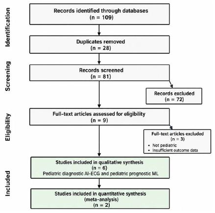
Figure 1: PRISMA flow diagram for AI studies for pediatric ventricular dysfunction.
PICO Framework
The review was designed using the Population-Intervention-Comparator-Outcome (PICO) framework [18].
Population: Children and adolescents (<18 years) with ventricular dysfunction, cardiomyopathy, CHD, ventricular remodeling, or HF.
Intervention: AI, ML, DL, neural network-based algorithms, AI-ECG, and imaging-based ML models.
Comparator: Conventional electrocardiography, echocardiography, clinician interpretation, or standard clinical risk stratification methods.
Outcomes: Diagnostic performance for ventricular dysfunction, ventricular remodeling, ventricular hypertrophy, ventricular dilation, cardiomyopathy, or HF, including sensitivity, specificity, diagnostic odds ratio (DOR), positive predictive value, and negative predictive value.
Eligibility Criteria
Studies were eligible if they: (1) evaluated AI, ML, or DL methods in pediatric cardiovascular populations; (2) focused on ventricular dysfunction, cardiomyopathy, ventricular remodeling, CHD, or HF; (3) reported original human data; and (4) provided diagnostic or prognostic performance metrics. Studies were excluded if they were review articles, editorials, conference abstracts without full manuscripts, animal studies, duplicate publications, or lack of sufficient outcome data for extraction. Adult-only studies were excluded unless they represented childhood disease survivorship populations or provided methodological insights directly relevant to pediatric cardiovascular care.
Data Extraction
Two reviewers (B.D, and S.D.) independently screened titles, abstracts, and full-text articles. Disagreements were resolved by consensus. Extracted variables included study design, publication year, patient population, sample size, underlying cardiac diagnosis, AI methodology, input data source, comparator methods, and diagnostic or prognostic outcomes. For diagnostic studies, sensitivity, specificity, and DOR were extracted whenever available. For prognostic studies, mortality, transplantation, HF progression, and remodeling outcomes were collected.
Risk of Bias Assessment
Risk of bias and applicability were independently assessed by the same investigators using the Quality Assessment of Diagnostic Accuracy Studies-Artificial Intelligence (QUADAS-AI) tool, an extension of QUADAS-2 specifically developed for AI-based diagnostic studies [19]. QUADAS-AI evaluates four domains: patient selection, index test, reference standard, and flow and timing, while incorporating AI-specific considerations related to dataset development, model validation, and clinical applicability. Studies were considered to have an overall low risk of bias and low concerns regarding applicability only when all domains were rated as low risk; any high or unclear rating resulted in an overall classification of potential bias or applicability concerns. Overall study quality was determined by consensus. Overall study quality was determined through consensus review.
Data Synthesis and Statistical Analysis
A quantitative synthesis was performed for pediatric AI-ECG studies reporting diagnostic performance for ventricular dysfunction. The primary diagnostic meta-analysis included studies by Mayourian et al. [11] and Anjewierden et al. [12] which provided extractable measures of sensitivity and specificity for LVSD, ventricular hypertrophy, ventricular dilation, and RVSD. Sensitivity, specificity, and diagnostic odds ratios were pooled using random-effects models after logit transformation. DOR was calculated as (True positive × True negative)/(False positive × False negative). Summary receiver operating characteristic (sROC) curves were generated to evaluate overall diagnostic performance. Studies by Garcia-Canadilla et al. [13] Güntürkün et al. [14] Lv et al. [15] and Yang et al. [20] were not included in the quantitative meta-analysis because they lack diagnostic accuracy outcomes suitable for quantitative pooling These studies were synthesized narratively.
Results
The characteristics of included studies are summarized in Table 1. Six studies met inclusion criteria for qualitative synthesis, including four diagnostic AI-ECG studies and two ML studies focused on pediatric cardiomyopathy phenotyping and HF risk prediction. Two independent pediatric diagnostic cohorts, reported by Mayourian et al. [11] and Anjewierden et al., [12] provided sufficient diagnostic accuracy data for quantitative synthesis. Studies by Garcia-Canadilla et al. [13] Güntürkün et al. [14] Lv et al. [15] and Yang et al. [20] were included in the narrative review because they focused primarily on prognostic modeling, phenotyping, mortality prediction, survivorship risk assessment, or methodological development rather than independent diagnostic accuracy.
Table 1: Characteristics of Included Studies.
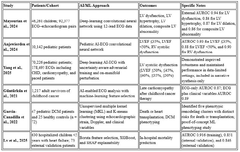
AI: Artificial Intelligence; AI-ECG: Artificial Intelligence–Enabled Electrocardiography; AUROC: Area Under the Receiver Operating Characteristic Curve; DCM: Dilated Cardiomyopathy; ECG: Electrocardiogram; HF: Heart Failure; LV: Left Ventricular; LVEF: Left Ventricular Ejection Fraction; ML: Machine Learning; RV: Right Ventricular; SHAP: SHapley Additive exPlanations; XGBoost: Extreme Gradient Boosting.
Mayourian et al. [11] developed and externally validated an AI-ECG model using 92,377 ECG-echocardiogram pairs from 46,261 children and demonstrated excellent performance for detecting LVSD, hypertrophy, and dilation, with externally validated area under the receiver operating characteristic curve (AUROC) values of 0.94, 0.84, and 0.87, respectively. Anjewierden et al. [12] evaluated 10,142 pediatric patients and reported AUROC values of 0.93 for severe LVSD (LVEF ≤35%), 0.88 for moderate LVSD (LVEF <50%), and 0.90 for RVSD. Yang et al. [20] subsequently expanded these observations in a large CHD cohort using >178,000 ECGs from over 70,000 pediatric patients and demonstrated that adversarial training strategies improved AI-ECG robustness and maintained diagnostic performance under data-limited conditions, further supporting the feasibility of large-scale pediatric AI-ECG screening. Although the Yang study [20] utilized a cohort that substantially overlapped with previous Boston Children’s Hospital datasets and did not report independent diagnostic accuracy outcomes suitable for meta-analysis, it provides important methodological validation for future deployment of AI-ECG systems.
Quantitative synthesis demonstrated high overall diagnostic performance for AI-ECG detection of pediatric ventricular abnormalities (Figure 2). Using a random-effects model, pooled sensitivity was 0.92 (95% CI 0.88-0.94), pooled specificity was 0.73 (95% CI 0.58-0.84), and the pooled diagnostic odds ratio was 34.22 (95% CI 11.08-105.72). Sensitivity remained consistently high across studies and disease phenotypes, whereas specificity demonstrated greater variability. The summary sROC curve further demonstrated excellent overall discriminatory performance (Figure 3).
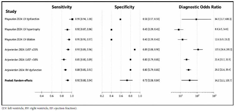
Figure 2: Forest Plot of AI-ECG Diagnostic Accuracy in Children.
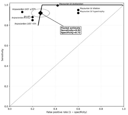
Figure 3: Summary Receiver Operating Characteristic (sROC) Curve for Artificial Intelligence–Enabled Electrocardiography in Pediatric Ventricular Dysfunction.
Risk-of-bias assessment using QUADAS-AI demonstrated predominantly low risk across patient selection, index test, reference standard, and flow/timing domains (Figure 4). The studies by Mayourian et al. [11] and Anjewierden et al., [12] which formed the basis of the quantitative meta-analysis, demonstrated consistently low risk across most domains. Studies focused on prognostic modeling or methodological development, including Garcia-Canadilla et al., [13] Güntürkün et al., [14] Lv et al., [15] and Yang et al., [20] demonstrated low risk for index test and reference standard domains but had unclear ratings regarding AI-specific methodology, external validation, and broader clinical applicability. No study was judged to have a high risk of bias in any major QUADAS-AI domain.
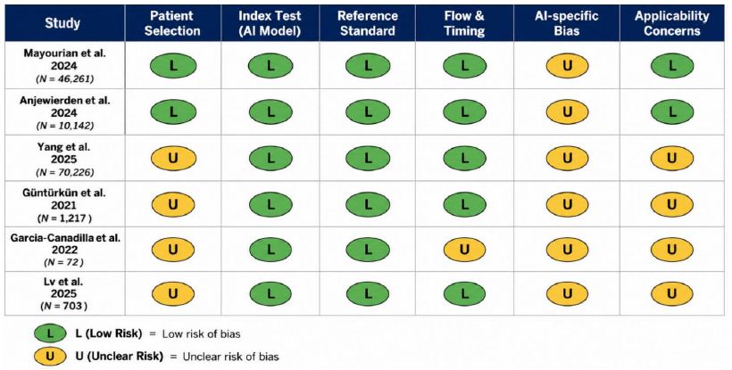
Figure 4: QUADAS-AI Assessment of Risk of Bias and Applicability Concerns in Included Studies.
Discussion
This systematic review demonstrates that AI‑ECG has matured into a reliable diagnostic tool for detecting ventricular dysfunction in children, with consistently high sensitivity across independent pediatric cohorts. Only two studies, Mayourian et al. [11] and Anjewierden et al. [12], provided sufficient diagnostic accuracy data for quantitative pooling, yet both contributed large, well‑characterized cohorts and demonstrated excellent performance for identifying LVSD and RVSD. The pooled estimates from these studies showed high sensitivity (0.92) and good overall discriminatory performance, although specificity varied more widely, reflecting differences in population characteristics, disease prevalence, and model thresholds.
Several additional studies contributed important methodological and prognostic insights despite not meeting criteria for quantitative synthesis. Yang et al. expanded prior work by demonstrating that adversarial training strategies improve model robustness and maintain diagnostic performance even under data‑limited conditions, supporting the feasibility of scalable pediatric AI‑ECG deployment [20]. Other ML studies, Garcia‑Canadilla et al., Güntürkün et al., and Lv et al. focused on phenotyping, mortality prediction, and survivorship risk assessment rather than diagnostic accuracy [13-15]. These investigations highlight the broader potential of AI to characterize disease heterogeneity, identify high‑risk subgroups, and support early risk stratification in pediatric cardiomyopathy and HF.
Risk‑of‑bias assessment using QUADAS‑AI demonstrated predominantly low risk across included studies. The two diagnostic accuracy studies underpinning the meta‑analysis showed consistently low risk across patient selection, index test, reference standard, and flow/timing domains. Studies focused on prognostic modeling or methodological development were generally low risk for index test and reference standard domains but had unclear ratings related to AI‑specific methodology, external validation, and generalizability. No study was judged to have high risk in any major domain.
Future Directions
The rapid evolution of AI is expected to extend the role of ECG‑based analytics in pediatric cardiovascular care, enhancing its utility as a scalable screening tool in resource‑limited settings and expanding applications to areas such as right ventricular functional assessment, where pediatric data remain limited. Building on early pediatric resynchronization studies demonstrating the prognostic relevance of electrical and mechanical dyssynchrony [21,22], future AI‑ECG models may detect early remodeling and stratify risk before overt ventricular dysfunction emerges. As multimodal integration of ECG, echocardiography, CMR, biomarkers, genomics, and electronic health record data advances [23], larger longitudinal datasets may enable AI systems to track disease progression, quantify response to therapy, and support the incorporation of ECG‑derived metrics as meaningful endpoints in pediatric HF research [24]. Adapting these AI‑enabled approaches to children may ultimately support precision pacing strategies tailored to individual anatomic substrates, ventricular geometry, and electromechanical activation patterns, an important need given that many pediatric patients with ventricular dysfunction do not meet traditional adult CRT criteria [25]. Federated learning frameworks offer a promising path to overcome the inherent rarity and heterogeneity of pediatric HF by enabling secure, multicenter model development while preserving patient privacy and institutional data ownership, a strategy increasingly recognized as essential for robust pediatric AI research. Together, these advances position AI‑ECG analytics to shift pediatric HF management from reactive treatment toward proactive, phenotype‑guided, and precision‑based care.
Conclusions
AI is rapidly transforming the detection, phenotyping, risk stratification, and management of pediatric ventricular dysfunction, cardiomyopathy, and HF. Current evidence demonstrates that AI-enhanced ECG provides high diagnostic accuracy for ventricular dysfunction and adverse remodeling, offering a scalable, low-cost screening tool that complements conventional imaging and may expand access to early diagnosis. ML approaches also show promise for cardiomyopathy phenotyping, prognostication, and surveillance of chemotherapy-related cardiotoxicity. However, widespread clinical implementation remains constrained by limited pediatric datasets, single-center studies, and methodological heterogeneity. Future efforts should prioritize multicenter validation, standardized reporting, prospective implementation studies, and integration of multimodal clinical, imaging, genomic, and physiologic data to enable robust, equitable, and precision-based care for children with ventricular dysfunction and heart failure.
References
- Bogle C, Colan SD, Miyamoto SD, Choudhry S, Baez-Hernandez N, et al. (2023) Treatment Strategies for Cardiomyopathy in Children: A Scientific Statement from the American Heart Association. Circulation 148: 174-195. [crossref]
- Irving C, Azeka E, Adorisio R, Blume ED, Bogle C, et al. (2025) The International Society for Heart and Lung Transplantation Guidelines for the Management of Pediatric Heart Failure (Update From 2014). J Heart Lung Transplant 44: e21-e71.
- Puri K, Singh H, Denfield SW, Cabrera AG, Dreyer WJ, et al. (2019) Missed Diagnosis of New-Onset Systolic Heart Failure at First Presentation in Children with No Known Heart Disease. J Pediatr 208: 258-264.e253.
- Amdani S, Conway J, George K, Martinez HR, Asante-Korang A, et al. (2024) Evaluation and Management of Chronic Heart Failure in Children and Adolescents with Congenital Heart Disease: A Scientific Statement from the American Heart Association. Circulation 150: e33-e50.
- Rosano GMC, Teerlink JR, Kinugawa K, Bayes-Genis A, Chioncel O, et al. (2025) The use of left ventricular ejection fraction in the diagnosis and management of heart failure. A clinical consensus statement of the Heart Failure Association (HFA) of the ESC, the Heart Failure Society of America (HFSA), and the Japanese Heart Failure Society (JHFS). Eur J Heart Fail 27: 1174-1187. [crossref]
- Ferreira ALC, Feitoza L, Benitez ME, Aziri B, Begic E, et al. (2025) Diagnostic accuracy of artificial-intelligence-based electrocardiogram algorithm to estimate heart failure with reduced ejection fraction: A systematic review and meta-analysis. Curr Probl Cardiol 50: 103004.
- Das BB, Deshpande SR, Choudhry S, Perumal G (2026) Predicting 1-Year Mortality After Pediatric Heart Transplantation Using Machine Learning: A Systematic Review and Meta-Analysis. JACC Adv 5: 102422. [crossref]
- Zhang S, Jiang J, Luo Y, Liu G, Hu S, et al. (2026) Artificial intelligence in electrocardiogram-based prediction of heart failure: a systematic review and meta-analysis. Frontiers in Cardiovascular Medicine 12.
- O’Sullivan D, Mayourian J, Anjewierden S, Liu K, Attia ZI, et al. (2026) Multicenter validation of AI-enabled ECG for pediatric biological sex prediction. NPJ Digit Med 9.
- Mayourian J, La Cava WG, de Ferranti SD, Mah D, Alexander M, et al. (2025) Expert-Level Automated Diagnosis of the Pediatric ECG Using a Deep Neural Network. JACC Clin Electrophysiol 11: 1308-1320. [crossref]
- Anjewierden S, O’Sullivan D, Mangold KE, Greason G, Attia IZ, et al. (2024) Detection of Right and Left Ventricular Dysfunction in Pediatric Patients Using Artificial Intelligence–Enabled ECGs. Journal of the American Heart Association 2024. [crossref]
- Mayourian J, Gearhart A, La Cava WG, Vaid A, Nadkarni GN, et al. (2024) Deep Learning-Based Electrocardiogram Analysis Predicts Biventricular Dysfunction and Dilation in Congenital Heart Disease. J Am Coll Cardiol 84: 815-828. [crossref]
- Garcia-Canadilla P, Sanchez-Martinez S, Martí-Castellote PM, Slorach C, Hui W, et al. (2022) Machine-learning–based exploration to identify remodeling patterns associated with death or heart-transplant in pediatric-dilated cardiomyopathy. The Journal of Heart and Lung Transplantation 41: 516-526. [crossref]
- Güntürkün F, Akbilgic O, Davis RL, Armstrong GT, Howell RM, et al. (2021) Artificial Intelligence-Assisted Prediction of Late-Onset Cardiomyopathy Among Childhood Cancer Survivors. JCO Clin Cancer Inform 5: 459-468. [crossref]
- Lv H, Sun F, Yuan T, Shen H, Baheti L, et al. (2025) Development and validation of a machine learning model for in-hospital mortality prediction in children under 5 years with heart failure. Frontiers in Pediatrics 13. [crossref]
- Ahmad T, Lund LH, Rao P, Ghosh R, Warier P, et al. (2018) Machine Learning Methods Improve Prognostication, Identify Clinically Distinct Phenotypes, and Detect Heterogeneity in Response to Therapy in a Large Cohort of Heart Failure Patients. Journal of the American Heart Association 7: e008081. [crossref]
- Page MJ, McKenzie JE, Bossuyt PM, Boutron I, Hoffmann TC, et al. (2021) The PRISMA 2020 statement: an updated guideline for reporting systematic reviews. Bmj 372: n71. [crossref]
- Eriksen MB, Frandsen TF (2018) The impact of patient, intervention, comparison, outcome (PICO) as a search strategy tool on literature search quality: a systematic review. Journal of the Medical Library Association 106. [crossref]
- Guni A, Sounderajah V, Whiting P, Bossuyt P, Darzi A, et al. (2024) Revised Tool for the Quality Assessment of Diagnostic Accuracy Studies Using AI (QUADAS-AI): Protocol for a Qualitative Study. JMIR Research Protocols 13: e58202. [crossref]
- Y Yang, LPJ Mayourian, JK Triedman, T Miller, WGL Cava (2025) Unknown article. arXiv preprint arXiv 2509: 19564.
- Dubin AM, Janousek J, Rhee E, Strieper MJ, Cecchin F, et al. (2005) Resynchronization therapy in pediatric and congenital heart disease patients: an international multicenter study. J Am Coll Cardiol 46: 2277-2283.
- Janoušek J, Gebauer RA, Abdul-Khaliq H, Turner M, Kornyei L, et al. (2009) Cardiac resynchronisation therapy in paediatric and congenital heart disease: differential effects in various anatomical and functional substrates. Heart 95: 1165-1171.
- Li X, Liu Y, Zhang X, Yang N, Xu T, et al. (2026) Artificial intelligence in heart failure. The Egyptian Heart Journal 78.
- Svennberg E, Han JK, Caiani EG, Engelhardt S, Ernst S, et al. (2025) State of the Art of Artificial Intelligence in Clinical Electrophysiology in 2025: A Scientific Statement of the European Heart Rhythm Association (EHRA) of the ESC, the Heart Rhythm Society (HRS), and the ESC Working Group on E-Cardiology. Europace 27.
- Schiller O, Dha N, Greene EA, Heath DM, Alexander ME, et al. (2015) Pediatric Dilated Cardiomyopathy Patients Do Not Meet Traditional Cardiac Resynchronization Criteria. J Cardiovasc Electrophysiol 26: 885-889.