Abstract
Neuroanatomical diagnosis is a crucial clinical assessment in animals with neurological disease. Given the complexity of the nervous system, however, it can be challenging for the primary care practitioner to recall and clinically integrate the plethora of neuroanatomical structures and their functions to form this important assessment. While it is sometimes necessary to have advanced knowledge of how discrete neuroanatomical elements of the nervous system as isolated entities, it is pragmatically important for the practicing clinician to have a “functional” understanding of how discrete neuroanatomical structures are integrated, interrelated, and coordinated into a functional unit. These functional units can then be used for neuroanatomical localization. In this review the important functional regions of the nervous system that are responsible for animals performing various normal functions will be discussed in the context of making a neuroanatomical diagnosis. Concurrently, classic clinical abnormalities associated with dysfunction of these functional units will be provided. Using this functional system, the clinician can make practical decisions about disease location with the nervous system. With accurate neuroanatomical diagnosis, the clinician can subsequently formulate an accurate differential diagnosis list followed by an appropriate diagnostic plan to ultimately identify the primary neurologic disease process.
Keywords
Clinical neurology, Clinical evaluation, Neuroanatomical diagnosis, Clinical signs
Introduction
During performance of the neurological examination in animals, abnormalities of nervous system function are identified and categorized. Each clinical abnormality provides a “clue” to the location of the dysfunction within nervous system. Fundamentally, an understanding of the neuroanatomic structures that are responsible for a particular neurologic examination finding are necessary when attempting to determine the location of the lesion resulting in the neurologic dysfunction. Most importantly, however, the practicing clinician needs to understand how these neuroanatomical structures coordinate as functional units to create clinically detectable outputs. Abnormalities found during the neurologic examination help to determine 1) that an abnormality exists in the nervous system and 2) the location of this abnormality within the nervous system. While the neuroanatomical structures are foundational, it is most important that the functional outputs from these anatomical structures which are evaluated clinically are identified and collated.
The determination of where the nervous system is abnormal is referred to as the neuroanatomic diagnosis. This assessment is the fundamental process that underpins clinical neurologic assessment in animals [1-13]. Determining the location of any abnormality in the nervous system is arguably the most important aspect of appropriate clinical evaluation of animals with neurologic disease. Only after determining the location of any abnormality can an appropriate differential diagnosis of disease processes affecting that region of the nervous system be logically generated. Once a list of possible disease processes is formulated, appropriate diagnostic tests to establish the specific disease processes can logically be decided upon, performed, and interpreted in this context.
Recalling specific locations of discrete neuroanatomical structures in the day-to-day clinical evaluations of affected patients, however, can be challenging. This difficulty in making neuroanatomical connections may be a component of a known neurophobia among medical professionals [14-17]. While the nervous system is exceedingly complex, the practicing clinician ultimately identifies the functional outputs of the nervous system through clinically detectable signs such as postures and movements. The astute clinician, then, needs to have a “functional” understanding of how the nervous system is organized and integrated to subsequently understand how disease manifests in clinical dysfunction. While it is sometimes necessary to possess knowledge of how discrete neuroanatomical elements of the nervous system function as isolated entities, it is most important for the practicing clinician to have an operational understanding of how these separate neuroanatomical entities are integrated, interrelated, and interacted as a functional unit. These functional units are interconnected forming functional relationships which are responsible for clinically detectable body functions. An understanding of neurologic functionality promotes an understanding of how information is transmitted or “flows” back and forth between structures within the nervous system and expressed as clinically observable activities. When abnormalities exist within the nervous system, these functional relationships are disrupted or disconnected resulting in clinical signs as evidence of this dysfunction.
Diseases involving individual functional units within the nervous system tend to result in a “classic” set of corresponding clinical signs [1-13]. One, some, or all these classic clinical signs may be present in any individual animal. For this discussion, disease may also be referred to as a lesion (a pathologic or traumatic discontinuity of tissue or loss of function of a part) or an abnormality (any deviation from or interruption of the normal structure or function of a body part). These terms are not to be taken to designate any specific disease process or etiology. Based on regions with similar functionality and the associated clinical signs, nervous system abnormalities can be pragmatically localized into one of twelve main anatomical/functional regions (Figure 1).
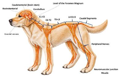
Figure 1: Image depicting the 12 Functional Regions (Zones) that can be used for neuroanatomical localization in dogs and cats.
For simplicity, these anatomical/functional regions will be referred to as “Zones” for this review. While attempting to be as specific as possible with a neuroanatomical diagnosis, a practical initial goal for most clinicians is to determine if disease within the nervous system is present in either the intracranial region (and associated cranial nerves), spinal cord segments, or the peripheral/neuromuscular junction/muscle complex. Answering a series of prompted questions is helpful in determining the neuroanatomical diagnosis during a clinical evaluation (Table 1).
Table 1: Clinical Questions associated with neuroanatomical localization.
| Important clinical questions relative to neuroanatomical diagnosis | |
| 1 | Is the abnormality rostral to, or caudal to, the foramen magnum? |
| 2 | If the abnormality is rostral to the foramen magnum, is the disease affecting the rostrotentorial (“forebrain”) structures, the brain stem, the cerebellum, or individual cranial nerves? |
| 3 | If the abnormality is caudal to the foramen magnum, does this disease involve the spinal cord, the peripheral nerve, neuromuscular junction, or muscle? |
| 4 | If the disease involves the spinal cord, which segment (s) (C1-C5, C6-T2, T3-L3, L4-S3, Caudal segments) |
| 5 | If the abnormality involves the peripheral nervous system, which nerve (s)? |
| 6 | Does the abnormality primarily/exclusively involve the neuromuscular junction? |
| 7 | Does the abnormality primarily/exclusively involve the muscle? |
| 8 | Do all the clinical signs localize to multiple regions within the nervous system? |
Question 1. Is the Abnormality Rostral to or Caudal to the Foramen Magnum?
Anatomical structures rostral to the foramen magnum include the intracranial nervous system (rostrotentorial, caudotentorial (brain stem, cerebellum)) and the specific cranial nerves [18]. Structures caudal to the foramen magnum include the spinal cord segments, specific peripheral nerves, neuromuscular junction, or muscular systems.
Clinical Signs Reflecting Intracranial Dysfunction
Clinical signs suggestive of intracranial disease include, but are not limited to, alterations in mental acuity, behavior abnormalities, cranial nerve abnormalities, seizures, various head posture abnormalities, propulsive circling movements, and conscious proprioceptive deficits in the limbs. Once it has been determined that an intracranial abnormality exists, further delineation of the location of the lesion may be possible based on the specific clinical signs present.
Question 2. Is the Abnormality Affecting the Rostrotentorial Structures, Brain Stem Structures, Cerebellum, or Individual Cranial Nerves?
The rostrotentorial (often referred to as the “forebrain”) structures (Zone 1) include the cerebral hemispheres, basal nuclei, diencephalon, rostral portion of the mesencephalon, and associated structures such as the meninges, blood vessels, and ventricles (lateral, third, and mesencephalic aqueduct). These structures are located rostral to an imaginary transverse plane extending from the tentorium cerebelli ventrally separating the intracranial nervous system into two components. The rostrotentorial nervous system structures are responsible for many functions requiring consciousness, awareness, and associated responses. Classic clinical signs of an abnormality of rostrotentorial structures are found in Table 2. The most common clinical signs of a rostrotentorial abnormality include alterations in their mental acuity or behavior, a menace response abnormality from poor or absent vision, facial sensation recognition abnormalities, true seizure activity, proprioceptive abnormalities in the limbs, a tendency to walk propulsively in a circular direction, “hemi-inattention” or “hemi-neglect”, head pressing, and or head turning. Unilateral lesions involving the rostrotentorial region predominantly result in clinical signs on the side of the body opposite (contralateral) to the abnormality.
Table 2: Classic Clinical Signs of Rostrotentorial Abnormalities.
| Classic Clinical Signs of Rostrotentorial (“Forebrain”) Abnormalities |
| Alterations in mental acuity and behavior (listless, lethargy, somnolence, stupor, coma) |
| “Head Pressing” |
| Hemi-inattention or Hem-neglect |
| Contralateral Menace Response Deficit with concurrent normal Facial Nerve Function and Pupillary Light Reflex (PLR) (unilateral abnormality) |
| Contralateral Facial Sensation Deficit (unilateral abnormality) |
| Contralateral Hemiparesis/Hemiplegia (unilateral abnormality) |
| Tetraparesis/Tetraplegia (bilateral abnormality) |
| Seizures |
| Head turning (usually in the direction of a unilateral abnormality) |
| Circling (usually in the direction of a unilateral abnormality) |
Importantly, while there is a constellation of clinical signs that may be present in any individual animal with a rostrotentorial disease, all these clinical signs may not be present in every affected animal. In some instances, for example, only a single clinical sign may be present to indicate an abnormality in these neuroanatomical structures. Additionally, because of the somatotopic organization of the nervous system, very discrete abnormalities may result in focal or localized clinical signs (for example, dysfunction of a single limb). The type and extent of any disease process ultimately will determine the specific associated clinical signs.
Clinical Signs of Caudotentorial (Brain Stem) Disease
The caudotentorial structures include the commonly defined brain stem (Zone 2), the cerebellum (Zone 3), and associated structures such as the meninges, blood vessels, and ventricles (mesencephalic aqueduct and fourth) [18]. The predominate clinical signs of a brain stem abnormality are found in Table 3. These include abnormal mental acuity and awareness, proprioceptive abnormalities in the limbs, abnormalities of Cranial Nerves III through XII, decerebrate posturing (midbrain abnormalities), and, in severe instances, respiratory irregularities or failure of respiratory function.
Table 3: Classic Clinical Signs of Caudotentorial Abnormalities (Brain Stem).
| Classic Clinical Signs of Brain Stem Abnormalities |
| Alterations in mental acuity and behavior (listless, lethargy, somnolence, stupor, coma) |
| Cranial nerve abnormalities (Cranial nerves III through XII) |
| Vestibular signs – head tilting, ataxia, wide-based posturing, nystagmus, positional strabismus |
| Ipsilateral Hemiparesis/Hemiplegia (unilateral abnormality) |
| Tetraparesis/Tetraplegia (bilateral abnormality) |
| Abnormalities of respiration |
Clinical Signs of Caudotentorial Disease Involving the Cerebellum
The cerebellum (Zone 3) is a unique structure of the central nervous system anatomically and functionally. Because of the important proprioceptive functions of the cerebellum, clinical signs of cerebellar disease are often characteristic as described as “uncoordinated” because of dysfunction of unconscious proprioception [1-13]. Unilateral lesions of the cerebellum result in ipsilateral clinical signs. Common clinical signs of cerebellar disease are found in Table 4. These include ataxia, dysmetria, Intention tremors, lack of balance, head tilting, nystagmus, broad or wide-based stance postures, a menace response deficit (ipsilateral to a unilateral cerebellar lesion) with normal vision, and, in severe instances, decerebellate posturing.
Table 4: Classic Clinical Signs of Caudotentorial Abnormalities (Cerebellum).
| Classic Clinical Signs of Cerebellar Abnormalities |
| Uncoordinated movements |
| Ataxia |
| Hypermetria |
| Falling |
| Intention tremor |
| Absent menace response with normal vision, normal palpebral reflex, and pupillary light reflex (ipsilateral to a unilateral abnormality) |
| Pupillary abnormalities (rare) |
Clinical Signs of Individual Cranial Nerves
Common clinical signs of individual cranial nerve abnormalities (Zone 4) are found in Table 5. The challenge with cranial nerve abnormalities is determining if the abnormality involves the central (primarily the cranial nerve nuclei) or peripheral components (ganglion and peripheral nerve axons) of the cranial nerve. If the central nervous system elements of the cranial nerves are abnormal, these are a component of the rostrotentorial (CN I and II) and brain stem (CN III – XII) localization schemata.
Table 5: Classic Clinical Signs of Cranial Nerve Abnormalities.
| Classic Clinical Signs of Cranial nerve Abnormalities | |
| CN I | Anosmia or dysosmia |
| CN II | Visual abnormalities; menace response abnormalities |
| CN III | Strabismus; ocular movement abnormalities (medial direction); pupillary dilation |
| CN IV | Strabismus; ocular movement abnormalities (rotational) |
| CN V | Facial sensation abnormalities; ocular sensation abnormalities; temporalis and masseter muscle abnormalities |
| CN VI | Strabismus; ocular movement abnormalities (lateral direction) |
| CN VII | Abnormalities of the muscles of facial expression; keratoconjunctivitis sicca |
| CN VIII | Head tilting; falling; nystagmus; positional strabismus |
| CN IX | Swallowing abnormalities; stridor |
| CN X | Swallowing abnormalities; stridor; abnormalities of gastrointestinal functionality |
| CN XI | Neck positional abnormalities (lateral direction) |
| CN XII | Tongue movement abnormalities; tongue atrophy |
Question 3. If the Abnormality is Caudal to the Foramen Magnum, is this Abnormality Affecting the Spinal Cord, Peripheral Nerve, Neuromuscular Junction, and or Muscle?
Neuroanatomical structures caudal to the foramen magnum include the spinal cord and the peripheral nerves as well as the associated neuromuscular junction and muscles [18]. Based on clinical signs such as proprioceptive abnormalities of limbs, spinal reflex function, and painful areas, the clinician can differentiate the abnormality into one of these two general functional systems.
Question 4. If the Abnormality Involves the Spinal Cord, Which Segments?
If the clinical signs suggest an abnormality within the spine or spinal cord, the next step is to determine the location relative to the various spinal segments of that abnormality. Based on the associated clinical signs, the clinician should be able to localize the abnormality to one or more of five functional regions within the spinal cord. Importantly, spinal cord localization schemata are based on spinal cord segments, not vertebral segments as these two separate anatomical entities do not align in all regions of the spine.
Clinical Signs and Localization of Spinal Cord Disease
Clinical signs of spinal cord disease vary with the location of the disease within the spinal cord. As pathways to and from the intracranial nervous system structures travel cranially and caudally through the spinal cord to influence limb function, abnormalities of limb conscious or general proprioception are often found with spinal cord disease. Once the affected limb (s) are identified, spinal reflex assessments are performed to determine if the abnormality involves the central or peripheral nervous system structures (historically referred to as upper motor neuron (UMN) or lower motor neuron (LMN) systems) [1-13]. After collecting this information, the examiner should be able to localize the lesion into one of five spinal cord areas.
The spinal cord is divided into functional areas based primarily on the presence of either central (historically “UMN”) or peripheral (historically “LMN”) clinical signs to the limbs. The presence or absence of these characteristic signs is based upon the concept of a local spinal reflex (that is, a function that does not require conscious control) and the normal control of these reflex functions from intracranial and central spinal cord nervous system. A central (“UMN”) is a neuron or group of neurons that does not exit the central nervous system, and influences, either positively or negatively, peripheral nerve (“LMN”) functionality. Clinical signs of central nervous system disease include proprioceptive abnormalities, increased or normal spinal reflexes, increased or normal limb muscle tone, and, if present, disuse atrophy (Tables 6 through 10).
Table 6: Classic Clinical Signs of Cervical Segment 1 to Cervical Segment 5 Abnormalities.
| Classic Clinical Signs of a Cervical Segment 1 to Cervical Segment 5 Abnormality |
| Limb movement abnormalities |
| Proprioceptive abnormalities when moving (ataxia, hypermetria, limb dragging) |
| Ipsilateral Hemiparesis/Hemiplegia (unilateral abnormality) |
| Tetraparesis/Tetraplegia (bilateral abnormalities) |
| Painful reactions (cervical region) |
| With severe abnormalities, impairment of respiration |
The peripheral nervous system and the effector organs (muscles) are components of what is commonly referred to as “the final common pathway.” This functional unit, however, also includes the cell bodies of the peripheral nervous system in the grey matter of the spinal cord. Clinical signs of peripheral nerve (“LMN”) disease include proprioceptive abnormalities, decreased or absent spinal reflexes, decreased or absent limb muscle tone, and neurogenic atrophy and may result from a spinal cord abnormality that involves the gray matter or reflex interneurons within the local spinal cord segments. As both central and peripheral nervous system abnormalities can result in proprioceptive deficits, evaluation of spinal reflex function becomes important in separating abnormalities of these two functional systems.
Lesions of the Cervical (C)1-C5 spinal segments (Zone 5) classically result in limb dysfunction such as ataxia, hypermetria, and tetraparesis or, if severe, tetraplegia (Table 6). Caudally projecting pathways from the respiratory nuclei of the brain stem to the peripheral nerves for respiration (phrenic nerve to the diaphragm and the intercostal nerves to the intercostal muscles) are located in the cervical spinal cord. Therefore, disease that interrupts these pathways may compromise respiratory functions. Respiratory function is usually only severely compromised in animals that lose voluntary movement (i.e., are tetraplegic). Often, animals will have a concurrent “whispery” bark due to inadequate tidal volumes and decreased projection of that air mass through the larynx and pharynx.
If the disease occurs unilaterally within the cervical spinal cord, hemiparesis may be found on the side ipsilateral to the abnormality as the spinal cord is functionally located caudal to the functional crossover within the intracranial nervous system. Cervical hyperesthesia is often present with many cervical spinal cord diseases but not, for example, in diseases associated with primary nervous system degeneration. One caveat: some animals with intracranial diseases may also have cervical hyperesthesia and this feature of intracranial disease should be considered when localizing an abnormality exclusively to the cervical spinal cord. Components of Horner’s syndrome (sympathetic dysregulation) may be present due to involvement of the caudally projecting sympathetic fibers in the lateral tectotegmental spinal tract from the intracranial central nervous system structures. With unilateral lesions, components of Horner’s syndrome can be found in the eye ipsilateral to the abnormality. Spinal reflex function should be normal or potentially exaggerated, as the peripheral nerves in the cervical and lumbar intumescence are not affected with an exclusive C1-C5 spinal cord segment abnormality.
Lesions of the C6- Thoracic (T)2 spinal segments (cervical intumescence – Zone 6) may also result in abnormalities of limb movements including ataxia, tetraparesis or tetraplegia similarly as with lesions of the C1-C5 spinal segments (Table 7). Spinal reflexes in the pelvic limb will be normal or exaggerated, while those in the thoracic limbs should be decreased to absent due to involvement of the motor neuron cell bodies and axons that make up the nerves to the thoracic limb. This decrease in thoracic limb reflex function is an important clinical feature for localization in these spinal segments.
Table 7: Classic Clinical Signs of Cervical Segment 6 to Thoracic Segment 2 Abnormalities.
| Classic Clinical Signs of a Cervical Segment 6 to Thoracic Segment 2 Abnormality |
| Limb movement abnormalities |
| Proprioceptive abnormalities when moving (ataxia, hypermetria, limb dragging) |
| Ipsilateral Hemiparesis/Hemiplegia (unilateral abnormality) |
| Tetraparesis/Tetraplegia (bilateral abnormalities) |
| Painful reactions (cervical; cranial thoracic regions) |
| Decreased to absent thoracic limb spinal reflexes |
| Decreased to absent thoracic limb muscle tone |
| Neurogenic Muscle atrophy of thoracic limb muscles |
| With severe abnormalities, impairment of respiration |
| Horner’s syndrome (miosis, ptosis, enophthalmos, elevated nictitating membrane) ipsilateral to a unilateral abnormality |
| Abnormalities of the cutaneous trunci reflex (due to a lateral thoracic nerve abnormality) |
If the abnormality is unilateral, hemiparesis is seen with similar reflex changes (decreased to absent thoracic limb spinal reflexes and normal to increased pelvic limb reflexes). Horner’s syndrome (sympathetic denervation) or only a component such as a miotic pupil may be present as the caudal cervical intumescence (T1 to T3 spinal segments) is the location of the preganglionic cell bodies lie and concurrently where the corresponding nerves exit the spinal cord and vertebral canal. With unilateral lesions, Horner’s syndrome will be found in the eye ipsilateral to the abnormality.
If the abnormality involves the C8-T1 spinal segments or nerves, the cutaneous trunci reflex may be abnormal. If the abnormality is unilateral, the ipsilateral cutaneous trunci muscle will not contract with stimulation on either side of the body. The nonaffected side, however, should still contract with stimulation on either side confirming the integrity of the T3-Lumbar (L)3 spinal region.
Abnormalities of the T3-L3 spinal segments (Zone 7) may result in abnormalities of limb movements including ataxia paraparesis or, if severe, paraplegia (Table 8). With unilateral abnormalities, a central nervous system monoparesis of the pelvic limb may be found. While the pelvic limbs are dysfunctional, spinal reflex function in the pelvic limbs will be normal or in some instances exaggerated. The thoracic limbs, however, should function normally.
Table 8: Classic Clinical Signs of Thoracic Segment 3 to Lumbar Segment 3 Abnormalities.
| Classic Clinical Signs of a Thoracic Segment 3 to Lumbar Segment 3 Abnormality |
| Limb movement abnormalities |
| Proprioceptive abnormalities when moving (ataxia, hypermetria, limb dragging) |
| Ipsilateral monoparesis/monoplegia (unilateral abnormality) |
| Paraparesis/Paraplegia (bilateral abnormalities) |
| Painful reactions (thoracolumbar region) |
| Abnormalities of the cutaneous trunci reflex |
| Urination (larger bladder; “firm” feeling; difficult to express; incontinence) and defection abnormalities (incontinence; normal anal tone) |
The cutaneous trunci reflex may be absent caudal to the location of the abnormality, especially with more severe spinal lesions. The level where the cutaneous trunci reflex cannot be elicited is usually within one to three spinal segments of the spinal cord abnormality. With severe spinal cord abnormalities in this location, Schiff-Sherrington posture (that is, hyper-extended thoracic limbs) may be seen. Hyperesthesia may be present on palpation or manipulation of the vertebral segments in this region. Focal hyperesthesia is a helpful neuroanatomical localizing sign in all regions of the spinal cord.
Urinary function may be abnormal with T3 to L3 spinal segment lesions. This is more common with abnormalities that impair voluntary movement. In these instances, the animal similarly cannot urinate voluntarily. The bladder is often large, has appropriate muscle tone on palpation, and can be difficult to manually express due to excessive tone in the urinary sphincters.
Abnormalities of the L4-Sacral (S) S3 spinal segments (lumbar intumescence – Zone 8) may also result in paraparesis/plegia (Table 9). Pelvic limb reflexes, however, will be decreased or absent which is the most important clinical distinction compared to spinal cord abnormalities occurring cranial to these segments. Depending upon which area of the intumescence is involved, specific reflexes may be abnormal. With an abnormality of the L4-L6 spinal segments (femoral nerve), the patella reflex is reduced or absent. Quadriceps muscle atrophy may be present from neurogenic atrophy or a combination of neurogenic and disuse atrophy if the animal has not been using the pelvic limbs for an extended period. Sensation on the distal medial side of the pelvic limb may be reduced or absent due to involvement of the sensory functions of the femoral nerve.
Table 9: Classic Clinical Signs of Lumbar Segment 4 to Sacral Segment 3 Abnormalities.
| Classic Clinical Signs of a Lumbar Segment 4 to Sacral Segment 3 Abnormality |
| Limb movement abnormalities |
| Ipsilateral monoparesis/monoplegia (unilateral abnormality) |
| Paraparesis/Paraplegia (bilateral abnormalities) |
| Proprioceptive abnormalities when moving pelvic limbs (ataxia, hypermetria, limb dragging) |
| Painful reactions (lumbosacral region) |
| Decreased to absent pelvic limb spinal reflexes |
| Decreased to absent pelvic limb muscle tone |
| Neurogenic Muscle atrophy of pelvic limb muscles |
| Urination (larger bladder; “flaccid” feeling; easy to express; incontinence) and defection abnormalities (dilated anal sphincter; incontinence) |
With abnormalities of the L6-S1,2,3 spinal segments (sciatic nerve), the withdrawal reflex is reduced or absent. The patellar reflex may appear exaggerated due to the loss of antagonist muscles to this reflex because of the sciatic involvement (referred to as “pseudohyperreflexia”). Atrophy of the muscles innervated by the sciatic nerve may be present. Atrophy can occur in any muscle innervated by the sciatic nerve but is often most obvious in the cranial tibial muscle.
Correlation between the vertebral and the spinal segments is important as well. In the lumbar area, the spinal cord segments are more cranially located compared to the corresponding vertebral segments. The spinal cord usually terminates in the cranial half of the L7 vertebrae in dogs [18]. In larger breeds of dogs, the spinal cord may terminate more cranially, and in smaller breeds, more caudally [18]. In cats, the spinal cord ends at the L7 or the sacral vertebrae [18]. In dogs, the sacral spinal cord segments tend to lie over L5 vertebra with some variability among individual breeds and individual animals [18]. In cats, the sacral spinal cord segments are found more distally (S2-S3 lie over L6 vertebrae) [18]. Generally, the L1 and L2 spinal cord segments are located within the L1 and L2 vertebrae, respectively. Spinal cord segments L3-L7 are located between L3 and L5 vertebrae.
With abnormalities of the S1-S3 spinal cord segments, bladder and colonic dysfunction may be present. The external anal sphincter may be dilated and have poor muscle tone for contraction. The bladder may be large, flaccid feeling on palpation, and easily expressed. Many animals with peripheral nerve innervation abnormalities or damage primarily to the detrusor muscle (detrusor muscle atrophy) will expel urine with minimal abdominal pressure.
With abnormalities of the caudal spinal cord segments (Zone 9), tail movement dysfunction and reduced or absent tail sensation are commonly present (Table 10). The tail often cannot be extended dorsally or is located in an abnormal position, ventrally or laterally. Hyperesthesia may be present on palpation or manipulation of the sacral or caudal vertebral segments.
Table 10: Classic Clinical Signs of Caudal Segment Abnormalities.
| Classic Clinical Signs of Caudal Segment Abnormalities |
| Tail movement abnormalities |
| Abnormal tail position |
| Painful reactions |
Clinical Signs and Localization of Peripheral Nerve, Neuromuscular Junction, and Muscle Disease
Question 5. If the abnormality involves the peripheral nerves (Zone 10), which nerves?
The peripheral nervous system includes any nerve myelinated by a Schwann cell [18]. Schwann cells begin myelinating peripheral nerves to the limbs at the intervertebral foramen. Using this strict definition, other components of the peripheral nervous system, such as the cell body of the motor neuron in the ventral grey of the spinal cord, would not be included in this strict definition of the peripheral nervous system. Functionally, however, an abnormality of the peripheral nerve or cell body, the neuromuscular junction (NMJ), an or the muscle can result in a similar constellation of clinical signs.
In animals, peripheral nerve (Zone 10), neuromuscular junction (Zone 11), and or muscle (Zone 12) disease primarily results in “weakness” of muscle function (Table 11 through 13). “Weakness” in this is used to indicate a muscle strength problem, however, often is equally used to describe movement dysfunction. If the animal can generate stepping movements, the limb movements are usually associated with shorter step distances, often referred to colloquially as “choppy”. Animals are sometimes described as “stiff” due to inability to advance an appropriate step distance and concurrently with a more extended limb posture. In other instances, the limbs may appear flaccid, have tremoring movements, and buckle primarily with weight bearing. Animals may stand with the limbs placed more centrally under the body. In some instances, this may result in a kyphotic spine or even cow-hocked posture. Animals with both peripheral nerve and muscle disease may also have abnormalities of limb posture. The limb joint angulation during the stance or swing phase of the gait cycle is abnormal, usually with an over-flexion during standing and lack of flexion during forward swing movements.
Table 11: Classic Clinical Signs of Peripheral Nerve Abnormalities.
| Classic Clinical Signs of a Peripheral Nerve Abnormality |
| Limb movement abnormalities |
| Proprioceptive abnormalities when moving pelvic limbs (limb dragging) |
| Ipsilateral monoparesis/monoplegia (unilateral abnormality) |
| Paraparesis/Paraplegia (bilateral abnormalities pelvic limbs only) |
| Tetraparesis/Tetraplegia (bilateral abnormalities all limbs) |
| Short inter-step distances/Shorten strides |
| Abnormal flexed or extended postures of the limbs |
| Exercise intolerance |
| Kyphotic posturing when exercising |
| Facial muscle weakness; decreased palpebral reflexes; excessive salivation |
| Decreased to absent limb spinal reflexes |
| Decreased to absent limb muscle tone |
| Neurogenic Muscle atrophy of limb muscles |
| Lateral recumbency (when severe) |
| Abnormal vocalization (indicative of poor air movement by the respiratory muscles) |
Abnormal muscle functionality may be seen in visceral structures such as the esophagus resulting in megaesophagus in animals with peripheral nerve disease. This may result in regurgitation, decreased swallowing, excessive salivation, gagging, or aspiration pneumonia. Peripheral nerve, neuromuscular junction, and or muscle involvement of the cranial nerves, such as the glossopharyngeal and vagus, may result in dysphagia, dysphonia, gagging, swallowing abnormalities, and coughing. Inspiratory stridor may be present with laryngeal paralysis seen in association with some peripheral nerve diseases. If the diaphragm and intercostal muscles are involved, decreases in effective respiration may result. Animals with respiratory muscle weakness tend to become tachypneic to compensate for decreased inspiratory volume. As the condition worsens, abdominal breathing, wherein the abdomen moves out during inspiration may be present.
Table 12: Classic Clinical Signs of Neuromuscular Junction Abnormalities.
| Classic Clinical Signs of a Neuromuscular Junction Abnormality |
| Limb movement abnormalities |
| Ipsilateral monoparesis/monoplegia (unilateral abnormality) |
| Paraparesis/Paraplegia (bilateral abnormalities pelvic limbs only) |
| Tetraparesis/Tetraplegia (bilateral abnormalities all limbs) |
| Short inter-step distances/Shorten strides |
| Exercise intolerance |
| Kyphotic posturing when exercising |
| Facial muscle weakness; decreased palpebral reflexes; excessive salivation |
Animals with peripheral nerve disease often have abnormalities of general or conscious proprioceptive testing (Table 11). When evaluating general or conscious proprioception in animals with suspected peripheral nerve, neuromuscular junction, and or muscle disease, it is important to evaluate the animal when full weight-bearing and again when the animal’s weight is supported by the examiner, as some diseases affect only the motor functionality of the nerve. With exclusive motor function abnormalities, when the animal’s weight is supported by the examiner, these animals can generate normal conscious proprioception or paw placement due to normal sensory function. With many peripheral nerve and central nervous system lesions, however, animals often have both motor and sensory weakness occurring concurrently. Conscious proprioceptive deficits will be present in these animals regardless of weight-bearing status in these animals.
Table 13: Classic Clinical Signs of Primary Muscle Abnormalities.
| Classic Clinical Signs of Primary Muscle Abnormalities |
| Limb movement abnormalities |
| Short inter-step distances/Shorten strides |
| Exercise intolerance |
| “Crabbing” |
| Kyphotic posturing when exercising |
| Facial muscle weakness; decreased palpebral reflexes; excessive salivation |
| Primary Myopathic Muscle atrophy of limb muscles |
Spinal reflexes are reduced or absent in animals with peripheral nerve disease. This is an important clue to the location of the abnormality in the peripheral nerve. Resting muscle tone and muscle resistance to passive range of motion in the limbs will be poor or absent.
With peripheral nerve disease involving motor nerve innervation, neurogenic atrophy of muscles occurs. This atrophy evolves relatively rapidly after a peripheral nerve injury (within 5 to 10 days) and results in significant amounts of muscle atrophy. Often the underlying bony structures become prominent due to the significant degree of atrophy. With single nerve disease, muscle atrophy may be in focal muscles or muscle groups corresponding to the distribution of the affected peripheral nerve innervation.
Individual peripheral nerves may be selectively involved with various pathological processes that involve the motor or sensory functional components. Primary sensory nerve disease often results in exaggerated uncoordinated movements resulting from the associated proprioceptive deficits. Animals with primary afferent or sensory neuropathies may have an awkward gait wherein they tend to “flap” the limbs and strike the ground on the more dorsal palmar and plantar regions of the paw and lower limb. Evaluation of the sensory distribution loss may be helpful in localization with sensory components of the peripheral nerve are abnormal. The sensory deficiencies can be present in the dermatomes and autonomous zones innervated by these nerves. In general, however, clinical assessment of skin innervation is inconsistent.
Question 6. Does the Abnormality Involve the Neuromuscular Junction (Zone 11)?
Animals with disease primarily involving the neuromuscular junction classically have overlapping clinical signs with peripheral nerve and primary muscle abnormalities. Short stepping is one of the more classical signs. With some neuromuscular diseases, muscle weakness is exacerbated with exercise (e.g., myasthenia gravis, hypoglycemia). Dogs and cats with neuromuscular junction, and muscle disease may lie down frequently. Cats particularly may rest their head on their thoracic limbs when lying down. Cats tend to have ventral neck flexion, or inability to lift their heads, presumably due to cervical muscle weakness. Animals with disease of the neuromuscular junction or muscle abnormalities may have proprioceptive deficits during weight bearing, but not when their weight is supported by the examiner as discussed previously. In diseases of the neuromuscular junction (e.g., myasthenia gravis) and muscle, spinal reflexes are classically normal as relatively minimal muscle strength is necessary to perform simple reflex functions.
Question 7. Does the Abnormality Primarily or Exclusively Involve the Muscle (Zone 12)?
Animals with disease primarily involving the muscle also have clinical signs overlapping with peripheral nerve and neuromuscular junction abnormalities. Weakness of muscle function is the most consistent clinical abnormality. Short stepping and muscle atrophy are clinical clues. Animals with primary muscle disease, however, should have normal conscious proprioceptive testing when the examiner supports the animal’s body weight.
Primary muscle abnormalities may result in dysfunction of visceral structures such as the esophagus resulting in megaesophagus similar to peripheral nerve disease [1-13]. This muscle weakness may result in regurgitation, swallowing abnormalities, excessive salivation, gagging, or aspiration pneumonia. Muscle involvement of muscles innervated by cranial nerves, such as the glossopharyngeal and vagus, may equally result in dysphagia, dysphonia, swallowing abnormalities, gagging, and coughing. If the diaphragm and intercostal muscles are involved, decreases in effective respiration may result.
With most primary diseases of muscle, muscle mass is classically decreased. In contrast, animals with myotonia or some other muscle disorders may have hypertrophy of their muscles. Their muscle groups may be well defined (prominent) anatomically. They similarly have short step distances when gaiting and often with extended limb postures. Equally, animals with muscle cramping or myotonia may have episodic bouts of extended limb postures. As with some neuromuscular diseases, muscle weakness may be exacerbated with exercise.
Multifocal or Diffuse Lesions of the Nervous System
Question 8. Do all the Clinical Signs Localize to a Focal Region in the Nervous System?
Some diseases can affect multiple regions of the nervous system simultaneously. Additionally, two or more different diseases can concurrently affect the nervous system in differing areas. If the clinical signs cannot be adequately explained by a single focal abnormality, then consideration should be given to a single disease with multifocal pathology or that there are multiple disease processes present affecting multiple functional units. Diffuse diseases such as inflammatory conditions would be an example.
Conclusion
The ability to make an accurate neuroanatomical diagnosis is the essential step in determining possible disease processes that affect that location within the animal’s nervous system. Correctly determining the location of any abnormality in the nervous system of animals is foundational to completing a logical differential diagnosis list and subsequently diagnostic plan. While seemingly complex, there are essentially twelve functional regions of the nervous system for neuroanatomical diagnosis. Based on the classic clinical signs associated with these twelve functional regions, an accurate neuroanatomical location of any neurologic abnormalities is possible. Once an accurate neuroanatomical diagnosis is identified, further clinical assessment can logically be performed to ultimately make a definitive diagnosis.
References
- De Lahunta A (1977) Veterinary Neuroanatomy and Clinical Neurology. W. B. Saunders.
- De Lahunta A, Glass E, Kent M. De Lahunta’s (2021) Veterinary Neuroanatomy and Clinical Neurology. 5th ed. Amsterdam: Elsevier.
- Bagley RS (2005) Fundamentals of Veterinary Clinical Neurology. Ames: Blackwell.
- Dewey CW, da Costa RC. Practical Guide to Canine and Feline Neurology, 3rd ed., Hoboken; Wiley-Blackwell.
- Hoerlein BF (1971) Canine Neurology: Diagnosis and Treatment. 2nd ed., W. B. Saunders.
- Palmer AC (1976) Introduction to Animal Neurology. 2nd ed., Blackwell Scientific Publications.
- Jenkins TW (1972) Functional Mammalian Neuroanatomy. 2nd ed., Lea & Febiger.
- King AS (1987) Physiological and Clinical Anatomy of the Domestic Mammals. Oxford UP.
- McGrath JT (1960) Neurologic Examination of the Dog. 2nd ed., Lea & Febiger.
- Braun KG (1994) Clinical Syndromes in Veterinary Neurology, 2d ed. St. Louis, Mosby.
- Chrisman CL (1991) Problems in Small Animal Neurology. Philadelphia, Lea & Febiger.
- Oliver JE, Hoerlei BF, Mayhew IG (1987) Veterinary Neurology. Philadelphia, W. B. Saunders.
- Oliver JE, Jr Lorenz, MD, Kornegay JN (1997) In Handbook of Veterinary Neurology, 3d ed. Philadelphia, W. B. Saunders, 341-402.
- Abkur T, Satti Z, Di Luca DG, Alhashyan I, Algethami H (2024) Neurophobia: challenges and solutions. Neurosciences (Riyadh) 29: 144-145.
- Jozefowicz RF (1994) Neurophobia: the fear of neurology among medical students. Arch Neurol 51: 328-329. [crossref]
- AlZahrani AA, AlQahtani BG, Bayazeed MA, Mahfouz ME (2025) Unveiling neurophobia: exploring factors influencing medical students, residents and non-neurologist physicians globally and its implications on neurology care – a systematic review and meta-analysis. BMJ Neurol Open 7: e001009.
- Murthy VD, Le L, Heater HD, Guess SC, Chen AV (2024) Investigation of Neurophobia amongst North American Veterinary Students and Development of a Veterinary Neurophobia Scoring Tool (VetNeuroQ). J Vet Med Educ 51: 819-833.
- Hermanson JW, de Lahunta A, Evans HE. Miller and Evans’ Anatomy of the Dog (5th ed.). Elsevier: St. Louis, USA, ISBN (Print): 9780323546010, pp. XI-XII.