DOI: 10.31038/IDT.2026711
Abstract
DNA aptamers were developed and sequenced against an envelope precursor polyprotein of Hantavirus (HV). The highest affinity aptamers from the final SELEX pool were determined by an enzyme-linked microplate assay against the recombinant envelope protein. Subsequent 3-D molecular models using two different docking platforms strongly suggest that the top aptamers will bind the exterior ectodomain of the envelope protein protruding from the viral lipid envelope and therefore could interfere with virus binding to host cell receptors, thus affording potential prophylaxis or therapy for Hanta-related hemorrhagic, pulmonary and renal syndromes.
Keywords
3-D Molecular modeling, Aptamer, Docking software, Hanta virus, Passive immunity
Introduction
Hanta viruses (HVs) including Sin Nombre virus are named after the Hantan river region in North and South Korea where an early outbreak occurred. HVs are spread mostly by rodent or rodent feces and urine exposure to humans, but not between humans. These viruses affect greater than 200,000 people worldwide although 90% of cases occur in China each year and can cause potentially lethal hantavirus pulmonary syndrome (HPS) and hemorrhagic fever with renal syndrome (HFRS) [1]. There are no effective government-approved treatments for HPS or HFRS except rest and treatment for the associated symptoms of fever, fatigue and severe muscle pain. However, at least two experimental monoclonal antibodies have demonstrated some efficacy by binding the external surface Gn and Gc viral proteins (cleavage products of the envelope polyprotein) to neutralize viral entry into host cells [2], thus opening the door for less expensive DNA aptamer-based passive immunity as well [3].
Materials and Methods
Recombinant Hanta Virus Envelope Polyprotein Target and Magnetic Bead Immobilization
The Hantavirus envelope recombinant polyprotein (Gn/Gc or G1/G2 precursor) target was produced by Bioclone Inc. (Dr. Peter Ding), San Diego, CA. It covered amino acids 50-450 from the Jurong TJK/06(RT50) Hantavirus strain (Swiss Protein number C7EMH7). Because the protein comes with a 6X histidine tail, the authors used NTA Nickel-coated magnetic beads from Thermo Fisher Inc. for immobilization of the target protein for SELEX aptamer development. By immobilizing at the tail end where the 6X histidine resides, most of the native protein was available to interact with the random DNA library to better select ectodomain aptamers versus randomly immobilizing the protein via tosyl leaving groups (common on magnetic microbeads) anywhere that a primary amine occurs in the protein. Figure 1 describes the SELEX template and PCR primers that were used for SELEX aptamer development. Otherwise, traditional magnetic bead-based SELEX methods as reported in the literature [3] were utilized and the final round 10 SELEX pool was sequenced using Illumina next generation sequencing by synthesis at Base Pair Biotechnologies Inc. (Pearland, TX, USA).
Figure 1: Schematic and DNA sequences of the 72 base SELEX template with randomized 36 bases (N36) region flanked by two fixed 18 base PCR primer binding ends and the Forward (F) and Reverse (R) primer sequences obtained from Integrated DNA Technologies (Coralville, IA, USA).
Results
Figure 2 documents successful 72 bp PCR amplicons from each of the final rounds of HV SELEX in an ethidium bromide-stained 2% agarose electrophoresis gel. The most frequent (i.e., top) 24 candidate aptamers that occurred at least 10 times or more in total (FCOUNT) in the final round 10 aptamer pool and greater than 6 times per million sequences (CPM) are reported in Table 1.
Figure 2: Ethidium bromide-stained 2% agarose electrophoretic gel image showing 72 bp aptamer amplicons from each of the final rounds of SELEX for HV envelope protein aptamer development.
The HV aptamer candidates were screened by ELISA-like (ELASA) assay as described previously [3] for affinity to the cognate HV envelope polyprotein. The ELASA relative affinity rankings by absorbance at 405 nm of each candidate aptamer from Table 1 are provided in Table 2 below and suggest the top aptamers to screen for inhibition of Hantavirus plaques in vitro [4], if that possibility ever exists. In particular, it appears that HV aptamers S2, S3, S7 and S9 with absorbance at 405 nm values greater than 2.0 are the best four candidates with which to start in vitro HV plaque inhibition studies in Vero cells [4].
Table 1: Top 24 Hantavirus Envelope Polyprotein Aptamer DNA Sequences and Frequency (Total Counts and Counts per Million Sequences).
Because Nanohmics developed a number of seemingly high affinity (Table 2) HV aptamers during this project, it followed that 3-D modeling of the top aptamer docking with the HV envelope polyprotein from the NCBI and Swiss Protein databases (number C7EMH7) was in order. The HV S1 aptamer emerged 115 times or 25 more total times than the nearest competitor in the NGS pool (Table 1) and was ranked in the top 7 aptamer candidates by ELISA-like assay vs. the envelope protein with an average absorbance at 405 nm of 1.9175 versus the top aptamer candidate (HV S7; A405 nm = 2.248) in terms of affinity. However, affinity is not always the best predictor of ability to block viral entry into host cells. So, HV S1 is also a logical candidate to begin modeling for future in vitro plaque assays.
Table 2: ELASA Plate Assay Rankings of the Top 24 Aptamers from NGS for Hantavirus Envelope Binding and Potential Inhibition (Average of Duplicate A405 nm Readings).
We were not able to find an established 3-D PDB model for the Hantavirus Jurong TJK/06(RT50) envelope protein, so we had to enter the amino acid sequence (cut and pasted) into a program within UniProt and then through a program called ModWeb from the Univ. of California at San Francisco (UCSF) to generate the 3-D envelope protein structure shown in Figure 3 below. Note the characteristic black “donut hole” in the middle of this envelope protein which helps to identify the polyprotein in 3-D docked images with candidate aptamers.
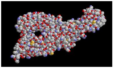
Figure 3: 3-D PBD model of the Hantavirus Jurong TJK/06(RT50) envelope protein generated by UniProt and ModWeb. Note the characteristic “donut hole” (black area in middle).
We utilized and compared results from HDock and ZDock internet programs as shown in Figures 4-6 below to determine where the HV S1 aptamer was preferentially binding on the HV envelope (E) polyprotein. From both the HDock and ZDock analyses, the HV S1 aptamer appears to prefer binding the thinner tapered segment of the HV E polyprotein. Unfortunately, according to Serris et al. [5], this end of the envelope protein may be inserted into the lipid envelope and not even available for aptamer binding, thus making the HV S1 aptamer potentially useless. However, some of the other top HV aptamer sequences subjected to 3-D docking analysis seemed to prefer binding the opposite end (ectodomain) of the HV envelope polyprotein making them better candidates (Figure 6).
Figure 4: Top 3-D docking ribbon structure for the HV1 aptamer (orange) with the yellow HV E polyprotein using HDock.
Figure 5: Top 3-D docking space-filled structure for the HV S1 aptamer (brown) with the green HV E polyprotein (green) using HDock software. Note again the “donut hole” in the E polyprotein and aptamer binding to the thinner end of the protein.
Figure 6: Top 3-D docking space-filled structure for the HV S1 aptamer binding the yellow ectodomain of the HV Envelope polyprotein using ZDock software rendered in RasMol software.
Discussion
Serris et al. [5] described the domain of Hantavirus (HV) envelope E polyprotein which is embedded in the viral envelope lipid membrane and the ectodomains that are outside of the viral coat and available for some particular aptamer binding. Figure 7 below illustrates how the 3-D ribbon structures of the HV envelope protein reported by Serris et al. [5] in panels A and B appear to match the general 3-D HV envelope structure that we used for aptamer docking studies. Note that Domain I in panels A and B appears to be the base domain that inserts into the viral lipid envelope and would be inaccessible to aptamers. However, the curved outer ectodomains appear to be accessible to aptamers and both of the top aptamer-HV E polyprotein docking models developed during this project show the S1 aptamer binding to these ectodomains as represented in panel D which could thus probably block binding to host cell receptors and inhibit or prevent plaque formation in vitro and infection in vivo. Naturally, all of this theoretical modeling needs to be tested empirically in vitro with plaque inhibition studies [4], but this publication provides a maximum of 24 candidate aptamers and some theoretical criteria for down selecting to the best ectodomain-binding candidates with HV neutralization potential in future studies.
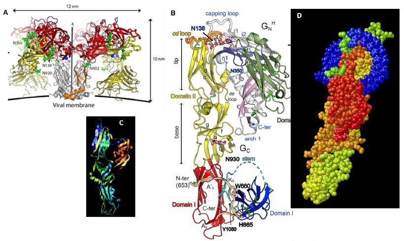
Figure 7: A and B – 3-D ribbon structures of Hantavirus envelope protein borrowed from Serris et al. [5] showing the protein orientation and insertion in the envelope/membrane. C – The authors’ similar 3-D ribbon structure and D – the authors’ predicted blue aptamer docked with the accessible outer ectodomain which could enable blocking of host cell receptor binding and block viral entry.
Conclusions
A total of 24 new DNA aptamer sequences against HV envelope polyprotein were generated and studied by static 3-D docking models that suggest binding of the HV envelope polyprotein external ectodomain by some of the candidates. If true, one or more of the reported aptamer DNA sequences could block or inhibit Hantavirus entry into host cells in vitro and in vivo, thus providing HV neutralization and passive immunity.
Acknowledgements
Funding was provided by US DoD SBIR Contract No. W91SR22P0007.
References
- Afzal S, Ali L, Batool A, Afzal M, Kanwal N, et al. (2023) Hantavirus: an overview and advancements in therapeutic approaches for infection. Frontiers in Microbiology 14: 2023. [crossref]
- Arikawa J, Schmaljohn AL, Dalrymple JM, Schmaljohn CS (1989) Characterization of Hantaan Virus Envelope Glycoprotein Antigenic Determinants Defined by Monoclonal Antibodies. Journal of General Virology 70: 615-624. [crossref]
- Bruno JG, Carrillo MP, Richarte AM, Phillips T, Andrews C, et al. (2012) Development, screening, and analysis of DNA aptamer libraries potentially useful for diagnosis and passive immunity of arboviruses. BMC Research Notes 5: 633. [crossref]
- Pádua M, Souza WM, Lauretti F, Figueiredo LT (2015) Development of a novel plaque reduction neutralisation test for hantavirus infection. Memorias do Instituto Oswaldo Cruz 110: 624-628. [crossref]
- Serris A, Stass R, Bignon EA, Muena NA, Manuguerra JC, et al. (2020) The Hantavirus Surface Glycoprotein Lattice and Its Fusion Control Mechanism. Cell 183: 442-456.e416. [crossref]