Abstract
The patient was a 51-year-old woman. After receiving acupuncture and moxibustion, she felt dyspnea. By examination she was diagnosed with right pneumothorax caused by acupuncture and moxibustion. We immediately inserted chest tube. In the beginning, her lung was not full expansion, so we considered Video-assisted thoracic surgery (VATS). But the right lung gradually expanded, and air leaks lowered. So she left hospital. Through this case, we think that acupuncture and moxibustion is risky. It is required that reducing the number of the patient of pneumothorax after acupuncture and moxibustion. We also think that enlightenment activities are necessary.
Keywords
Pneumothorax, Acupuncture, Moxibustion, Dyspnea, Chest tube, Video-assisted thoracic surgery
Introduction
Acupuncture is the therapy using specific needles and stimulation for physical points called “keiketu or tsubo”. We reported a case of pneumothorax after acupuncture and moxibustion.
Case Study
Patient was a 51-year-old woman
Chief complaint was dyspnea
Medical history has no special notes
No Allergy
Never smoked
History of Present Illness
After receiving acupuncture and moxibustion on X-1 day, right chest pain appeared on the way home. She also had a cough and difficulty breathing. In addition, she could not sleep on that day. When she visited a nearby doctor on X day, she was diagnosed with right pneumothorax, and was referred to our department on the same time.
Presenting Symptoms
Height was 155.5 cm. Weight was 47.5 kg. Blood pressure was 106/68 mmHg. Heart Rate is 83/min. Body Temperature was 36.4°C. SpO2 was 99% (room air). However, dyspnea and shortness of breath worsened over time. There were many acupuncture and moxibustion on the anterior chest and back. And right lung sound was decreased.
Examination Finding
Blood test showed no anemia or coagulation abnormalities. Chest X-ray showed right severe pneumothorax (Figure 1). And CT-scan showed pneumothorax, but there were no lesions caused pneumothorax, such as bulla or bleb (Figure 2).
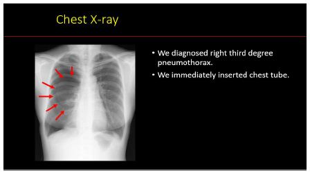
Figure 1: Illustrates the overall workflow.
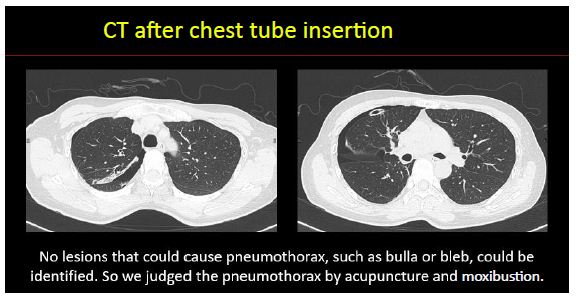
Figure 2: Outlines the retrieval-to-generation flow.
Clinical Course
We diagnosed right severe pneumothorax caused by acupuncture and moxibustion, and immediately inserted chest tube. On X+1 day, her lung was not full expansion, so we considered Video-assisted thoracic surgery (VATS). But the right lung gradually expanded, and air leaks lowered. So, on X+5 day, we removed chest tube. On X+6 day, she left the hospital (Figure 3). And after discharge from the hospital, she is doing well.
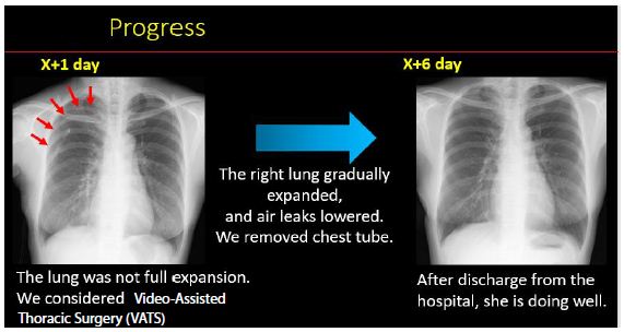
Figure 3: Progress.
Discussion
In our case, acupuncture and moxibustion caused right severe pneumothorax. There are other cases of pneumothorax after receiving acupuncture and moxibustion, also the case of bilateral pneumothorax has been reported [1]. And the patients of bilateral pneumothorax often have a fetal course, furthermore the patients sometimes die [2,3]. In addition to pneumothorax, adverse events such as infections [4,5] and organ damages including the heart [6] have been reported. As other adverse events, neurological damage [7], skin damage [8] and chylothorax [9] have been known.
As examples of pulmonary disease, which is easy to cause pneumothorax by acupuncture and moxibustion, asthma, emphysema and pulmonary fibrosis are mentioned [1]. Also, it has been reported that the safety length of acupuncture penetration is 8-26 mm in the anterior chest and 20 mm on the back 1. In our case, because we heard that it was approximately 40 mm deep from the patient, and we could not find a primary illness which could cause pneumothorax on CT scan, we predicted that over safety length of acupuncture penetration cause pneumothorax. Although our patient has been doing well by insertion chest tube, it is generally known that pneumothorax caused by acupuncture is more severe than other pneumothorax because of the inflow of air into the chest cavity through the acupuncture hole and the larger hole in the lung [1]. Also, there are reports that the use of lung ultrasound is useful for rapid diagnosis and therapeutic intervention for pneumothorax at the bedside [10,11]. In our case, the patient had a unilateral pneumothorax, and tension pneumothorax or hemopneumothorax could not be detected, so a chest X-ray could be taken. But if there was an urgent need, there was a way to consider the use of a lung echocardiogram. In addition, if it was a tension pneumothorax, it would have been necessary to consider degassing based only on examination findings. In order to reduce pneumothorax after acupuncture and moxibustion like our case, we think that enlightenment activities for people are also important. For example, one way is to create a consent form and a explanation sheet used in medical practice.
Conclusion
We experienced a case of pneumothorax after acupuncture and moxibustion. In order to reduce the number of the patient of pneumothorax after acupuncture and moxibustion, we think that enlightenment activities are necessary by using a consent form and a explanation sheet used in medical practice.
Conflict of Interest
There are no companies, organizations or groups with COI status to disclose related to this paper.
References
- Shindo H, Yamagishi T, Nagaoka T, Tsuboi K, et al. (2020) Acupuncture-related bilateral pneumothorax: a case report. KANTO Journal of Japanese Association for Acute Medicine 41: 271-273.
- Akcam TI, Kavurmaci O, Ergonul AG, Aydin S, Turhan K, et al. (2008) Analysis of the patients with simultaneous bilateral spontaneous pneumothorax. Clin Respir J 12: 1207-1211. [crossref]
- Jian J, Shao Y, Wan L, Zhang M, Liu N, et al. (2018) Autopsy diagnosis of acupuncture-induced bilateral tension pneumothorax using whole-body postmortem computed tomography: A case report. Medicine (Baltimore) 97: e13059. [crossref]
- Kim D, Lee S (2017) An autopsy case of fatal acute peritonitis complicated by illegal acupuncture therapy. Forensic Sci Int 276: e13-e15. [crossref]
- Alexandre AR, Raimundo P (2018) Epidural, paravertebral and bilateral psoas abscess after lumbar acupuncture. BMJ Case Rep 11: e228047. [crossref]
- Llamas Fuentes R, Valle Alonso J, Larrasa Soriano S (2020) Cardiac tamponade and right ventricle perforation after an acupuncture procedure. Med Clin (Barc) 154: 416-417. [crossref]
- Eghbal K, Ghaffarpasand F (2016) An Acute Cervical Subdural Hematoma as the Complication of Acupuncture: Case Report and Literature Review. World Neruosurg 95: 616. e11-616.e13. [crossref]
- Park MY, Lee JS, Jin HJ, You HS, Kim GW, Ko HC, et al. (2018) Localized argyria: troublesome side effect of acupuncture. J Eur Acad Dermatol Venereol 32: e62-e65. [crossref]
- Furuse N, Yamashita H (2021) Literature Review on Adverse Events (2016-2019) Associated with Acupuncture and Moxibustion 71: 245-264.
- Harriott A, Mehta N, Secko M, Romney ML (2014) Sonographic diagnosis of bilateral pneumothorax following an acupuncture session. J Clin Ultrasound 42: 27-29. [crossref]
- Frankel HL, Kirkpatrick AW, Elbarbary M, Kirkpatrick AW, Su E, et al. (2015) Guidelines for the Appropriate Use of Bedside General and Cardiac Ultrasonography in the Evaluation of Critically Ill patients-Part Ⅰ: General Ultrasonography. Critical Care Med 43: 2479-2502. [crossref]