Abstract
We present the results of our Raman studies on natural lonsdaleite crystals in fluorite of the two tin deposits Sadisdorf and Zinnwald in the E-Erzgebirge/Germany. The Raman spectra, specially from Zinnwald, can deconvoluted into three Raman-active vibrational modes: E2g, A1g, and E1g. For the origin, transport via supercritical fluid or melting from the mantle region into the crust is necessary.
Keywords
Lonsdaleite, REE-rich fluorite, Raman spectroscopy, Sadisdorf and Zinnwald tin-tungsten deposits, Supercritical fluid
Introduction
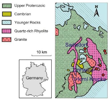
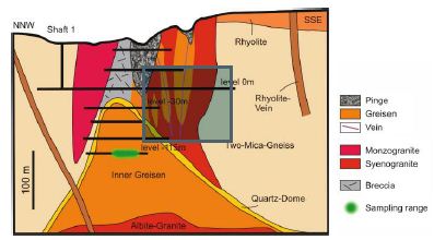
The Variscan Erzgebirge (German side) and Krušné hory (Czech side) contain many tin-tungsten deposits of vein and greisen type. Famous deposits are that from Zinnwald, Cinnovec, Krupka, Sadisdorf, Ehrenfriedersdorf, and Geyer. For the regional geology and description of single deposits, extensive literature is present [1-5] and references in these). From a repetition of that, we will abstain because our described observations are new and do not fit into the old genetic view, forcing us to adopt a new approach for the whole region. In the abandoned Sadisdorf Sn-W-(Cu) deposits, there are vein and greisen- type mineralizations (Figures 1a and 1b).
The frequently present breccia pipes here are analogous to the famous Schneckenstein breccia in the W-Erzgebirge, which is very characteristic of this mineralization type [6]. During the study of thin sections from the Sadisdorf deposit, peculiar violet fluorite aggregates attract attention. These aggregates contain many black, sometimes bent needles, as well as also spherical to elliptical carbon solids. A similarity to the other described new findings of spheric high-pressure minerals is very obvious (see, for example, Thomas et al., 2023) [7]. This publication also contains a schematic geological map of the Erzgebirge-Vogland Zone with the Variscan granites, etc.

Figure 1a: Location and simplified geological map of the Sadisdorf and Zinnwald deposit/ E-Erzgebirge.

Figure 1b: Schematic cross-section through the Sadisdorf deposit with the approximate origin place of the studied sample. The green color marks the origin of the Sadisdof sample.
Sample Material
Sadisdorf
The rock material is from the endocontact of the tin-tungsten- copper deposit Sadisdorf in eastern Erzgebirge/Germany [1,8]. The sample thin section (SD-H01B) is from a fine-granular white quartz-topaz rock with violet fluorite, taken during the exploration campaign Sc-ore 2012/2013 – the violet fluorite is, according to Raman spectroscopy, very REE-rich. Besides the primary mineral quartz and rarer topaz, there are many small, mostly opaque mineral grains of wolframite, cassiterite, columbite, and others. The large fluorite aggregates are not isomorphic but more in tube or irregular form. Smaller fluorite crystals are isometrically. These large tubular fluorite aggregates contain many black and transparent needles, grains, and curved crystals (Figure 2) and are generally fluid inclusion-free. If present, then they are secondary late formations.
The curved black crystal (Figure 2) contains many nano-diamonds. The straight black needles are prevailing graphite-like material. Graphite is very often present in the Variscan tin mineralization, and it is also often present in cassiterite. Graphite has mostly not been paid attention to in the past.

Figure 2: Curved graphite aggregate with nano-diamonds in REE-rich fluorite. All black points are graphite-like stuff and contain nano-diamonds.
Zinnwald
For comparison, we used fluorite grains included in tabular zinnwaldite crystals from the Zinnwald deposit (deep Bünau gallery) in the E-Erzgebirge/Germany, which is not far from the Sadisdorf deposit (taken by the first author in 1984). This violet fluorite sample contains numerous sharp black disk-like bodies of lonsdaleite-diamond-graphite (Figure 3). Some of these form half- moon-like bodies (Figure 4) where the sharp edges lie parallel to the crystallographic{100} planes.
It follows from Figures 3 and 4 that the black bodies obviously form hemispheres on the fluorite surfaces. The general impression is that this fluorite is an early formation and has a more plastic behavior at the formation time. That spoke for a high-temperature and high- pressure formation from a fluorine-rich melt containing such C-aggregates.

Figure 3: Small disk-like bodies of lonsdaleite in fluorite from Zinnwald/E-Erzgebirge (top view).

Figure 4: Side view of such bodies, as shown in Figure 3, arranged to the growth zones of the fluorite host.
Methodology: Microscopy and Raman Spectroscopy
For the study of the lonsdaleite-diamond-graphite-bearing samples and their paragenetic main minerals, we use the Zeiss JENALAB pol as well as the Raman spectrometer EnSpectr R532 combined with the Olympus BX43 microscope both for transmitted and reflected light and equipped with a rotating stage. Note here that the incident laser light is always polarized – in our case, N – S (see Tuschel, 2012) [9]. Generally, we used an Olympus long-distance LMPLFL100x objective lens. For the identification of different minerals, we used the RRUFF and the Hurai et al. Raman mineral databases [10,11]. As references, we applied a water-clear diamond crystal from Brazil (1331.63 ± 0.60 cm-1 and a semiconductor-grade silicon single-crystal (520.70 ± 0.15 cm-1). For this study, we used laser energies from 0.9 to 50 mW on the sample. Because the minerals lonsdaleite, diamond, and nano- diamond contain black graphite particles and are metastable in the new upper crust surrounding, heating by the laser energy can partially destroy these minerals in the extreme case or shift the characteristic peak position of Raman bands [12,13] to lower values.
Results
Sadisdorf
The sizeable bent carbon crystal (Figure 2) contains many small nano-diamonds. The diamond main band lies at 1331.8 ± cm-1 (11 different crystals). The FWHM (Full Width at Half Maximum) is 83.1 ± 13.9 cm-1. Needle-formed diamonds, generally black, give the prominent diamond peak a value of 1333.6 ± 5 cm-1 (5 needles). In violet REE-rich fluorite of the rock, there are a lot of black needles and sphäric or elliptic crystals. Some needles are twisted or bent. Most of them contain graphite or carbonaceous material (see Beyssac et al., 2002) as well as nano-diamonds. There are also whisker-like transparent and some thick, short, transparent prismatic crystals. As a total surprise, the rare whiskers and these short prismatic crystals are, according to Raman spectroscopy, lonsdaleite (Lon). Such crystals are present exclusively only in fluorite. Figure 5 shows two such prismatic lonsdaleite crystals, and Figure 6 depicts a lonsdaleite whisker. The lonsdaleite crystal (Lon) in the center of Figure 5 is 14 x 2 µm large.

Figure 5: Lonsdaleite (Lon) crystals in REE-rich fluorite
By the Raman spectrum (band at 1325 cm-1), these transparent crystals are monocrystalline lonsdaleite (see Shumilova et al., 2011) [14] and, according to Bhargava et al. (1995) [15], hexagonal diamond. By the form (long-prismatic), the hexagonal diamond has a high probability of being lonsdaleite (Figure 6).
With Raman spectroscopy, we obtained the following data for the five lonsdaleite crystals: 1318 ± 3.8 cm-1. Opposite to fluorite, the black points (mainly carbon) in quartz are ore minerals like wolframite, columbite, and different sulfides. Obviously, bulk rocks (quartz and topaz) are the result of varying evolutions. Fluorite looks like remnants of a high-temperature melt. According to Seiranian et al. (1974), the eutectic points in the CaF2– YF3 system occur at 60 and 91 % (mol/mol) and 1120 and 1106 °C. Similar systems (CaF2-BaF2) have analog high temperatures (Figure 8) [16].
Zinnwald
In the violet fluorite from Zinnwald, there are a lot of black bodies (Figures 3 and 4). Some are large enough to perform systematic Raman studies. In the beginning, we used 20 mW on the sample. The corresponding Raman spectrum (Figure 9) resembles the monophase lonsdaleite (Figure 3 in Shumilova et al., 2011) [14]. To prevent heating of the lonsdaleite sample by always presenting black carbon, we used low laser energy on the sample (0.9 mW of the 532 nm laser). The authors Goryainov et al., 2018 [17] used the excitation of a UV laser with a wavelength of 325 nm and low intensity of 1 mW on the sample.
Interpretation
The synthesis of hexagonal lonsdaleite succeeded in 1966 by Bundy and Kasper (1967) – [18] at 1000°C and 130 kbar. In nature, lonsdaleite was found first (1967) in the Canyon Diablo meteorite by Frondel and Marvin (cited in Shumilova et al., 2011) [14] and in the Kumdykol diamond deposit (North Kazakhstan) by Shumilova et al., 2011) [14]. The Raman bands at 1318 to 1324 cm-1 are evident and characteristic of the hexagonal diamond phase in the Sadisdorf material (see also Misra et al., 2006) [19]. Lonsdaleite in the fluorite shows a certain metastability due to laser irradiation, perceptible by the increase of the G bands at about 1580 to 1600 cm-1 (Figures 6 and 8) and the black coloring of the nearly colorless lonsdaleite crystals during the Raman measuring. The metastability of high-pressure minerals coming from mantle deeps and staying at high temperatures for a long time in low- pressure regions (upper crust) is very characteristically [13,20-22]. One exception is moissanite here, which is very stable. Because lonsdaleite is a high-pressure and high-temperature mineral, its formation in the deposit level (≤ 3km; see Thomas and Klemm, 1997) [23] is usually not possible. Therefore, the formation of the lonsdaleite-bearing fluorite occurred at significantly greater depths and came from mantle depths via supercritical fluids or melts into the crustal level. However, the formation of lonsdaleite whisker is, at the moment, a mystery.

Figure 6: Raman spectrum of both lonsdaleite crystals in Figure 5. The broad Raman band at about 1325 cm-1 results from a small component of diamond (a two-phase crystal of lonsdaleite-diamond [14].

Figure 7: Lonsdaleite (Lon) whisker in fluorite. The whisker has a length of 45 µm and a thick of 0.9 µm.

Figure 8: Raman spectrum of the whisker-like (Figure 7) monophase lonsdaleite crystal (characteristic band at 1319 cm-1). The bands at 321 cm-1 and lower values come from the matrix fluorite.

Figure 9: Raman spectrum of lonsdaleite in fluorite from Zinnwald/E-Erzgebirge taken at 20 mW on the sample.
Discussion
According to Németh et al., 2014 [24] lonsdaleite does not exist as a discrete material. That is in contradiction to our observations. Figures 5 and 7 clearly show prismatic crystals with a Raman spectrum corresponding, according to Shumilova et al. (2011) [14], to natural lonsdaleite. Of course, the formation of lonsdaleite is unclear. However, this material is existent as prismatic crystals. The lonsdaleite whisker (Figure 7) shows the same Raman spectrum. Although it is well-known that cubic crystals can also form whiskers (for example, GaP-whiskers grown from a non-stochiometric Sn-melt produced by the first author in 1970 (unpublished results). Shiell et al. (2016) [25] report the synthesis of almost pure nanocrystalline lonsdaleite in a diamond anvil cell at 100 GPa and 400°C from glassy carbon. That means lonsdaleite is, in contrast to Németh et al. (2014) [24], a discrete material. Our findings of macroscopic lonsdaleite in fluorite from Sadisdorf underline this statement. That means at least that at the formation of lonsdaleite, an enormous pressure has worked. Conceivable is the transport from mantle regions via supercritical fluids or melts or a tremendous pressure impact during the breccia formation. The lonsdaleite crystals in violet fluorite from Zinnwald/E- Erzgebirge are more frequent and more stable than the lonsdaleite from Sadisdorf. Therefore, we could perform more Raman measurements under different conditions. At the high intensity of the laser (about 20 mW on the sample), a very strong graphite line at 1581 cm-1 appears. The clear differentiation between diamond and lonsdaleite is uncertain (line at 1328 cm-1). To avoid heating the lonsdaleite sample using the laser, we used a low-intensity laser excitation (0.9 mW on the sample) in analogy to Goryainov et al., 2018) [17], which used a 325 nm UV laser with 1 mW on the sample. Figure 10 shows the results of our measurements. We see clearly three Gaussian components at (1251.3 ± 9.4 cm-1), (1310.6 ± 3.9), and (1350.4 ± 9.9 cm-1), with the FWHMs of 57.4, 58,8, and 67.0 cm-1, respectively – 10 measurements each. These three experimental lines, according to Goryainov et al., 2018 [17], can be assigned to the theoretical Raman lines E2g, A1g, and E1g obtained through ab initio calculations [17]. Independent of the interpretation of our described carbon phases as lonsdaleite or hexagonal diamonds as inclusions in upper crustal minerals, transport via supercritical phases from the mantle region to the crust is necessary. Our here-presented results increase the number of high- pressure and high-temperature minerals (diamond, nano-diamonds, moissanite, stishovite, coesite, kumdykolite, beryl-II, cristobalite-II, cristobalite X-I, and CaCl2-type cassiterite) in the Variscan granites and tin mineralizations in the crustal Erzgebirge region [26].

Figure 10: Raman spectrum of a lonsdaleite crystal (diameter ~ 24 µm, thickness ~14 µm) in fluorite from Zinnwald/E-Erzgebirge, taken with 0.9 mW on the sample and a measuring time of 1000 s.
Acknowledgment
For the longstanding and often controversial discussions of the interaction between mantle and crust, the first author thanks Otto Leeder (1933-2014) from the Mining Academy Freiberg.
References
- Baumann L, Kuschka E, Seifert T (2000) Lagerstätten des Enke. Pg: 300.
- Hösel G (1994) DasZinnerz-Lagerstättengebiet Ehrenfriedersdorf/Erzgebirge. Bergbau in Sachsen. Bd.1, Pg: 195.
- Leopardi D, Gutzmer J, Lehmann B, Burisch M (2024) The spatial and temporal evolution of the Sadisdorf Li-Sn-(W,Cu) magmatic-hydrothermal greisen and vein system, Eastern Erzgebirge, Germany Economic Geology 110: 771-803.
- Seltmann R, Kampf H, Möller P (eds) (1994) Metallogeny of Collisional Orogens focused on the Erzgebirge and comparable metallogenetic settings. Czech Geological Survey, Prague 1994, Pg: 448.
- Weinhold G (2002) die Zinnerz-Lagerstätte Altenberg/Osterzgebirge. Bergbau in Bd. 9, 283 p.
- Rösler HJ, Baumann L, Jung W (1968) Postmagmatic mineral deposits of the northern edge of the Bohemian Massif (Erzgebirge-Harz). International Geological Congress, XXIII Session, guide to Excursion 22 AC, ZGI: 57 p.
- Thomas R, Davidson P, Rericha A, Recknagel U (2023) Ultrahigh-pressure mineral inclusions in a crustal granite: Evidence for a novel transcrustal transport mechanism. Geosciences 13: 1-13.
- Schröcke H (1954) Zur Paragenese erzgebirgischer Zinnlagerstätten. Neues Jb Mineral Abh 87: 33-109.
- Tuschel D (2012) Raman crystallography, in theory and in Spectroscopy 27: 2-6.
- Lafuente B, Downs, RT, Yang H, Stone N (2015) The power of database: the RRUFF project. In: Armbruster T, Danisi RM (eds.). Highlights in mineralogical Berlin, 1-30.
- Hurai V, Huraiova M, Slobodnik M, Thomas R (2015) Geofluids – Developments in Microthermometry, Spectroscopy, Thermodynamics, and Stable Isotopes. Elsevier, Pg:489.
- Tuschel D (2016) Raman Spectroscopy 31: 8-13.
- Hemley RJ, Prewitt CT, Kingma KJ (1994) High-pressure behavior of silica. Reviews in Mineralogy 29: 41-81.
- Shumilova TG, Mayer E, Isaenko SI (2011) Natural monocrystalline Doklady Earth Sciences, 441: 1552-1554.
- Bhargava S, Bist HD, Sahli, S, Aslam M, Tripathi HB (1995) Diamond polytypes in the chemical vapor deposited diamond films. Appl Phys Lett 67: 17061708.
- Seiranian KB, Fedorov P, Garashina LS, Molev GV, Karelin VV (1974) Phase diagram of the system CaF2-YF3. Journal of Crystal Growth 26: 61-64.
- Goryainov SV, Likhacheva Y, Ovsyuk NN (2018) Raman scattering in Journal of Experimental and Theoretical Physics 127: 20-24.
- Bundy FP, Kasper JS (1967) Hexagonal diamond – a new form of carbon. The J of Chemical Physics 46: 3437-3446.
- Misra A, Tyagi PK, Yadav BS, Rai P, Misra DS (2006) Hexagonal diamond synthesis on h-GaN strained Applied Physics Letters 89: 071911-171911-3.
- Gigl PD, Dachille F (1968) Effect of pressure and temperature on the reversal transitions of stishovite. Meteoritics 4: 123-136.
- Thomas R (2023a) Growth of SiC whiskers in beryl by a natural supercritical VLS Aspects in Mining and Mineral Science 11: 1292-1297.
- Thomas R, Davidson P, Rericha A, Recknagel U (2022) Discovery of stishovite in the prismatine-bearing granulite from Waldheim, Germany: A possible role of supercritical fluids of ultrahigh-pressure origin. Geosciences 196: 1-13.
- Thomas R, Klemm W (1997) Microthermometric study of silicate melt inclusions in Variscan granites from SE Germany: Volatile content and entrapment Journal of Petrology 38: 1753-1765.
- Németh P, Garvie LAJ, Aoki T, Dubrovinskaia N, Dubrovinsky, Buseck, PR (2014) Lonsdaleite is faulted and twinned cubic diamond and does not exist as a discrete Nature Communications 5: 1-5.
- Shiel TB, McCulloch DG, Bradby JE, Habed B, Boehler R, McKenzoe DR ((2016) Nanocrystalline hexagonal diamond formed from glassy carbon. Scientific Reports 6: 1-8.
- Thomas R (2023b) The CaCl2-to-rutile phase transition in SnO2 from high to low pressure in Geology, Earth and Marine Sciences 6: 1-4
- Beyssac O, Goffé B, Chopin C, Rouzaud, JN (2002) Raman spectra of carbonaceous material in metasediments: a new J metamorphic Geol 20: 859-871.